
Oral Vestibule
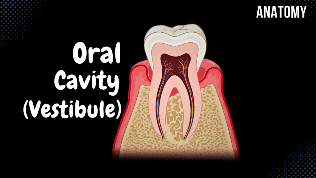
Oral Vestibule (Lips, Cheeks, Teeth, Gums) Official Links Instagram Youtube Jki-discord Notes & Illustrations Quizzes Summary & Transcript Notes ☆ Member Only Go to PDF Notes Illustrations ☆ Member Only Go to Illustrations 12345678910 Oral Vestibule – QUIZ Test your understanding with 10 random multiple-choice questions from the question bank. You're in the preview mode. Note: All elements work correctly on the front end. 1 / 10 The vestibule of the mouth lies between which two boundaries? A) Tongue and hard palate B) Hard palate and pharynx C) Lips/cheeks and teeth/gums D) Tongue and teeth The vestibule lies between the lips/cheeks and the teeth/gums. 2 / 10 What role does the gingival margin play in the oral vestibule? A) Elevates the soft palate B) Connects lips to the gums C) Facilitates mastication D) Protects the periodontal tissues It protects the underlying periodontal tissues from bacteria. 3 / 10 Which glands are responsible for saliva production within the lips? A) Buccinator muscle B) Labial glands C) Parotid duct D) Gingival papillae Labial glands produce saliva within the lips. 4 / 10 What tissue forms the inner lining of the oral vestibule? A) Tunica mucosa B) Buccinator fascia C) Stratified squamous epithelium D) Subcutaneous tissue The inner lining is composed of tunica mucosa. 5 / 10 What defines the gingival sulcus within the oral vestibule? A) Layer of connective tissue B) Protective margin C) Duct opening D) Groove between gum and tooth It is the groove between the gum and the tooth. 6 / 10 The oral vestibule is located between which two sets of structures? A) Palate and pharynx B) Lips/cheeks and tongue C) Tongue and teeth D) Lips/cheeks and teeth/gums It lies between the lips/cheeks and the teeth/gums. 7 / 10 Which layer contains the parotid duct within the cheeks? A) Submucosal connective tissue B) Subcutaneous layer C) Buccopharyngeal fascia D) Tunica mucosa The parotid duct is found within the buccopharyngeal fascia. 8 / 10 The frenulum labii superioris is associated with which structure? A) Cheeks B) Upper lip C) Tongue D) Lower lip It is associated with the upper lip, attaching it to the gums. 9 / 10 The parotid duct opens into the oral vestibule near which tooth? A) Second upper molar B) Canine C) First upper molar D) Third upper molar The parotid duct opens near the second upper molar tooth. 10 / 10 Which type of mucosa lines the oral vestibule? A) Tunica mucosa B) Gingival margin C) Connective tissue D) Submucosal layer Tunica mucosa lines the oral vestibule. Your score is The average score is 0% Description General Overview of the Digestive System This video covers the general structures of the digestive system, including the oral cavity, pharynx, oesophagus, stomach, small intestine, large intestine, and accessory digestive organs. 1. General Structures of the Digestive System: Main Digestive Tract: Oral Cavity Pharynx Oesophagus Stomach Small Intestine Large Intestine Accessory Digestive Organs: Teeth Tongue Salivary Glands Liver Pancreas Gallbladder 2. External Structures of the Mouth: Upper Lip (Labium Superior) Lower Lip (Labium Inferior) Oral Angle (Labial Commissure) Nasolabial Sulcus (Sulcus Nasolabialis) Philtrum Mentolabial Sulcus (Sulcus Mentolabialis) Oral Fissure (Rima Oris) 3. Division of the Oral Cavity: Oral Vestibule (Vestibulum Oris) External Borders: Lips and Cheeks Internal Borders: Teeth and Gums Oral Cavity Proper (Cavitas Oris Propria) 4. Anatomy of the Lips: Frenulum of the Upper Lip (Frenulum Labii Superioris) Frenulum of the Lower Lip (Frenulum Labii Inferioris) Labial Glands (Glandulae Labialis) 5. Anatomy of the Cheeks: Buccinator Muscle (Musculus Buccinator) Buccopharyngeal Fascia Buccal Fat Pad (Bichat’s Fat Pad) Layers of the Skin (Cutis) Tunica Mucosa Parotid Duct (Ductus Parotideus) Papilla of the Parotid Duct (Papillae Ductus Parotidei) 6. Anatomy of the Teeth: Basic Tooth Structures: Crown of the Tooth (Corona Dentis) Root of the Tooth (Radix Dentis) Neck of the Tooth (Cervix Dentis) Periodontium Gomphosis (Dentoalveolar Joint) Dental Pulp (Pulpa Dentis) Root Canal (Canalis Radicis Dentis) Apical Foramen (Foramen Apicis Dentis) Dentin (Dentinum) Enamel (Enamelum) Cement (Cementum) Types of Teeth: Milk Teeth (Dentes Decidui) – 20 teeth, appear between 6-24 months. Permanent Teeth (Dentes Permanentes) – 32 teeth. Wisdom Teeth (Dentes Serotinus) – Appear between 17-24 years. 7. Tooth Arrangement: European system: Teeth are divided into 4 quadrants. Each quadrant contains: 2 Incisors (Dentes Incisivi) 1 Canine (Dentes Canini) 2 Premolars (Dentes Premolares) 3 Molars (Dentes Molares) Total: 8 teeth per quadrant × 4 quadrants = 32 permanent teeth. Milk Teeth: 2 Incisors, 1 Canine, 2 Molars per quadrant. 8. Anatomy of the Gums (Gingiva): Alveolar Mucosa – Covers the root of the tooth. Gum Proper – Attached to the periosteum. Gingival Papillae (Papillae Gingivales) Gingival Margin (Margo Gingivalis) Gingival Sulcus (Sulcus Gingivalis) 9. Sources: Memorix Anatomy, 2nd Edition by Hudák Radovan, Kachlík David, and Volný Ondřej. Biorender. University notes and lectures. Transcript Introduction0:04What’s up, Meditay here, Let’s talk about the anatomy of the oral cavity.0:07Now since this is my first video of the digestive system, I wanna spend a quick minute giving you a0:12little overview of the whole digestive system. And to do that, we’ll use this chocolate0:16cheesecake to highlight al the structures it’s gonna go through within your digestive0:20system. The reason why I chose a cheesecake is because it most probably starts you salivating0:26because you probably wanna eat it. That implies that the Oral cavity is the first part of the0:30digestive system. After the oral cavity is the Pharynx. Then when you swallow the food,0:36it’s going to go through the esophagus, and then all the way down to your stomach.0:41After it’s been processed by the hydrochloric acid in the stomach, it’s then going to enter0:46the small intestine, which consists of the duodenum, then the jejunum, and then the0:50ileum. And after the Ileum, it’s going to enter the Large Intestine, which consists of the caecum0:56and the colon, and then the rectum. And by the time it gets to here,0:59this is how the cheesecake’s gonna look like. And the fuller the rectum gets,1:03the higher you feel the urge to poop. So those are all the structures
Lungs
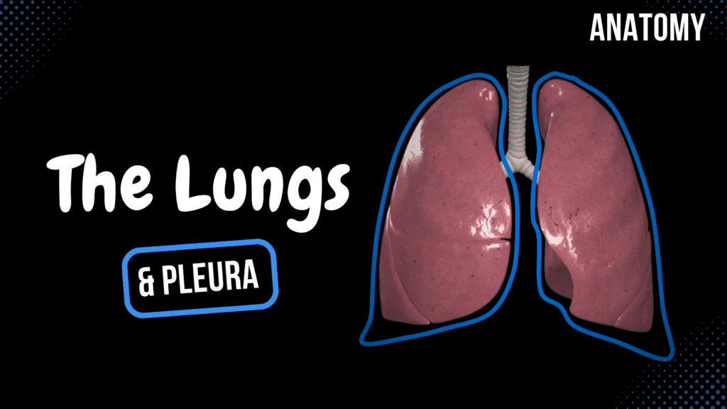
Lungs (Function, Parts, Pleura & Recesses) Official Links Instagram Youtube Jki-discord Notes & Illustrations Quizzes Summary & Transcript Notes ☆ Members Only Go to PDF Notes Illustrations ☆ Members Only Go to Illustrations 12345678910 Lungs – QUIZ Test your understanding with 10 random multiple-choice questions from the question bank. You're in the preview mode. Note: All elements work correctly on the front end. 1 / 10 Which pleural recess is located between the diaphragm and ribs? A) Vertebromediastinal recess B) Phrenicomediastinal recess C) Costomediastinal recess D) Costodiaphragmatic recess The costodiaphragmatic recess is found between the diaphragm and ribs. 2 / 10 What is the largest lobe of the right lung? A) Middle lobe B) Superior lobe C) Inferior lobe D) Lingula The inferior lobe of the right lung is the largest due to its position and size. 3 / 10 Which lobe of the right lung contains the lateral and medial segments? A) Inferior lobe B) Middle lobe C) Superior lobe D) Superior lingular lobe The middle lobe of the right lung contains the lateral and medial segments. 4 / 10 What is the highest structure in the hilum of the right lung? A) Pulmonary vein B) Pulmonary artery C) Pulmonary ligament D) Bronchus The bronchus is the highest structure in the hilum of the right lung. 5 / 10 What is the apex of the lung (Apex pulmonis)? A) The hilum of the lung B) The apex of the lung C) The diaphragmatic surface D) The base of the lung The apex of the lung is the superior, pointed part that extends into the cervical pleura above the first rib. 6 / 10 Which structure separates the visceral pleura from the parietal pleura? A) Cardiac notch B) Costal pleura C) Apex D) Pleural cavity The pleural cavity separates the visceral pleura from the parietal pleura. 7 / 10 Which structure forms the “tongue-like” projection on the superior lobe of the left lung? A) Lingula B) Inferior margin C) Apex of the lung D) Mediastinal surface The lingula of the left lung is a projection analogous to the middle lobe of the right lung. 8 / 10 Which lobe contains the apicoposterior segment (S1+2) in the left lung? A) Superior lobe B) Middle lobe C) Inferior lobe D) Lingula The apicoposterior segment (S1+2) is part of the superior lobe of the left lung. 9 / 10 How many segments does the left lung typically have? A) 6 B) 8-Sep C) 12 D) 10 The left lung typically has 8-9 bronchopulmonary segments. 10 / 10 What is the role of the pulmonary veins? A) Supply oxygen to lung tissues B) Return oxygenated blood to the heart C) Carry deoxygenated blood to the lungs D) Drain lymph from the lungs Pulmonary veins carry oxygenated blood from the lungs to the left atrium of the heart. Your score is The average score is 0% Description Functions and Anatomy of the Lungs This video covers the functions, parts, segments, and pleura of the lungs, as well as the surrounding mediastinum. 1. Functions of the Lungs: Essential organ of respiration. Facilitates gas exchange (O2 uptake and CO2 removal). Muscles of Inspiration: Sternocleidomastoideus. External Intercostal Muscles. Diaphragm. Muscles of Expiration: Internal Intercostal Muscles. Abdominal Muscles. Functional Unit of the Lungs: Alveolar Sacs (Sacculi Alveolares): Site of gas exchange. 2. Parts and Surfaces of the Lungs: Apex of Lung (Apex Pulmonis) – superior-most part. Base of Lung (Basis Pulmonis) – rests on the diaphragm. Costal Surface (Facies Costalis) – faces the ribs. Diaphragmatic Surface (Facies Diaphragmatica) – contacts the diaphragm. Mediastinal Surface (Facies Mediastinalis) – faces the mediastinum. Hilum of Lung (Hilum Pulmonis) – entry point for bronchi, vessels, and nerves. Pulmonary Ligament (Ligamentum Pulmonale) – stabilizes lung position. Root of Lung (Radix Pulmonis) – contains pulmonary vessels and bronchi. Hilum Orientation Mnemonic: Right Lung: BRIGHT IS RIGHT – Highest structure is the Bronchus, followed by Pulmonary Arteries, then Pulmonary Veins. Left Lung: Highest structure is the Pulmonary Artery, followed by the Bronchus, then Pulmonary Veins. Margins of the Lungs: Inferior Margin (Margo Inferior): Separates costal and diaphragmatic surfaces. Anterior Margin (Margo Anterior): Forms a distinct border. Cardiac Notch (Incisura Cardiaca Pulmonis Sinistri): Indentation in the left lung. Lingula of Left Lung (Lingula Pulmonis): Tongue-like projection of the left superior lobe. 3. Pulmonary Lobes: Oblique Fissure (Fissura Obliqua) – divides superior and inferior lobes. Horizontal Fissure (Fissura Horizontalis) – divides right lung into three lobes. Lobes of the Lungs: Right Lung: 3 lobes – Superior, Middle, Inferior. Left Lung: 2 lobes – Superior, Inferior. 4. Pulmonary Segments: Right Lung (10 Segments): Superior Lobe: Apical, Posterior, Anterior. Middle Lobe: Lateral, Medial. Inferior Lobe: Superior, Basal Medial, Basal Anterior, Basal Lateral, Basal Posterior. Left Lung (8-9 Segments): Superior Lobe: Apicoposterior, Anterior, Superior Lingular, Inferior Lingular. Inferior Lobe: Superior, Basal Anterior, Basal Lateral, Basal Posterior, (±Basal Medial). 5. Pleura of the Lungs: Visceral Pleura (Pleura Visceralis): Covers the lungs directly. Parietal Pleura (Pleura Parietalis): Costal Part. Diaphragmatic Part. Mediastinal Part. Pleural Part. Pleural Cavity (Cavitas Pleuralis): Space between visceral and parietal pleura. Pleural Recesses: Costodiaphragmatic Recess (Recessus Costodiaphragmaticus). Costomediastinal Recess (Recessus Costomediastinalis). Vertebromediastinal Recess (Recessus Vertebromediastinalis). Phrenicomediastinal Recess (Recessus Phrenicomediastinalis). 6. Mediastinum: Superior Mediastinum (Mediastinum Superius): Above the heart. Inferior Mediastinum (Mediastinum Inferius): Divided into: Anterior Mediastinum. Middle Mediastinum. Posterior Mediastinum. 7. Sources: Memorix Anatomy, 2nd Edition by Hudák Radovan, Kachlík David, and Volný Ondřej. Complete Anatomy by 3D4Medical. Biorender. University notes and lectures. Snell’s Clinical Anatomy, 10th Edition. Transcript Introduction0:03Hey, what’s up, Meditay here. Let’s talk about the anatomy of the respiratory system.0:07In this segment, we will be talking about the anatomy of the Lungs and the pleura. Alright, so0:12the respiratory system consists of all the organs involved in breathing. These are the Nasal Cavity,0:17Pharynx, Larynx, Trachea, Bronchi, and the Lungs.0:21Now let’s look detailed into the anatomy of the Lungs.0:25So In this video, we’re first gonna go through the functions of the lungs.0:29Then we’re gonna go through the parts, and surfaces, and margins of the lungs.0:34After that, we’ll
Trachea, Bronchial Tree and Alveolar Tree
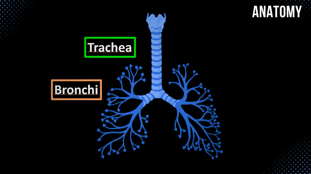
Trachea, Bronchial Tree and Alveolar Tree (Parts, Structures and Walls) Official Links Instagram Youtube Jki-discord Notes & Illustrations Quizzes Summary & Transcript Notes ☆ Members Only Go to PDF Notes Illustrations ☆ Members Only Go to Illustrations 12345678910 Trachea and Bronchi – QUIZ Test your understanding with 10 random multiple-choice questions from the question bank. You're in the preview mode. Note: All elements work correctly on the front end. 1 / 10 What structural feature differentiates the left and right main bronchi? A) Presence of smooth muscle B) Absence of cartilage C) Orientation and diameter D) Length of the bronchi The right bronchus is shorter, wider, and more vertical. 2 / 10 How many lobar bronchi are present in the left lung? A) 4 B) 3 C) 2 D) 1 There are two lobar bronchi in the left lung: superior and inferior. 3 / 10 What structural layer is unique to the bronchial wall but absent in the trachea? A) Tela submucosa B) Fibromusculocartilaginous layer C) Tunica adventitia D) Smooth muscle layer The fibromusculocartilaginous layer is unique to the bronchial wall. 4 / 10 Which part of the bronchial wall contains cartilage plates? A) Fibromusculocartilaginous layer B) Mucosa C) Adventitia D) Submucosa The fibromusculocartilaginous layer contains cartilage plates in the bronchi. 5 / 10 What type of cartilage makes up the tracheal rings? A) Fibrocartilage B) Articular cartilage C) Hyaline cartilage D) Elastic cartilage Hyaline cartilage forms the tracheal rings, providing structural support. 6 / 10 Where does the trachea bifurcate into the primary bronchi? A) T2 B) T4-T5 C) T6 D) T8 The trachea bifurcates at the level of T4-T5 (sternal angle). 7 / 10 Which epithelium lines the trachea? A) Transitional B) Pseudostratified columnar C) Simple cuboidal D) Simple squamous The trachea is lined by pseudostratified ciliated columnar epithelium. 8 / 10 What is the approximate length of the trachea in adults? A) 9-15 cm B) 5-7 cm C) 7-9 cm D) 15-20 cm The trachea is about 9-15 cm long in adults, depending on individual anatomy. 9 / 10 At which vertebral levels does the trachea typically extend? A) T1-T6 B) C6-T4 C) T4-T7 D) C7-T5 The trachea extends from C6-C7 to T4-T5. 10 / 10 What type of epithelium lines the trachea? A) Stratified squamous B) Pseudostratified columnar C) Simple cuboidal D) Transitional The trachea is lined by pseudostratified ciliated columnar epithelium. Your score is The average score is 0% Description Trachea and Bronchial Tree: Anatomy and Structure This video covers the anatomy, layers, and function of the trachea, bronchi, and alveolar tree, along with a comparison of their structural differences. 1. Trachea (Windpipe): Divides into bronchi at the level of T4-T5. Length: 9-15 cm. Diameter: 2-2.5 cm. Skeletopy: Extends from C6-C7 to T4-T5. Parts of the Trachea: Tracheal Cartilages (Cartilagines Tracheales): C-shaped cartilages maintaining airway patency. Annular Ligaments (Ligamenta Anularia): Connect tracheal rings. Membranous Part (Paries Membranaceus): Posterior part containing smooth muscle. Tracheal Bifurcation (Bifurcatio Tracheales): Division into right and left main bronchi. Carina of Trachea (Carina Tracheae): Internal ridge at the bifurcation, directs airflow. Layers of the Trachea: Tunica Mucosa: Contains tracheal lymphoid nodules and tracheal glands (Noduli Lymphoidei Tracheales and Glandulae Tracheales). Tela Submucosa: Supports the mucosal layer. Tunica Adventitia: Outermost connective tissue layer. 2. Bronchi: Main Bronchi (Bronchi Principales): Right Main Bronchus (Bronchus Principalis Dexter): Shorter, wider, and more vertical. Left Main Bronchus (Bronchus Principalis Sinister): Longer, narrower, and more oblique. Enter the lungs through the hilum of the lungs (hilum pulmonis). Lobar Bronchi (Bronchi Lobares): Right Lung: 3 Lobar Bronchi – Superior, Middle, Inferior. Left Lung: 2 Lobar Bronchi – Superior, Inferior. Segmental Bronchi (Bronchi Segmentales): Further divisions supplying bronchopulmonary segments. Foreign Body Aspiration: Foreign objects are more likely to fall into the right main bronchus due to its vertical orientation. 3. Bronchial Tree (Arbor Bronchialis): Left & Right Principal Bronchi: Enter through the pulmonary hilum. Lobar Bronchi (Bronchi Lobares): Secondary bronchi. Segmental Bronchi (Bronchi Segmentales): Tertiary bronchi. Terminal Bronchi (Bronchi Terminales): Smallest non-respiratory bronchi. Epithelium Changes: Respiratory epithelium transitions to cuboidal epithelium in smaller bronchioles. 4. Alveolar Tree (Arbor Alveolaris): Terminal Bronchioles (Bronchi Terminales): Last part of conducting airways. Respiratory Bronchioles: Primary, Secondary, and Tertiary Respiratory Bronchioles. Alveolar Ducts (Ductus Alveolares): Transport air to alveolar sacs. Alveolar Sacs (Sacculi Alveolares): Clusters of alveoli for gas exchange. 5. Comparison of Walls: Trachea, Bronchus, and Bronchiole Tracheal Wall: Tunica Mucosa: Lined with pseudostratified ciliated epithelium. Tela Submucosa: Contains mucus-secreting glands. Tracheal Cartilage: C-shaped rings. Smooth Muscles (Membranous Part): Adjusts airway diameter. Tunica Adventitia: External connective tissue layer. Bronchial Wall: Tunica Mucosa: Lined with pseudostratified ciliated epithelium. Tela Submucosa: Contains mucus-secreting glands. Fibromusculocartilaginous Layer (Tunica Fibromusculocartilaginea): Cartilage plates replace C-shaped rings. Tunica Adventitia: External connective tissue layer. Bronchiolar Wall: Tunica Mucosa: Lined with simple columnar or cuboidal epithelium. Tela Submucosa: Lacks cartilage. Tunica Muscularis: Well-developed smooth muscle layer. Tunica Adventitia: External connective tissue layer. 6. Sources: Memorix Anatomy, 2nd Edition by Hudák Radovan, Kachlík David, and Volný Ondřej. Biorender. University notes and lectures. Transcript Introduction0:03Hey, What’s up. Meditay here. Let’s talk about the anatomy of the respiratory system.0:08In this segment, we will be talking about the anatomy of the Trachea and the Bronchi.0:12Alright, so the respiratory system consists of all the organs involved in breathing.0:17These are the Nose and the nasal cavity, Pharynx, Larynx, Trachea, Bronchi and the0:22Lungs. In our last two videos, we covered the anatomy of the nasal cavity and the Larynx.0:28Now let’s do the anatomy of the Trachea and Bronchi.0:32So In this video, we’re going to cover the anatomy of the Trachea, which includes the0:36parts that make up the trachea, and the layers of the tracheal wall. Then we’re gonna cover0:41the bronchial tree and the Alveolar tree and then we’re going to compare the Layers of the Tracheal,0:47Bronchial, and Bronchiolar wall to really understand the anatomical differences0:52of structures as you get closer to the lungs. Alright, so here we see the anterior view ofTopography of the Trachea0:57the chest. The Larynx is up here, and inferior to it, you’ll see
Larynx
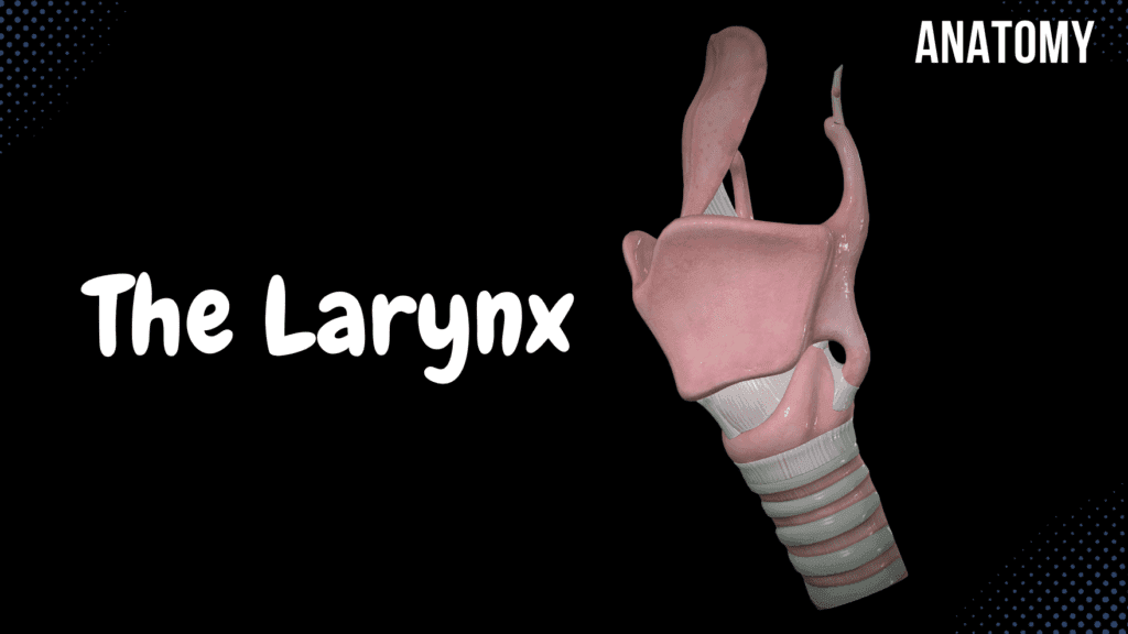
Larynx (Voice Box) – Cartilage, Ligaments, Joints, Wall, Cavity Official Links Instagram Youtube Jki-discord Notes & Illustrations Quizzes Summary & Transcript Notes ☆ Members Only Go to PDF Notes Illustrations ☆ Members Only Go to Illustrations 12345678910 Larynx – QUIZ Test your understanding with 10 random multiple-choice questions from the question bank. You're in the preview mode. Note: All elements work correctly on the front end. 1 / 10 Which structure separates the laryngeal vestibule from the vocal folds? A) Aryepiglottic fold B) Quadrangular membrane C) Vocal folds D) Vestibular folds The vestibular folds (false vocal cords) separate the laryngeal vestibule from the vocal folds. 2 / 10 The superior horn of the thyroid cartilage is connected to which structure? A) Hyoid bone B) Arytenoid cartilage C) Epiglottis D) Cricoid cartilage The superior horn of the thyroid cartilage is connected to the hyoid bone via the thyrohyoid ligament. 3 / 10 What part of the cricoid cartilage articulates with the inferior horn of the thyroid cartilage? A) Articular facet B) Posterior ridge C) Superior lamina D) Arch The articular facet on the cricoid cartilage articulates with the inferior horn of the thyroid cartilage. 4 / 10 What is the main blood supply to the mucosa of the larynx above the vocal folds? A) Recurrent laryngeal artery B) Superior laryngeal artery C) Inferior thyroid artery D) Cricothyroid artery The superior laryngeal artery supplies the mucosa above the vocal folds. 5 / 10 What is the role of the quadrangular membrane? A) Supports aryepiglottic folds B) Anchors vocal cords C) Protects the airway D) Forms the vestibular ligament The quadrangular membrane forms the vestibular ligament, which contributes to the vestibular fold. 6 / 10 Which cartilage provides structural support to the aryepiglottic folds? A) Cuneiform cartilage B) Corniculate cartilage C) Thyroid cartilage D) Arytenoid cartilage The cuneiform cartilage provides structural support to the aryepiglottic folds. 7 / 10 Which muscle attaches to the muscular process of the arytenoid cartilage? A) Posterior cricoarytenoid B) Cricothyroid muscle C) Aryepiglottic muscle D) Thyroarytenoid muscle The posterior cricoarytenoid muscle attaches to the muscular process of the arytenoid cartilage. 8 / 10 The thyroid cartilage laminae meet at the midline to form which structure? A) Laryngeal prominence B) Thyroid tubercle C) Epiglottic fold D) Cricothyroid ligament The thyroid cartilage laminae meet at the midline to form the laryngeal prominence (Adam’s apple). 9 / 10 Which muscle is responsible for abduction of the vocal cords? A) Lateral cricoarytenoid B) Posterior cricoarytenoid C) Thyroarytenoid muscle D) Cricothyroid muscle The posterior cricoarytenoid muscle abducts the vocal cords by rotating the arytenoid cartilages. 10 / 10 Which cartilage is the only complete ring in the larynx? A) Arytenoid cartilage B) Cuneiform cartilage C) Cricoid cartilage D) Thyroid cartilage The cricoid cartilage forms a complete ring and is located below the thyroid cartilage. Your score is The average score is 0% Description Larynx: Anatomy, Cartilage, Ligaments, and Function This video covers the anatomy of the larynx, its cartilage structures, ligaments, and functions related to phonation and respiration. 1. Larynx Orientation: Located between the hyoid bone and the trachea. Situated in front of the esophagus. Skeletopy: Extends from C4-C5 to C6-C7. Functions of the Larynx: Acts as an air passage. Produces sound through phonation. 2. Cartilage of the Larynx: Unpaired Cartilages (3): Epiglottis. Thyroid Cartilage. Cricoid Cartilage. Paired Cartilages (3): Arytenoid Cartilage. Corniculate Cartilage. Cuneiform Cartilage. 3. Details of Laryngeal Cartilages: Thyroid Cartilage (Cartilago Thyroidea): Right and left laminae (Lamina Dextra and Lamina Sinistra). Laryngeal Prominence (Adam’s Apple) (Prominentia Laryngea). Superior and inferior horns (Cornua Superiora and Cornua Inferiora). Cricoid Cartilage (Cartilago Cricoidea): Arch (Arcus). Plate (Lamina). Arytenoid and thyroid articular surfaces (Facies Articularis Arytenoidea & Facies Articularis Thyroidea). Epiglottis: Behind thyroid cartilage and hyoid bone. Prevents food from entering the airway during swallowing. Arytenoid Cartilage (Cartilago Arytenoidea): Triangular in shape with apex and base. Vocal Process: Anterior process that attaches to the vocal cords. Muscular Process: Posterior process for muscle attachment. Corniculate Cartilage (Cartilago Corniculata): Sits on top of the arytenoid cartilage. Serves as an attachment for muscles. Cuneiform Cartilage (Cartilago Cuneiforme): Located in the aryepiglottic fold. Forms the cuneiform tubercle. 4. Laryngeal Ligaments & Joints: Connections in the Larynx (Juncturae Laryngis): Continuous Articulation (Synarthroses): Cartilaginous (Synchondroses): Between corniculate cartilage and apex of arytenoid cartilage. Fibrous (Syndesmoses): Thyrohyoid Membrane (Membrana Thyrohyoidea). Cricothyroid Membrane (Membrana Cricothyroidea). Cricotracheal Ligament (Ligamentum Cricotracheale). Thyroepiglottic Ligament (Ligamentum Thyroepiglottica). Hyoepiglottic Ligament (Ligamentum Hyoepiglotticum). Discontinuous Articulation (Synovial): Cricothyroid Articulation (Articulatio Cricothyroidea). Cricoarytenoid Articulation (Articulatio Cricoarytenoidea). 5. Laryngeal Wall Layers: Tunica Mucosa: Vestibular Fold: Lined by respiratory epithelium. Vocal Fold: Lined by stratified squamous epithelium. Contains laryngeal glands and lymph nodules. Tela Submucosa: Fibroelastic Membrane (Membrana Fibroelastica Laryngis): Quadrangular Membrane: Forms the vestibular ligament. Lateral Cricothyroid Ligament (Conus Elasticus): Free margin forms the vocal ligament. Muscles of the Larynx: Controls Laryngeal Inlet: Opens and narrows the entrance. Controls Rima Glottidis: Adjusts airflow. Acts on the Vocal Cord: Cricothyroid Muscle: Tenses the vocal cord. Vocalis Muscle: Decreases tension in the vocal cord. Tunica Adventitia: Dense connective tissue for structural support. 6. Laryngeal Cavity: Laryngeal Vestibule (Vestibulum Laryngis). Glottis: Rima Glottidis: Opening between the vocal folds. Anterior 3/5: Intermembranous part. Posterior 2/5: Intercartilaginous part. Laryngeal Ventricles (Ventriculus Laryngis). Infraglottic Cavity. 7. Sources: Memorix Anatomy, 2nd Edition by Hudák Radovan, Kachlík David, and Volný Ondřej. Biorender. University notes and lectures. Transcript Introduction0:00Hey what’s up.0:02Meditay here.0:03Let’s talk about the anatomy of the respiratory system.0:08In this segment, we will be talking about the anatomy of the Larynx.0:12Alright, so the respiratory system consist of all the organs involved in breathing.0:17These are the Nose, Pharynx, Larynx, Trachea, Bronchi and the Lungs.0:23In our last video, we covered the anatomy of the nasal cavity.0:27Now let’s do the anatomy of the Larynx.0:29So in this video, we’ll start with orientation by looking at the anterior and posterior view0:35of the Larynx.0:36Then we’ll talk briefly about what cartilages make up the larynx.0:40As well as the ligaments and joints that hold the whole thing together.0:44Then we’ll do the walls of the Larynx and
Pharynx Anatomy
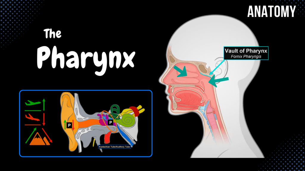
Pharynx Anatomy (Parts, Layers, Muscles) Official Links Instagram Youtube Jki-discord Notes & Illustrations Quizzes Summary & Transcript Notes ☆ Members Only Go to PDF Notes Illustrations ☆ Members Only Go to Illustrations 12345678910 Pharynx – QUIZ Test your understanding with 10 random multiple-choice questions from the question bank. You're in the preview mode. Note: All elements work correctly on the front end. 1 / 10 What structure connects the nasopharynx with the middle ear? A) Torus tubarius B) Auditory tube C) Oropharyngeal isthmus D) Pharyngeal recess The auditory tube (Eustachian tube) connects the nasopharynx with the middle ear for pressure equalization and drainage. 2 / 10 Which nerve provides motor innervation to the stylopharyngeus muscle? A) Hypoglossal nerve B) Vagus nerve C) Glossopharyngeal nerve D) Facial nerve The glossopharyngeal nerve (CN IX) provides motor innervation to the stylopharyngeus muscle. 3 / 10 At which vertebral level does the laryngopharynx transition to the esophagus? A) C3 B) C4 C) C1 D) C6 The laryngopharynx continues into the esophagus at the level of the C6 vertebra. 4 / 10 Which muscle is the highest of the pharyngeal constrictors? A) Superior constrictor B) Middle constrictor C) Palatopharyngeus D) Inferior constrictor The superior pharyngeal constrictor is the highest of the constrictor muscles and narrows the pharyngeal space during swallowing. 5 / 10 What type of epithelium lines the laryngopharynx? A) Transitional epithelium B) Pseudostratified ciliated C) Simple columnar D) Stratified squamous The laryngopharynx is lined with stratified squamous non-keratinized epithelium to protect it from mechanical damage during swallowing. 6 / 10 What is the role of the piriform recess in the laryngopharynx? A) Guides food to esophagus B) Opens the auditory tube C) Amplifies sound D) Anchors the vocal cords The piriform recess channels food and liquids away from the airway and toward the esophagus. 7 / 10 Which tonsil is located near the auditory tube opening? A) Lingual tonsil B) Pharyngeal tonsil C) Tubal tonsil D) Palatine tonsil The tubal tonsil is located near the auditory tube opening in the nasopharynx. 8 / 10 What is the function of the laryngeal inlet? A) Equalize pressure B) Protect airway C) Resonate sound D) Propel food to esophagus The laryngeal inlet allows air to pass into the larynx and protects the airway during swallowing. 9 / 10 What type of epithelium is found in the nasopharynx? A) Pseudostratified ciliated B) Columnar epithelium C) Stratified squamous D) Transitional epithelium The nasopharynx is lined with pseudostratified columnar ciliated epithelium with goblet cells for respiratory function. 10 / 10 What is the clinical significance of the pharyngeal recess? A) Anchors pharyngeal wall B) Drains mucus C) Site for tumor growth D) Site of tonsil formation The pharyngeal recess is a potential site for tumor growth or infection due to its location in the nasopharynx near the auditory tube opening. Your score is The average score is 0% Description Pharynx: Anatomy, Layers, and Muscles This video covers the parts of the pharynx, the layers of the pharyngeal wall, and the muscles involved in swallowing and phonation. 1. Pharynx Overview: Muscular tube measuring 12 to 15 cm in length. Located behind the nasal and oral cavities, connecting to the esophagus. Divisions: Nasopharynx (Pars Nasalis) Oropharynx (Pars Oralis) Laryngopharynx (Pars Laryngis) 2. Nasopharynx (Pars Nasalis): Located at the level of C1-C2. Structures: Vault of Pharynx (Fornix Pharyngis). Attachment Points: Pharyngeal Tubercle of the Occipital Bone (Tuberculum Pharyngeum). Petrooccipital Fissure (Petrooccipital Synchondrosis). Inferior Surface of Petrous Part (Temporal Bone). Medial Lamina of Pterygoid Process. Openings: Choana (Internal Nose). Auditory Tube (Tuba Auditiva). Pharyngeal Opening of the Auditory Tube (Ostium Pharyngeum Tubae Auditivae). Cushion of the Auditory Canal (Torus Tubarius). Pharyngeal Recess (Recessus Pharyngeus). Tonsils: Pharyngeal Tonsils / Adenoids (Tonsilla Pharyngealis). Tubal Tonsils (Tonsilla Tubaria). Auditory Tube Overview: Connects the nasopharynx to the middle ear. Function: Equalizes pressure in the middle ear. Drains fluids from the middle ear. 3. Oropharynx (Pars Oralis): Located at the level of C3-C4. Bordered by the soft palate superiorly and the epiglottis inferiorly. Communicates with the oral cavity via the oropharyngeal isthmus (Isthmus Faucium). 4. Laryngopharynx (Pars Laryngis): Located at the level of C5-C6. Continues into the larynx through the laryngeal inlet (Aditus Laryngis). Contains the piriform fossa (Recessus Piriformis), an important structure in swallowing. 5. Layers of the Pharyngeal Wall: Tunica Mucosa: Lined by different epithelium based on location: Nasopharynx: Respiratory epithelium (pseudostratified columnar with cilia and goblet cells). Oropharynx & Laryngopharynx: Stratified squamous non-keratinized epithelium. Tela Submucosa: Contains connective tissue, blood vessels, lymphatic vessels, and glands. Tunica Muscularis: Composed of two muscle layers for peristalsis. Internal Circular Muscle Layer (Stratum Circulare). Outer Longitudinal Muscle Layer (Stratum Longitudinale). Tunica Adventitia: Connective tissue covering the pharynx externally. 6. Muscles of the Pharynx: External Pharyngeal Muscles (Pharyngeal Constrictors) Superior Pharyngeal Constrictor (Musculus Constrictor Pharyngis Superior). Middle Pharyngeal Constrictor (Musculus Constrictor Pharyngis Medius). Inferior Pharyngeal Constrictor (Musculus Constrictor Pharyngis Inferior). Internal Pharyngeal Muscles (Pharyngeal Elevators) Stylopharyngeus Muscle (Musculus Stylopharyngeus). Palatopharyngeus Muscle (Musculus Palatopharyngeus). Salpingopharyngeus Muscle (Musculus Salpingopharyngeus). 7. Clinical Relevance: Pharyngeal Tonsil Enlargement (Adenoid Hypertrophy): Can obstruct nasal airflow and cause sleep apnea. Auditory Tube Dysfunction: Leads to middle ear infections (otitis media). Piriform Recess: Common site where food or foreign objects get lodged. 8. Sources: Memorix Anatomy, 2nd Edition by Hudák Radovan, Kachlík David, and Volný Ondřej. Biorender. University notes and lectures. Transcript Introduction0:03What’s up.0:04Meditay here, and in this video, we’re gonna go through the anatomy of the Pharynx.0:09So in the last video, we went through the anatomy of the Oral Cavity.0:12Now the step after the oral cavity is the Pharynx, as you see here.0:17So in this video, we’re first going to look at the parts of the Pharynx.0:20So we’re gonna go detailed into the anatomical structures associated with the Nasopharynx,0:25the Oropharynx, and the Laryngopharynx.0:28After that, we’ll cut the Pharynx and look at the layers of the pharyngeal wall.0:33And then we’re gonna go through the muscles that act on the Pharynx.Parts of the Pharynx0:37Now, let’s start by holding the Pharynx and pulling it out.0:41You’ll see a kind of
Nasal Anatomy
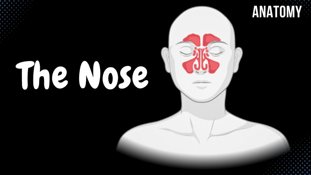
Nasal Anatomy (Cartilage, Nasal Cavity, Sinuses, Meatuses, Nasal Mucosa) Official Links Instagram Youtube Jki-discord Notes & Illustrations Quizzes Summary & Transcript Notes ☆ Member Only Go to PDF Notes Illustrations ☆ Member Only Go to Illustrations 12345678910 Nasal Anatomy – QUIZ Test your understanding with 10 random multiple-choice questions from the question bank. You're in the preview mode. Note: All elements work correctly on the front end. 1 / 10 What is the function of the choanae? A) Support nasal cartilage B) Drain sinuses C) Separate nasal chambers D) Connect to nasopharynx The choanae connect the nasal cavity to the nasopharynx, allowing airflow between these structures. 2 / 10 What is the function of the nasal conchae (turbinates)? A) To produce mucus B) To support olfactory nerves C) To humidify the air D) To anchor nasal cartilage The conchae create air turbulence in the nasal cavity, increasing contact with mucosa for warming, humidifying, and filtering the air. 3 / 10 The nasal vestibule is lined by which type of epithelium? A) Olfactory epithelium B) Skin with vibrissae C) Respiratory epithelium D) Pseudostratified epithelium The nasal vestibule is lined with skin containing vibrissae (coarse hairs) to trap debris from entering the respiratory tract. 4 / 10 Which sinus is most commonly affected in sinusitis? A) Ethmoidal sinus B) Sphenoid sinus C) Frontal sinus D) Maxillary sinus The maxillary sinus is most frequently affected in sinusitis due to its drainage pathway into the middle meatus. 5 / 10 The superior meatus lies directly below which nasal structure? A) Middle nasal concha B) Inferior nasal concha C) Superior nasal concha D) Nasal septum The superior meatus lies directly below the superior nasal concha (turbinate). 6 / 10 Which nerve provides general sensory innervation to the nasal cavity? A) Maxillary nerve (CN V2) B) Glossopharyngeal nerve (CN IX) C) Olfactory nerve (CN I) D) Facial nerve (CN VII) The maxillary nerve (CN V2), a branch of the trigeminal nerve, supplies general sensory innervation to the nasal cavity’s lower and middle regions. 7 / 10 Which lymph nodes receive drainage from the nasal cavity? A) Preauricular lymph nodes B) Submandibular lymph nodes C) Axillary lymph nodes D) Popliteal lymph nodes The submandibular lymph nodes primarily drain the nasal cavity, along with contributions to the retropharyngeal and deep cervical nodes. 8 / 10 What is the largest paranasal sinus? A) Sphenoid sinus B) Maxillary sinus C) Frontal sinus D) Ethmoidal sinus The maxillary sinus is the largest of the paranasal sinuses and drains into the middle meatus. 9 / 10 Which artery is the primary supplier to the posterior nasal septum? A) Greater palatine artery B) Superior labial artery C) Anterior ethmoidal artery D) Sphenopalatine artery The sphenopalatine artery, a branch of the maxillary artery, supplies the posterior septum and lateral wall of the nasal cavity. 10 / 10 Which structure marks the boundary between the nasal vestibule and the nasal cavity proper? A) Limen nasi B) Middle meatus C) Superior nasal concha D) Choanae The limen nasi marks the boundary where the epithelial lining transitions from skin to mucous membrane. Your score is The average score is 0% Description External and Internal Anatomy of the Nose This video covers the external structures of the nose, the anatomy of the nasal cavity, the functions and openings of sinuses, and the mucosa lining of the nasal cavity. We will also discuss the clinical relevance of conditions such as sinusitis. 1. External Structures of the Nose: Root of the Nose (Radix Nasi) – Uppermost part near the forehead. Dorsum of Nose (Dorsum Nasi) – Extends from root to apex. Apex of Nose (Apex Nasi) – Tip of the nose. Ala of Nose / Wing of Nose (Ala Nasi) – Lateral rounded portion surrounding the nostrils. Nostrils (Nares) – Openings for airflow into the nasal cavity. 2. Nasal Cartilages: Nasal Bones (Os Nasale Dextrum/Sinistrum). Lateral Nasal Cartilages (Cartilago Nasi Lateralis). Accessory Nasal Cartilage (Cartilagines Nasales Accessoriae). Major Alar Cartilage (Cartilagines Alares Majores). Minor Alar Cartilage (Cartilagines Alares Minores). Septal Nasal Cartilage (Cartilago Septi Nasi). Alar Fibrofatty Tissue (Tela Fibroadiposa Alaris). Bony Septal Part (Pars Ossea). 3. Anatomy of the Nasal Cavity: Located above the palate and in front of the pharynx. Divisions: Nasal Vestibule (Vestibulum Nasi). Nasal Cavity Proper (Cavitas Nasi Proprium). Limen Nasi – Boundary between vestibule and nasal cavity proper. Nasal Cavity Proper: Olfactory Part (Pars Olfactoria) – Specialized for smell. Respiratory Part (Pars Respiratoria) – Warms, humidifies, and filters air. Olfactory Structures: Olfactory Tract (Tractus Olfactorius). Olfactory Bulb (Bulbus Olfactorius). Olfactory Nerves (Fila Olfactoria). Respiratory Structures: Superior Conchae (Superior Turbinate). Middle Conchae (Middle Turbinate). Inferior Conchae (Inferior Turbinate). Meatuses: Superior Meatus Middle Meatus Inferior Meatus Spheno-Ethmoidal Recess 4. Sinuses and Meatuses: Sphenoid Sinus: Opens into the sphenoethmoidal recess. Frontal Sinus: Opens into the middle meatus. Ethmoidal Sinus: Anterior and Middle Ethmoidal Air Cells → Middle Meatus. Posterior Ethmoidal Air Cells → Superior Meatus. Maxillary Sinus: Opens into the middle meatus. Nasolacrimal Duct: Opens into the inferior meatus. Functions of Sinuses: Decrease relative mass of the skull. Enhance voice resonance. Humidify and warm inhaled air. Produce mucus to prevent nasal dryness. 5. Nasal Mucosa: Olfactory Mucosa: Olfactory Epithelium – Detects odors. Sustentacular Cells – Support cells; damaged in COVID-19, leading to anosmia. Basal Cells – Stem cells for regeneration. Bowman’s Glands – Produce mucus in the tela submucosa. Respiratory Mucosa: Epithelium: Pseudostratified columnar with cilia. Goblet Cells: Produce mucus. Tela Submucosa: Contains mixed mucous glands. 6. Clinical Relevance: Sinusitis Definition: Inflammation of the sinuses due to mucus accumulation and blockage. Causes: Common cold. Allergic rhinitis. Nasal polyps. Deviated nasal septum. Sources: Memorix Anatomy, 2nd Edition by Hudák Radovan, Kachlík David, and Volný Ondřej. Complete Anatomy by 3D4Medical. Biorender. University notes and lectures. Transcript Introduction0:01Hey what’s up, meditay here.0:04Let’s talk about the anatomy of the respiratory system.0:07In this segment, we will be talking about the Nasal anatomy.0:11Alright, so the respiratory system consist of all the organs involved in breathing.0:17These are the Nose, Pharynx, Larynx, Trachea, Bronchi and
Ureter, Urinary Bladder & Urethra

Ureter, Urinary Bladder & Urethra (Structures & Walls) Official Links Instagram Youtube Jki-discord Notes & Illustrations Quizzes Summary & Transcript Notes ☆ Member Only Go to PDF Notes Illustrations ☆ Member Only Go to Illustrations 12345678910 Ureter Urinary Bladder and Urethra – QUIZ Test your understanding with 10 random multiple-choice questions from the question bank. You're in the preview mode. Note: All elements work correctly on the front end. 1 / 10 Which muscle layer forms the detrusor muscle in the urinary bladder? A) Tela submucosa B) Tunica adventitia C) Tunica mucosa D) Tunica muscularis The tunica muscularis, consisting of three muscle layers, forms the detrusor muscle. 2 / 10 What is the function of umbrella cells in the urinary bladder? A) Regulate urine pH B) Transport urine C) Allow stretching of bladder D) Produce mucous Umbrella cells are specialized for stretching and protecting the bladder lining. 3 / 10 What prevents backflow of urine from the bladder into the ureters? A) Detrusor muscle contraction B) Oblique entry of ureters C) Transitional epithelium D) Urethral sphincters The oblique entry of the ureters into the bladder and the ureteric orifices prevent backflow. 4 / 10 Which structure forms the base of the urinary bladder? A) Apex of bladder B) Trigone of bladder C) Vesical crest D) Fundus of bladder The trigone of the bladder forms its base, bordered by the ureteric orifices and internal urethral orifice. 5 / 10 Which part of the urinary bladder contains no folds in the mucosa? A) Apex of bladder B) Body of bladder C) Fundus of bladder D) Trigone of bladder The trigone of the bladder has no folds in the mucosa. 6 / 10 Which layer of the ureter wall provides structural support? A) Tunica mucosa B) Tunica adventitia C) Tela submucosa D) Tunica muscularis The tunica adventitia provides structural support to the ureter wall. 7 / 10 Which part of the male urethra passes through the corpus spongiosum? A) Urethral crest B) Spongy urethra C) Membranous urethra D) Prostatic urethra The spongy urethra runs through the corpus spongiosum of the penis. 8 / 10 Which structure in the male urethra contains openings for the ejaculatory ducts? A) Spongy urethra B) Membranous urethra C) Prostatic urethra D) Urethral crest The prostatic urethra contains the openings of the ejaculatory ducts. 9 / 10 What forms the internal urethral sphincter? A) Lamina propria B) Submucosa of urethra C) Middle circular muscle fibers D) Outer longitudinal muscle fibers The internal urethral sphincter is formed by the middle circular layer of the tunica muscularis. 10 / 10 Where is the ureter’s upper narrowing located? A) Intramural part B) Junction with renal pelvis C) At the line terminalis D) At the ureteric orifice The ureter’s upper narrowing is at the junction with the renal pelvis. Your score is The average score is 0% Description Ureter, Urinary Bladder, and Male/Female Urethra This video covers the anatomy and function of the ureter, urinary bladder, and male and female urethra, including their structures, layers, and clinical significance. PS! At 6:23, I incorrectly stated that the Median Umbilical Ligament is a remnant of the umbilical arteries. It is, in fact, a remnant of the fetal urachus. 1. Ureter Function: Transports urine from the renal pelvis to the bladder via peristaltic movement. Course of the Ureter: Enters the lesser pelvis and crosses structures: In Males: Crosses the ductus deferens. In Females: Passes between the broad ligament (Ligamentum Latum Uteri) and the cardinal ligament (Ligamentum Cardinale Uretri). Opens into the bladder as the ureteric orifices (Ostium Ureteris). Parts of the Ureter: Abdominal Part (Pars Abdominalis). Pelvic Part (Pars Pelvica). Intramural Part (Pars Intramuralis). Diameter: 7-8 mm. Narrowings of the Ureter: Upper: Junction with the renal pelvis. Middle: Line terminalis. Lower: Intramural part. Layers of the Ureter Wall: Tunica Mucosa: Inner lining. Muscularis: Lower 1/3: Three muscle layers (inner longitudinal, middle circular, outer longitudinal). Upper 2/3: Two muscle layers (inner longitudinal, outer circular). Tunica Adventitia: Outer connective tissue. 2. Urinary Bladder (Vesica Urinaria) Function: Stores 250-500 ml of urine. Location: Empty bladder lies behind the pubic symphysis; a full bladder extends above it. Parts of the Bladder: Apex of Bladder (Apex Vesicae). Body of Bladder (Corpus Vesicae). Fundus of Bladder (Fundus Vesicae). Trigone of Bladder (Trigonum Vesicae). Right and Left Ureteric Orifices (Ostium Ureteris Dextrum et Sinistrum). Internal Urethral Orifice (Ostium Urethrae Internum). Interureteric Crest (Plicae Interureterica). Layers of the Bladder Wall: Tunica Mucosa: Lined with transitional epithelium. Tela Submucosa: Loose connective tissue; no folds in the trigone. Tunica Muscularis (Detrusor Muscle): Inner longitudinal layer. Middle circular layer (forms the internal urethral sphincter). Outer longitudinal layer. Tunica Adventitia or Serosa: Adventitia if facing the pelvis. Serosa if facing the peritoneum. 3. Male Urethra (Urethra Masculina) Length: ~18-20 cm. Parts of the Male Urethra: Prostatic Urethra (Pars Prostatica): Contains the urethral crest, seminal colliculus, and prostatic utricle. Receives openings of ejaculatory and prostatic ducts. Membranous Urethra (Pars Membranacea): Between the prostate and the bulb of the penis. Surrounded by the external urethral sphincter. Spongy Urethra (Pars Spongiosa): Located inside the corpus spongiosum. Receives the duct of the bulbourethral gland. Narrowings of the Male Urethra: Internal Urethral Orifice. Membranous Part. External Urethral Orifice. Enlargements of the Male Urethra: Prostatic Urethra. Spongy Urethra. Navicular Fossa. 4. Female Urethra (Urethra Femina) Length: ~4 cm. Location: Opens posterior to the clitoral glans. Walls of the Female Urethra: Tunica Mucosa: Contains urethral lacunae. Tunica Muscularis: Internal Urethral Sphincter. External Urethral Sphincter. 5. Clinical Relevance Kidney Stones (Urolithiasis): Commonly lodged at ureter narrowings. Urinary Tract Infections (UTIs): More common in females due to shorter urethra. Benign Prostatic Hyperplasia (BPH): Can obstruct the prostatic urethra, causing urinary retention. Sources: Biorender. University notes and lectures. Transcript Introduction0:01[Music]0:04what’s up0:04mediterra here let’s talk about the0:06anatomy of the urinary system0:07in this segment we will be talking about0:09the anatomy of the ureter the urinary0:11bladder and the urethra in both male and0:13female0:14so the urinary system consists of all0:16the organs involved in handling the0:18urine0:19and these are the kidneys the ureter
Kidneys
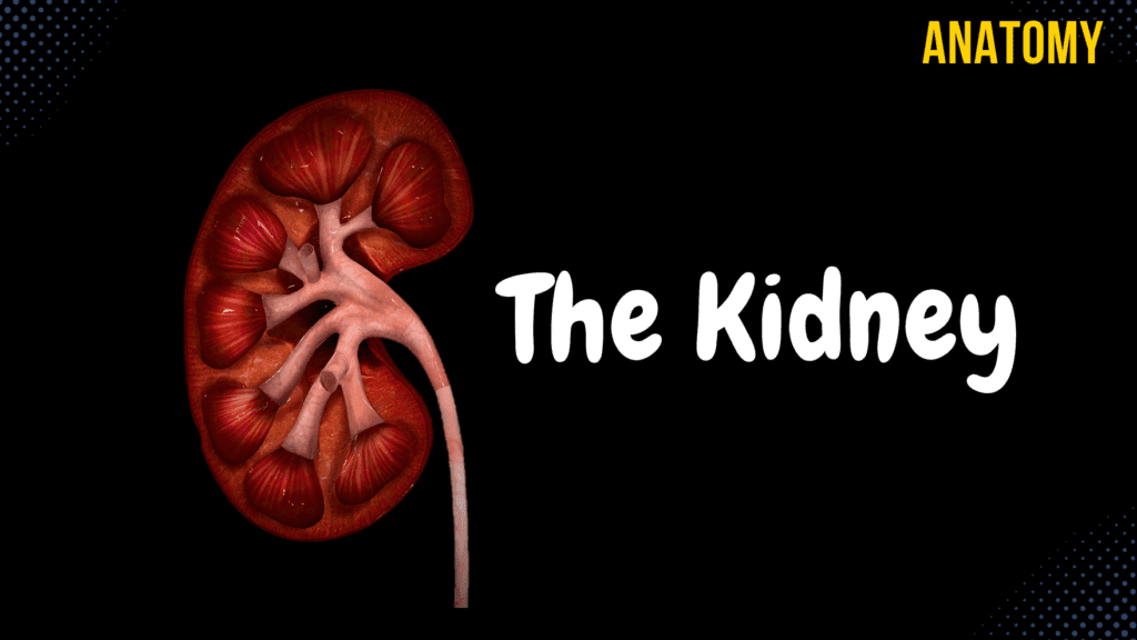
Kidneys (Functions, Structures, Coverings, Nephron) Official Links Instagram Youtube Jki-discord Notes & Illustrations Quizzes Summary & Transcript Notes ☆ Member Only Go to PDF Notes Illustrations ☆ Member Only Go to Illustrations 12345678910 Kidneys – QUIZ Test your understanding with 10 random multiple-choice questions from the question bank. You're in the preview mode. Note: All elements work correctly on the front end. 1 / 10 Which structure is found between the renal pyramids? A) Medullary rays B) Renal papillae C) Renal columns D) Renal lobes Renal columns are extensions of the cortex that separate the renal pyramids. 2 / 10 What is the apex of a renal pyramid called? A) Medulla B) Renal column C) Cortex D) Renal papilla The apex of a renal pyramid is called the renal papilla, where urine flows into the minor calyces. 3 / 10 What is the approximate thickness of the renal cortex? A) 15-20 mm B) 11-15 mm C) 4-11 mm D) 2-5 mm The renal cortex is approximately 4-11 mm thick. 4 / 10 Which anatomical structure collects urine from multiple collecting ducts? A) Renal pelvis B) Renal pyramid C) Renal papilla D) Minor calyx The minor calyx collects urine from multiple collecting ducts and funnels it into the major calyces. 5 / 10 Which structure anchors the kidneys to surrounding tissues? A) Perinephric fat B) Renal fascia C) Paranephric fat D) Fibrous capsule The renal fascia anchors the kidney and adrenal gland to surrounding structures. 6 / 10 What is the function of the renal pelvis? A) Filters plasma B) Anchors the kidney C) Produces hormones D) Funnels urine into the ureter The renal pelvis collects urine from the major calyces and funnels it into the ureter. 7 / 10 What is the terminal part of the nephron that delivers urine into the renal pelvis? A) Collecting duct B) Loop of Henle C) Renal corpuscle D) Proximal convoluted tubule The collecting duct collects urine from the nephron and empties into the renal pelvis. 8 / 10 Which structure of the kidney contains renal corpuscles? A) Renal medulla B) Renal cortex C) Renal sinus D) Renal pyramid The renal cortex contains renal corpuscles, proximal and distal convoluted tubules. 9 / 10 What forms the apex of the renal pyramid? A) Renal column B) Renal papilla C) Renal hilum D) Renal lobe The renal papilla forms the apex of the renal pyramid and drains urine into the minor calyces. 10 / 10 What is the primary site of plasma filtration in the kidney? A) Loop of Henle B) Collecting duct C) Bowman’s capsule D) Glomerulus Plasma filtration occurs in the glomerulus, a network of capillaries in the renal corpuscle. Your score is The average score is 0% Description Kidney Anatomy: Topography, Structures, and Function This video covers a comprehensive overview of the kidneys, including their topography, functions, external and internal structures, and nephron anatomy. 1. Topography of the Kidney The right kidney is slightly lower than the left. Both kidneys start at T12. Left kidney: Ends at L2. Right kidney: Ends at L3. 2. Functions of the Kidney Plasma Filtration: Through nephrons. Excretion: Removal of metabolic waste. Acid-Base Homeostasis: Regulates pH balance. Hormone Production: Erythropoietin, Renin. Vitamin D Metabolism: Activation of Vitamin D. 3. External Structures Weight: 120-200 grams. Size: 10-13 cm long, 5-6 cm wide, 4 cm thick. Inferior Pole (Extremitas Inferior). Superior Pole (Extremitas Superior): Suprarenal gland located on top. Lateral Border (Margo Lateralis). Medial Border (Margo Medialis): Contains the hilum (Hilum Renalis). 4. Coverings of the Kidney Fibrous Capsule (Capsula Fibrosa). Perinephric Fat (Capsula Adiposa). Renal Fascia (Fascia Renalis): Anterior Layer: Prerenal Layer (Lamina Prerenalis) / Fascia of Toldt. Posterior Layer: Retrorenal Layer (Lamina Retrorenalis) / Fascia of Zuckerkandl. Both layers fuse laterally. Peritoneum. 5. Internal Structures Renal Sinus Sinus Renalis: Fat-filled space inside the kidney. Renal Pelvis (Pelvis Renalis): Central collecting region. Renal Calyces (Calyces Renales): Divided into: Minor Renal Calyces (Calyx Renales Minor). Major Renal Calyces (Calyx Renales Major). Renal Medulla Renal Pyramids (Pyramides Renales). Renal Lobes (Lobi Renales). Renal Papillae (Papillae Renales): Tip of pyramids. Renal Medullary Rays (Radii Medullares): Contain nephrons’ loops. Loop of Henle and Collecting Ducts. Openings of Papillary Ducts (Foramina Papillaria). Renal Cortex Thickness: 4-11 mm. Renal Columns (Columna Renalis): Extension of the cortex into the medulla. Contains Renal Corpuscles, Proximal & Distal Convoluted Tubules. 6. Renal Lobe & Nephron Renal Corpuscle (Corpusculum Renis) Glomerulus: Network of capillaries. Afferent Arteriole (Vas Afferens): Brings blood in. Efferent Arteriole (Vas Efferens): Drains blood out. Glomerular Capsule (Capsula Glomeruli): Encases glomerulus. Nephron Tubules Proximal Convoluted Tubule (Tubulus Proximalis): Reabsorbs nutrients. Has a convoluted part and a straight part. Loop of Henle (Ansa Nephroni): Descending Limb. Ascending Limb. Distal Convoluted Tubule (Tubulus Distalis): Fine-tunes ion balance. Collecting Duct (Ductus Colligentes): Final urine concentration. Papillary Duct (Ductus Papillares): Empties into minor calyx. 7. Clinical Relevance Polycystic Kidney Disease (PKD): Genetic disorder causing multiple cysts in the kidney. Kidney Stones (Nephrolithiasis): Crystallized minerals causing obstruction. Acute Kidney Injury (AKI): Sudden loss of kidney function. Chronic Kidney Disease (CKD): Progressive decline in kidney function. Sources Used: Memorix Anatomy (2nd Edition) – Hudák Radovan, Kachlík David, Volný Ondřej. Complete Anatomy by 3D4Medical. Biorender. University notes and lectures. Transcript Introduction0:03What’s up.0:04Meditay here.0:05Let’s talk about the anatomy of the urinary system.0:07In this segment, we will be talking about the anatomy of the Kidneys.0:10Alright, so the Urinary system consists of all the organs involved in handling the urine.0:15These are the Kidneys, the Ureter, the Urinary bladder, and the urethra.0:20Our goal is to cover the anatomy of all these structures you see here, step by step, and0:25we’ll start with the Kidneys In this video, we’re first going to talk about0:28the functions of the kidneys.0:30Then we’ll talk about the external structures and the coverings of the kidneys.0:34After that, we’ll open up the kidney and cover the internal structures.0:39When we’re done with the kidneys, we’ll talk about the general anatomy of the nephron,0:42which is the functional unit of the kidney.Topography of Kidney0:45Alright, so here you see
Male Genital System
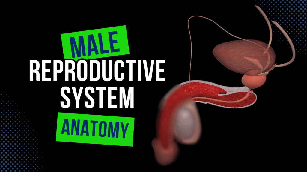
Male Genital System (Internal & External) Official Links Instagram Youtube Jki-discord Notes & Illustrations Quizzes Summary & Transcript Notes ☆ Member Only Go to PDF Notes Illustrations ☆ Member Only Go to Illustrations 12345678910 Male Genital System – QUIZ Test your understanding with 10 random multiple-choice questions from the question bank. You're in the preview mode. Note: All elements work correctly on the front end. 1 / 10 What structure carries sperm from the epididymis to the ejaculatory duct? A) Pampiniform plexus B) Ductus deferens C) Tunica vaginalis D) Prostate gland The ductus deferens transports sperm from the epididymis to the ejaculatory duct. 2 / 10 What structure anchors the penis to the pubic symphysis? A) Tunica albuginea B) Fundiform ligament C) Dartos fascia D) Suspensory ligament The suspensory ligament anchors the penis to the pubic symphysis. 3 / 10 What is the main role of Sertoli cells? A) Regulate blood supply B) Secrete testosterone C) Produce ejaculate fluid D) Regulate spermatogenesis Sertoli cells provide structural support and regulate spermatogenesis in the seminiferous tubules. 4 / 10 What is the name of the structure where the ductus deferens and seminal gland ducts join? A) Spongy urethra B) Prostatic urethra C) Membranous urethra D) Ejaculatory duct The ductus deferens and seminal gland ducts join to form the ejaculatory duct. 5 / 10 Which artery supplies blood to the ductus deferens? A) Artery to ductus deferens B) External iliac artery C) Pampiniform plexus D) Testicular artery The artery to the ductus deferens arises from the internal iliac artery and supplies the ductus deferens. 6 / 10 What is the function of the urethral crest in the prostate? A) Facilitates ejaculation B) Produces sperm C) Provides structural support D) Stores sperm The urethral crest provides structural support to the prostatic urethra. 7 / 10 What layer of the testis is responsible for the formation of septa and lobules? A) Cremaster muscle B) Tunica albuginea C) Vascular layer D) Internal spermatic fascia The tunica albuginea is a dense connective tissue layer that extends inward to form septa, dividing the testis into lobules. 8 / 10 Which artery primarily supplies the testis? A) Testicular artery B) Pampiniform plexus C) External iliac artery D) Internal iliac artery The testicular artery arises from the abdominal aorta and supplies the testis. 9 / 10 What is the primary function of the prostate gland? A) Facilitates sperm maturation B) Produces nourishing fluid for sperm C) Produces hormones D) Regulates testicular function The prostate gland produces a fluid that nourishes and protects sperm. 10 / 10 What does the seminal gland secrete? A) Mucus B) Spermatogenic fluid C) Testosterone D) Fructose-rich fluid The seminal gland secretes a fluid rich in fructose and prostaglandins to nourish sperm. Your score is The average score is 0% Description Complete Overview of Male Genital Organs (Internal & External) This video provides a comprehensive breakdown of male internal and external genital anatomy, covering structures, histology, functions, and clinical relevance. 1. Internal Genital Organs Testis Key Features: Male reproductive gland responsible for sperm production. Leydig Cells: Produce testosterone. Sertoli Cells: Structural support and aid spermatogenesis. Prenatal Development: Originates in the lumbar region. Descends and takes abdominal layers, forming scrotal layers. External Structures: Upper Pole (Extremitas Superior). Lower Pole (Extremitas Inferior). Anterior Border (Margo Anterior). Posterior Border (Margo Posterior). Lateral Surface (Facies Lateralis). Medial Surface (Facies Medialis). Internal Structures: Tunica Albuginea. Vascular Layer (Tunica Vasculosa). Septa of Testis (Septula Testis). Lobules of Testis (Lobuli Testis). Seminiferous Tubules (Tubuli Seminiferi Contorti & Recti). Rete Testis. Efferent Ductules (Ductuli Efferentes Testis). Epididymis Composed of tightly packed tubules; stores sperm. Head (Caput Epididymidis), Body (Corpus Epididymidis), Tail (Cauda Epididymidis). Coverings & Fixation of Testes & Epididymis Tunica Vaginalis: Visceral (Epiorchium) & Parietal Layer (Periorchium). Fascial Layers: Internal Spermatic Fascia, Cremaster, External Spermatic Fascia. Dartos Fascia (Tunica Dartos). Spermatic Cord (Funiculus Spermaticus) Contains Ductus Deferens, Testicular Artery, Pampiniform Plexus, Lymph Vessels. Ductus Deferens Pathway for sperm transport. Divided into Scrotal, Funicular, Inguinal, & Pelvic Parts. Seminal Gland (Glandula Vesiculosa) Produces ejaculate fluid. Joins ductus deferens to form Ejaculatory Duct. Prostate (Prostata) Structure: Base & Apex. Posterior & Anterior Surfaces. Prostatic Urethra. Histological Zones: Peripheral, Central, Transitional, & Anterior Fibromuscular Zones. Posterior Features: Urethral Crest, Seminal Colliculus, Prostatic Utricle (Fetal Remnant). Openings of Ejaculatory Ducts. Bulbo-Urethral Glands (Glandulae Bulbourethrales) Empties into the spongy urethra. Male Urethra (Urethra Masculina) Prostatic, Membranous, and Spongy Urethra. 2. External Genital Organs Penis External Structures: Root (Bulb & Crura). Body (Corpora Cavernosa & Corpus Spongiosum). Glans Penis (Corona, Collum, Prepuce, Frenulum). Internal Structures: Corpora Cavernosa & Corpus Spongiosum. Tunica Albuginea. Buck’s Fascia (Deep Fascia of Penis). Scrotum Contains layers derived from abdominal structures: Tunica Vaginalis, Internal & External Spermatic Fasciae, Cremaster Muscle, Dartos Fascia. 3. Clinical Relevance Testicular Torsion: Twisting of the spermatic cord leading to ischemia. Benign Prostatic Hyperplasia (BPH): Enlargement of the prostate affecting urination. Cryptorchidism: Failure of testis to descend. Sources Used: Memorix Anatomy (2nd Edition) – Hudák Radovan, Kachlík David, Volný Ondřej. Complete Anatomy by 3D4Medical. Biorender. Snell, Richard S. (2012). Clinical Anatomy by Regions (9th Edition). University notes and lectures. Transcript Introduction0:03What’s up.0:04Let’s go ahead and cover the male genital system.0:07We will cover the most important aspect of the male genitals, and hopefully, in the end,0:12you’ll have a pretty good understanding about this topic0:15Alright.0:16The male genital organs are divided into the internal genital organs and the external genital0:22organs.0:23We’ll cover both of these in detail, And we’ll start with the internal genitalInternal Genital Organs0:27organs So the internal genital organs consist of0:31the testes, epididymis, ductus deferens, seminal gland, Ejaculatory Duct, Prostate, Male urethra,0:40and the bulbourethral glands.0:43Alright, let’s follow this arrangement, starting with the testis.Testis0:47Now, of the whole male reproductive system, the primary functional cells the male has0:54are Leydig and Sertoli cells.0:56And both of them are found in the testes.1:00So the testicle or testis is the male reproductive gland or gonad in all animals, not just humans.1:07Leydig cells produce testosterone, which is a steroid hormone that binds to intracellular1:14receptors and regulates protein synthesis.1:18And what
Female Genital System
Female Genital System (Internal & External) Official Links Instagram Youtube Jki-discord Notes & Illustrations Quizzes Summary & Transcript Notes ☆ Member Only Go to PDF Notes Illustrations ☆ Member Only Go to Illustrations 12345678910 Female Genital System – QUIZ Test your understanding with 10 random multiple-choice questions from the question bank. You're in the preview mode. Note: All elements work correctly on the front end. 1 / 10 What is the purpose of the broad ligament in the female genital system? A) Facilitate uterine contractions B) Supply blood to ovaries C) Separate abdominal organs D) Support internal organs The broad ligament is a double fold of peritoneum that supports the uterus, uterine tubes, and ovaries, helping maintain their position. 2 / 10 Which lymph nodes drain the external genitalia in females? A) Para-aortic B) Lumbar C) Internal iliac D) Superficial inguinal The superficial inguinal lymph nodes primarily drain the external genitalia, including the labia majora and minora. 3 / 10 What is the function of the zona pellucida in oocyte development? A) Provides nutrients B) Supports the corpus luteum C) Facilitates sperm binding D) Produces follicular fluid The zona pellucida surrounds the oocyte and facilitates sperm binding, protecting the developing oocyte. 4 / 10 Which veins primarily drain the blood from the ovaries? A) Vaginal venous plexus B) Ovarian veins C) Uterine venous plexus D) Pudendal veins The right ovarian vein drains into the inferior vena cava, while the left ovarian vein drains into the left renal vein. 5 / 10 What is the histological structure of the vaginal epithelium? A) Stratified squamous B) Simple columnar C) Simple cuboidal D) Transitional epithelium The vaginal epithelium is composed of stratified squamous non-keratinized epithelium, which stores glycogen. 6 / 10 What is the histological lining of the uterine cavity? A) Transitional epithelium B) Simple columnar C) Simple cuboidal D) Stratified squamous The uterine cavity is lined by simple columnar epithelium that forms the endometrium. 7 / 10 What type of follicle is ready for ovulation? A) Primary follicle B) Graafian follicle C) Secondary follicle D) Primordial follicle The Graafian follicle, or tertiary follicle, contains the secondary oocyte and is ready for ovulation. 8 / 10 What are the main functions of the ovaries? A) Produce oocytes and hormones B) Transport gametes C) Regulate blood supply D) Provide structural support The ovaries are responsible for gametogenesis and the production of hormones such as estrogen and progesterone. 9 / 10 What are the structural layers of the uterine wall? A) Endometrium, myometrium, perimetrium B) Myocardium, epimysium C) Epimetrium, perimysium D) Serosa, stroma The uterine wall is composed of the endometrium (mucosa), myometrium (muscular layer), and perimetrium (serosa). 10 / 10 What are the functional layers of the endometrium during the menstrual cycle? A) Endometrial, stromal layers B) Mucosal, serosal layers C) Functional, basal layers D) Perimetrium, myometrium The functional layer of the endometrium undergoes cyclic changes and is shed during menstruation, while the basal layer regenerates it. Your score is The average score is 0% Description Complete Overview of Female Internal and External Genital Organs This video provides a detailed anatomical breakdown of the internal and external genital structures, including their histology, functions, and clinical relevance. Small Correction: At 7:06, the ovarian artery and vein run within the suspensory ligament of the ovary, not the ligament of the ovary as originally stated. 1. Internal Genital Organs Ovary Inner Structures: Tunica Albuginea (Tunica albuginea ovarii) Ovarian Cortex (Cortex ovarii) Ovarian Medulla (Medulla ovarii) Follicles External Structures: Mesovarian Border (Margo mesovaricus) Free Border (Margo liber) Tubal Extremity (Extremitas tubaria) Uterine Extremity (Extremitas uterina) Medial Surface (Facies medialis) Lateral Surface (Facies lateralis) Fixation of the Ovary: Ligament of Ovary (Ligamentum ovarii proprium) Suspensory Ligament of Ovary (Ligamentum suspensorium ovarii) Mesovarium Growth and Maturation of Follicles Primordial Follicle: Contains primary oocyte, single layer of follicular cells. Primary Follicles: After puberty (FSH stimulation), granulosa cells form. Secondary Follicle: Zona pellucida, follicular fluid, theca folliculi, estrogen production. Graafian Follicle: Mature follicle containing a secondary oocyte, released during ovulation. Corpus Luteum: Hormone-secreting structure post-ovulation. Corpus Albicans: Remnant of corpus luteum. Uterine Tube (Tuba Uterina) Parts of the Uterine Tube: Infundibulum (Infundibulum tubae uterinae) Abdominal Ostium (Ostium abdominale) Fimbriae (Fimbriae tubae uterina) Ovarian Fimbria (Fimbria ovarica) Ampulla (Ampulla tubae uterinae) Isthmus (Isthmus tubae uterinae) Uterine Part (Pars uterina) – Uterine Ostium (Ostium uterinum) Walls of Uterine Tube: Tunica Mucosa Tunica Muscularis Tunica Serosa Mesosalpinx Uterus Parts: Positioned in anteflexion. Fundus of Uterus (Fundus uteri). Body of Uterus (Corpus uteri). Intestinal (Posterior) Surface (Facies intestinalis). Vesical (Anterior) Surface. Cervix (Portio supravaginalis cervicis and vaginal part). Internal Structures (Histology): Endometrium: Functional and basal layers, simple columnar epithelium, uterine glands. Myometrium: Smooth muscle layers, vascularized middle layer. Perimetrium: Outer serous layer. Fixation of the Uterus Peritoneal Folds: Broad Ligament (Ligamentum latum uteri) Mesovarium Mesosalpinx Mesometrium Parametrial Ligaments: Cardinal Ligament (Ligamentum cardinale uteri). Vesico-uterine Ligament. Pubocervical Ligament. Uterosacral Ligaments. Recto-uterine Ligament. Round Ligament of Uterus (Ligamentum teres uteri). 2. Vagina Vaginal Orifice (Ostium Vaginae). Vestibule (Vestibulum Vaginae). Vaginal Canal (Canalis Vaginalis). Vaginal Fornices (Anterior, Posterior, and Lateral). Vaginal Wall (Histology): Mucosa – Forms transverse folds. Muscularis Layer. Clinical Relevance: Imperforate Hymen: May require medical intervention. Recto-uterine Pouch (Pouch of Douglas): Important for fluid accumulation. 3. External Genital Organs Vestibule: Lesser and Greater Vestibular Glands (Bartholin’s Glands). Labia Minora (Labia Minora Pudendi). Clitoris: Glans, Body, Crura, Prepuce. Bulb of Vestibule (Bulbus Vestibuli). Labia Majora (Labia Majora Pudendi). Mons Pubis. Sources Used: Memorix Anatomy (2nd Edition) – Hudák Radovan, Kachlík David, Volný Ondřej. Complete Anatomy by 3D4Medical. Biorender. Transcript Introduction0:03What’s up. Meditay Here. Let’s finally talk about the female genital system. Now, this is going to0:08be a longer video than usual. It’s not because the female genital system is necessarily difficult,0:14but it’s because I want to cover everything in detail so that you understand the fundamentals of the female genital system. I’ve put some timestamps on the bottom of the video,0:23so you can use that if it’s any specific topic you came here for. Alright. So the