
Peritoneum
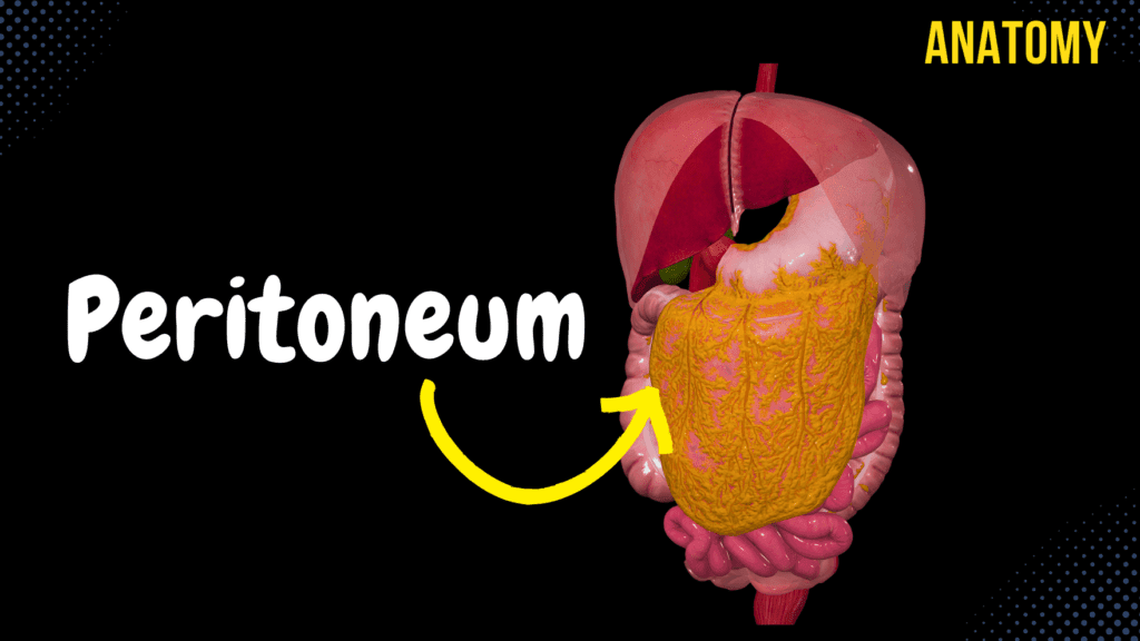
Peritoneum (Parts, Lesser & Greater Omentum, Mesentery, Peritoneal Cavity) Official Links Instagram Youtube Jki-discord Notes & Illustrations Quizzes Summary & Transcript Notes ☆ Members Only Go to PDF Notes Illustrations ☆ Members Only Go to Illustrations 12345678910 Peritoneum – QUIZ Test your understanding with 10 random multiple-choice questions from the question bank. You're in the preview mode. Note: All elements work correctly on the front end. 1 / 10 What is the clinical significance of the omental (epiploic) foramen? A) Site of Ligament Attachment B) Pathway for Arteries C) Communication Between Sacs D) Junction of Viscera The omental foramen allows communication between the greater and lesser sacs of the peritoneal cavity. 2 / 10 What type of fluid is contained within the peritoneal cavity? A) Pancreatic Juice B) Bile C) Mucus D) Serous Fluid Serous fluid lubricates the peritoneal cavity to reduce friction between organs. 3 / 10 Which level of the peritoneal cavity contains the omental bursa? A) Lower Level B) Middle Level C) Entire Peritoneal Cavity D) Upper Level The omental bursa is located in the upper level of the peritoneal cavity. 4 / 10 What is the role of the root of the mesentery? A) Separate Peritoneal Levels B) Anchor Intestines to Wall C) Aid in Digestion D) Produce Lymph The root of the mesentery anchors the small intestine to the posterior abdominal wall and allows passage for vessels. 5 / 10 What is the anatomical term for the peritoneal cavity pouch between the uterus and rectum in females? A) Rectouterine Pouch B) Retrocecal Pouch C) Vesicouterine Pouch D) Rectovesical Pouch The rectouterine pouch (pouch of Douglas) lies between the uterus and rectum. 6 / 10 What is the peritoneal attachment for the small intestine? A) Mesentery B) Greater Omentum C) Lesser Omentum D) Retroperitoneum The mesentery provides attachment for the small intestine. 7 / 10 Which part of the peritoneum lines the abdominal wall? A) Mesentery B) Parietal Peritoneum C) Visceral Peritoneum D) Greater Omentum The parietal peritoneum lines the abdominal and pelvic walls. 8 / 10 What is the primary function of the omental bursa? A) Separate Lobes B) Reduce Friction and Facilitate Movement C) Support Vessels D) Absorb Nutrients The omental bursa reduces friction and facilitates organ movement. 9 / 10 What is the primary lymphatic drainage of the peritoneal cavity? A) Paracolic Nodes B) Hepatic Nodes C) Mesenteric Lymph Nodes D) Splenic Nodes The mesenteric lymph nodes provide drainage for the peritoneal cavity. 10 / 10 What structure connects the liver to the anterior abdominal wall? A) Gastrophrenic Ligament B) Coronary Ligament C) Falciform Ligament D) Hepatoduodenal Ligament The falciform ligament connects the liver to the anterior abdominal wall. Your score is The average score is 0% Description This video is about the anatomy of the peritoneum. Parts of the Peritoneum: Parietal Peritoneum (peritoneum parietale) – Lines the internal surface of the abdominal and pelvic wall. Visceral Peritoneum (peritoneum viscerale) – Covers the walls of organs. Peritoneal Cavity (cavitas peritonealis) – The space between the parietal and visceral peritoneum. Visceral Peritoneum: How does the Visceral Peritoneum Cover the Organs? Intraperitoneal Viscera: Organs completely covered by the peritoneum. Mesoperitoneal Viscera: Partly covered by the peritoneum (on three different sides). Retroperitoneal Viscera: Covered by the peritoneum only on one side. The Course of the Peritoneum: Parietal peritoneum continues into the visceral peritoneum through: Falciform Ligament (ligamentum falciforme hepatis) Coronary Ligament (ligamentum coronarium hepatis) Right Triangular Ligament (ligamentum triangulare dextrum) Left Triangular Ligament (ligamentum triangulare sinistrum) These ligaments cover the liver. Lesser Omentum: Consists of all ligaments under the liver but above the lesser curvature of the stomach. Lesser Omentum: Hepatogastric Ligament (ligamentum hepatogastricum) Hepatoduodenal Ligament (ligamentum hepatoduodenale) Both start at the porta hepatis of the liver. Greater Omentum: Gastrophrenic Ligament (ligamentum gastrophrenicum) Gastrosplenic Ligament (ligamentum gastrosplenicum) Gastrocolic Ligament (ligamentum gastrocolicum) Course: Extends downward in front of the abdominal organs until reaching the terminal line of the pelvis (linea terminalis), then turns around and attaches to the omental tenia (tenia omentalis) on the transverse colon. Ends at the mesocolic tenia (tenia mesocolica) and transitions into the transverse mesocolon (mesocolon transversum). Mesentery: Transverse Mesocolon (mesocolon transversum) Root of Mesentery (radix mesenterii) Mesentery completely encloses the intestines, provides structural support, and forms pathways for blood vessels. Peritoneal Cavity (Cavitas Peritonealis): The peritoneal cavity is divided into three levels: Upper Level (above the transverse mesocolon) Middle Level (below the transverse mesocolon but above the terminal line of the pelvis) Lower Level (in the lesser pelvis) Upper Level: Contains 2 recesses and 1 bursa. Recess: A small opening. Bursa: A fluid-filled sac. Structures in the upper level: Subphrenic Recess (recessus subphrenicus) Subhepatic Recess (recessus subhepaticus) Omental Bursa (bursa omentalis) Walls/Borders of the Omental Bursa: The Omental Bursa contains the Omental Foramen/Epiploic Foramen (foramen omentale/foramen epiploicum), bordered by the Vestibule of the Omental Bursa (vestibulum bursa omentalis). Recesses of the Bursa Omentalis: Superior Recess (recessus superior) Inferior Recess (recessus inferior) Splenic Recess (recessus splenicus) Middle Level of the Peritoneal Cavity: Superior Duodenal Recess (recessus duodenalis superior) Inferior Duodenal Recess (recessus duodenalis inferior) Superior Ileocecal Recess (recessus ileocaecalis superior) Inferior Ileocecal Recess (recessus ileocaecalis inferior) Retrocecal Recess (recessus retrocaecalis) Intersigmoid Recess (recessus intersigmoideus) Lower Level of the Peritoneal Cavity: In this level, the term excavatio means a pouch. Female: Rectouterine Pouch (excavatio rectouterina) Vesicouterine Pouch (excavatio vesicouterina) Male: Rectovesical Pouch (excavatio rectovesicalis) Sources: Memorix Anatomy 2nd Edition by Hudák Radovan, Kachlík David, Volný Ondřej Biorender University Notes and Lectures Transcript Introduction0:03What’s up. Meditay here. And in this video, we’re gonna go through the anatomy of the peritoneum,0:08which is the serous membrane that lines the inside of the abdominal wall,0:12as well as surrounding various organs we have inside the abdominal cavity.0:17So in this video, we’ll first cover the parts of the peritoneum.0:21Then we’ll go through the lesser and greater omentum and the mesentery.0:25After that, we’ll look at the different structures you’ll find in the peritoneal cavity.0:30So now, now that we’ve covered the anatomy of the organs in the abdominal cavity,0:35We need something
Bile Pathway, Gall Bladder & Pancreas
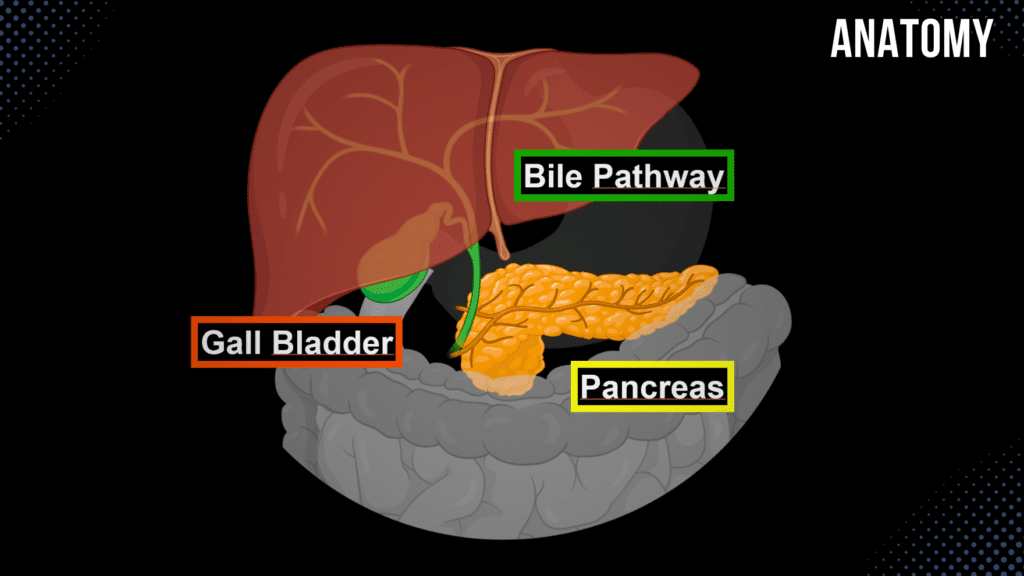
Bile Pathway, Gall Bladder & Pancreas Official Links Instagram Youtube Jki-discord Notes & Illustrations Quizzes Summary & Transcript Notes ☆ Member Only Go to PDF Notes Illustrations ☆ Member Only Go to Illustrations 12345678910 Bile and Pancreas – QUIZ Test your understanding with 10 random multiple-choice questions from the question bank. You're in the preview mode. Note: All elements work correctly on the front end. 1 / 10 What is the primary function of the pancreatic islets (Islets of Langerhans)? A) Hormone Secretion B) Emulsify Lipids C) Secrete Digestive Enzymes D) Neutralize Acid The islets secrete hormones such as insulin and glucagon for blood sugar regulation. 2 / 10 What separates the uncinate process from the body of the pancreas? A) Superior Mesenteric Vessels B) Hepatic Vein C) Splenic Artery D) Inferior Vena Cava The superior mesenteric vessels pass between the uncinate process and the body of the pancreas. 3 / 10 Which duct carries bile from the liver to the gallbladder? A) Cystic Duct B) Hepatic Duct C) Common Bile Duct D) Pancreatic Duct The cystic duct carries bile to the gallbladder for storage. 4 / 10 Which artery primarily supplies the body and tail of the pancreas? A) Inferior Mesenteric Artery B) Splenic Artery C) Celiac Trunk D) Superior Mesenteric Artery The splenic artery provides branches to the body and tail of the pancreas. 5 / 10 What is the anatomical position of the pancreas? A) Suprarenal Region B) Retroperitoneal C) Hypogastric Region D) Intraperitoneal The pancreas is located retroperitoneally at L1-L2 vertebral levels. 6 / 10 What is the anatomical term for the major pancreatic duct? A) Common Hepatic Duct B) Duct of Wirsung C) Duct of Santorini D) Accessory Pancreatic Duct The main pancreatic duct is also called the duct of Wirsung. 7 / 10 What is the role of the accessory pancreatic duct? A) Emulsify Fats B) Minor Papilla Drainage C) Produce Hormones D) Store Enzymes The accessory duct allows pancreatic juice to flow through the minor duodenal papilla. 8 / 10 What is the endocrine product of beta cells in the pancreas? A) Insulin B) Glucagon C) Pancreatic Polypeptide D) Somatostatin Beta cells in the Islets of Langerhans secrete insulin. 9 / 10 Which hormone stimulates the release of bile into the duodenum? A) Insulin B) Gastrin C) Secretin D) Cholecystokinin Cholecystokinin (CCK) stimulates bile release from the gallbladder. 10 / 10 What type of epithelium lines the mucosa of the gallbladder? A) Simple Cuboidal B) Pseudostratified Columnar C) Simple Columnar D) Stratified Squamous Simple columnar epithelium lines the gallbladder mucosa. Your score is The average score is 0% Description This video covers the anatomy of the bile pathway, gall bladder, and pancreas. Function of Bile: Bile contains 97% water, less than 1% bile salts, bilirubin, and fats. Emulsification of fats. Bile Pathway: Bile is produced by the liver. Biliary Ducts: Right Hepatic Duct (Ductus Hepaticus Dexter) Left Hepatic Duct (Ductus Hepaticus Sinister) Common Hepatic Duct (Ductus Hepaticus Communis) Gall Bladder (Vesica Biliaris) Cystic Duct (Ductus Cysticus) Bile Duct (Ductus Choledochus) – 7 cm long Pancreatic Duct (Ductus Pancreaticus) Hepatopancreatic Ampulla (Ampulla Hepatopancreatica) Major Duodenal Papilla (Papilla Duodeni Major) Hepatopancreatic Sphincter (Sphincter Ampullae Hepatopancreatica) – also known as the Sphincter of Oddi Gall Bladder: Fossa for the Gall Bladder (Fossa Vesicae Biliaris) Fundus of the Gall Bladder (Fundus Vesicae Biliaris) Body of the Gall Bladder (Corpus Vesicae Biliaris) Neck of the Gall Bladder (Collum Vesicae Biliaris) Wall of the Gall Bladder: Tunica Mucosa: Mucosal Folds of the Gall Bladder (Plicae Tunicae Mucosa Vesicae Biliaris) Spiral Folds (Plica Spiralis) Mucosal Glands (Glandulae Mucosae) Tunica Muscularis: Circular Muscle Fibers (Stratum Circulare) Tunica Adventitia Tunica Serosa (Part of the Hepatoduodenal Ligament) Pancreas: 14-18 cm long, 2-3 cm wide, weighing 60-100 grams. Functions of the Pancreas: Exocrine Function: Produces pancreatic juice (Succus Pancreaticus) containing digestive enzymes and bicarbonate. Endocrine Function: Releases hormones directly into the bloodstream, including insulin and glucagon. Parts of the Pancreas: Head of the Pancreas (Caput Pancreatis) Body of the Pancreas (Corpus Pancreatis) Tail of the Pancreas (Cauda Pancreatis) Topography of the Pancreas: Skeletopy: L1-L2 Holotopy: Epigastric Region, Left Hypochondriac Region Pancreatic Duct System: Main Pancreatic Duct (Ductus Pancreaticus) Accessory Pancreatic Duct (Ductus Pancreaticus Accessorius) Major Duodenal Papilla (Papilla Duodeni Major) Minor Duodenal Papilla (Papilla Duodeni Minor) Sources: Memorix Anatomy 2nd Edition by Hudák Radovan, Kachlík David, Volný Ondřej Biorender University Notes and Lectures Transcript Introduction0:03What’s up. Meditay here, and in this video, we’re gonna go through the anatomy of the Gallbladder,0:08Bile Pathway, and the Pancreas. Which are organs we call accessory organs.0:12Accessory organs are not a part of the digestive tract, but they have0:16an essential role in the actual digestion. That’s why they’re called accessory organs.0:21So, In this video, We’re first going to go through the Bile pathway, as you see here.0:25We’re gonna cover the Parts of the bile pathway, what the function of Bile essentially is,0:30and the layers of the bile duct. After that, we’ll go through the anatomy of the Gallbladder,0:35this one. Basically, talk through the different parts of it, its layers, and coverings. And then,0:41we’ll go through the anatomy of the pancreas, basically talk about its function, its topography,0:45and structures associated with it. Cool. So let’s start with the Bile pathway first.Function of Bile0:51Let’s now start by looking at an anterior view of the bile pathway. And then tilt our model a0:56little bit, so we see the entrance into the Liver. So what we see now is the Liver, gall bladder,1:03pancreas, and a perfectly drawn duodenum going around the pancreatic head as you see here.1:09So the Bile Pathways is shown here, and let’s now follow the Bile from beginning to end to really1:15see what’s going on. It all starts up here, at the Liver, because and the Liver produces the Bile.1:21Now I’m not gonna go in too much detail into the physiology of Bile since I wanna1:24focus on anatomy for now, but the Bile contains roughly around 97% water and under 1% bile salts,1:32and some slight percent of bilirubin and
Liver Anatomy
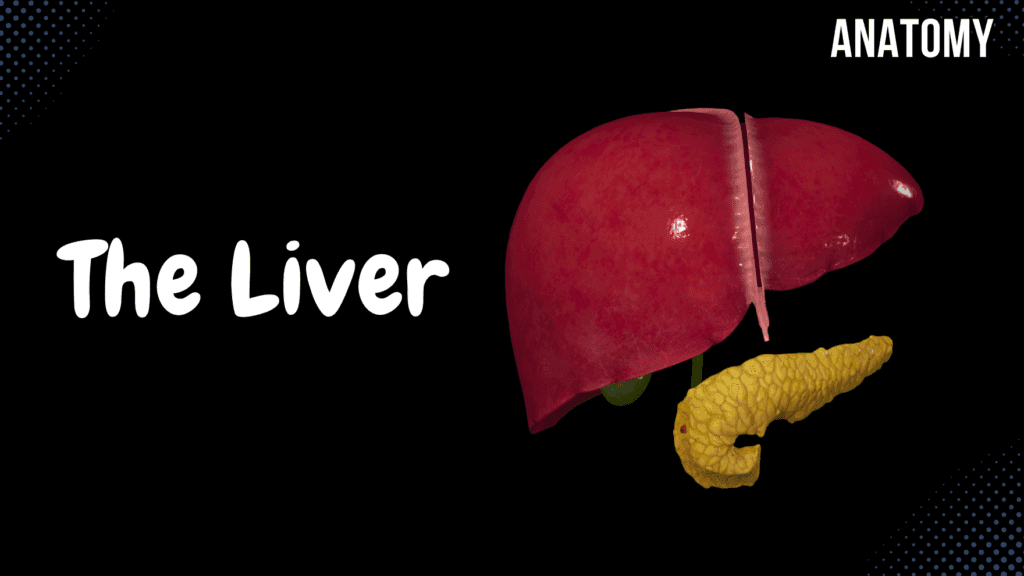
Liver Anatomy (Function, Topography, External Structures, Ligaments) Official Links Instagram Youtube Jki-discord Notes & Illustrations Quizzes Summary & Transcript Notes ☆ Member Only Go to PDF Notes Illustrations ☆ Member Only Go to Illustrations 12345678910 Liver – QUIZ Test your understanding with 10 random multiple-choice questions from the question bank. You're in the preview mode. Note: All elements work correctly on the front end. 1 / 10 Which lobe of the liver is anatomically smallest? A) Left Lobe B) Quadrate Lobe C) Caudate Lobe D) Right Lobe The caudate lobe is the smallest anatomical lobe of the liver. 2 / 10 Which surface of the liver contains the bare area? A) Diaphragmatic Surface B) Visceral Surface C) Inferior Surface D) Posterior Surface The posterior surface of the liver contains the bare area. 3 / 10 What is the main digestive function of the liver? A) Glucose Storage B) Cholesterol Synthesis C) Bile Production D) Detoxification The liver produces bile, which is essential for fat digestion and absorption. 4 / 10 What structure carries bile out of the liver? A) Right Hepatic Artery B) Portal Vein C) Hepatic Vein D) Common Hepatic Duct The common hepatic duct transports bile from the liver to the bile duct. 5 / 10 What is the significance of the hepatoduodenal ligament? A) Forms Epiploic Foramen B) Suspends Liver C) Contains Coronary Ligaments D) Contains the Portal Triad The hepatoduodenal ligament encloses the portal triad (hepatic artery, portal vein, and bile duct). 6 / 10 What type of blood is carried by the hepatic artery? A) Deoxygenated Blood B) Oxygen-Poor Blood C) Oxygenated Blood D) Nutrient-Rich Blood The hepatic artery carries oxygenated blood to the liver. 7 / 10 Which region does the liver primarily occupy in the abdomen? A) Right Iliac Region B) Right Hypochondriac Region C) Left Lumbar Region D) Umbilical Region The liver is primarily located in the right hypochondriac, epigastric, and left hypochondriac regions. 8 / 10 Which veins combine to form the portal vein? A) Superior Mesenteric and Splenic Veins B) Inferior Mesenteric Vein C) Hepatic Veins D) Renal Vein The portal vein is formed by the confluence of the superior mesenteric vein and splenic vein. 9 / 10 What is the significance of the ligamentum teres? A) Hepatic Lobe Divider B) Attachment to Diaphragm C) Fetal Vein Remnant D) Lymph Node Containment The ligamentum teres is a remnant of the fetal umbilical vein, found within the falciform ligament. 10 / 10 What is the porta hepatis? A) Entry for vessels B) Hepatic Sinusoids C) Lobe Division D) Capsule of the Liver The porta hepatis is the gateway for the hepatic artery, portal vein, and bile ducts entering and leaving the liver. Your score is The average score is 0% Description Liver Function Filters blood from the digestive tract via the portal system Detoxification of harmful substances Produces bile for digestion Cholesterol synthesis Stores fat and glycogen Regulates blood sugar levels Topography of the Liver (Hepar) Holotopy: Right Hypochondriac Region, Epigastric Region, Left Hypochondriac Region Skeletopy: Superior Border: Starts at the 10th rib, extends to the 5th intercostal space at the sternum, and ends at the 6th intercostal space Inferior Border: Starts at the 10th rib, extends to the level of the 8-9th rib, and continues to the 6th intercostal space Posteriorly: T9-T11 vertebrae Syntopy: Superior Border: Diaphragmatic Surface Inferior Border (Visceral Surface): Pylorus of the stomach Colon Duodenum Right kidney Stomach Esophagus External Surface of the Liver Anterior Surface: Falciform Ligament (Ligamentum Falciforme) Right and Left Lobes Inferior Margin (Margo Inferior) Posterior Surface: Posterior Margin (Margo Posterior) Bare Area (Area Nuda) Right Sagittal Groove (Sulcus Sagittalis Dexter) Groove for Inferior Vena Cava (Sulcus Venae Cavae) Fossa for the Gallbladder (Fossa Vesicae Biliaris) Left Sagittal Groove (Sulcus Sagittalis Sinister) Round Ligament of the Liver (Ligamentum Teres Hepatis) Venous Ligament of the Liver (Ligamentum Venosum) Transverse Groove (Porta Hepatis) Division of the Liver Lobes of the Liver (Anatomical Division): Right Lobe Left Lobe Caudate Lobe Quadrate Lobe Couinaud Classification (Surgical Classification) Porta Hepatis Common Hepatic Duct Hepatic Portal Vein Proper Hepatic Artery Hepatic Plexus Hepatic Lymph Nodes Hepatoduodenal Ligament (Ligamentum Hepatoduodenale) Coverings of the Liver Tunica Fibrosa Visceral Peritoneum (Tunica Serosa) Ligaments Around the Liver Ligaments Connecting to the Diaphragm: Falciform Ligament (Ligamentum Falciforme Hepatis) Coronary Ligament (Ligamentum Coronarium Hepatis) Right Triangular Ligament (Ligamentum Triangulare Dexter) Left Triangular Ligament (Ligamentum Triangulare Sinister) Ligaments Connecting to Other Organs: Hepatogastric Ligament (Ligamentum Hepatogastricum) Hepatoduodenal Ligament (Ligamentum Hepatoduodenale) Hepatorenal Ligament (Ligamentum Hepatorenale) Round Ligament of the Liver (Ligamentum Teres Hepatis) Sources Memorix Anatomy 2nd Edition by Hudák Radovan, Kachlík David, Volný Ondřej Complete Anatomy by 3D4Medical Biorender University Notes and Lectures Transcript Introduction0:03What’s up. Meditay here, and in this video, we’re gonna go through the anatomy of the Liver.0:08So now that we’ve gone through all the digestive system structures, along with the three accessory0:13organs up here in the mouth, we’ll do the next structure, which is the Liver, since it has an0:18essential role in digestion. Our aim in this video is to understand the complete anatomy0:24and the orientation of the Liver. And to do that, We’re first going to cover the Function and the0:28Topography of the Liver. And then we’re going to look at the external structures of the Liver0:34by going through the lobes, margins, and grooves we have on the surface of the Liver,0:39and then we’re going to talk about porta Hepatis, along with how the Liver is fixated.0:43Basically, going through the covering and the ligaments you’ll find around the Liver.The Function of the Liver0:48So the Liver, or The Hepar in Latin, is the largest internal organ in the body.0:53It lies on the right side of the belly and weighs approximately 1.5 kilograms, so it’s pretty heavy.0:59The Liver’s main job is to filter the blood coming from the digestive tract before passing it to the1:05rest of the body. And it does that through the portal system. And even if you’re not familiar1:10with the portal system yet, it is quite important to understand this
Large Intestine Anatomy
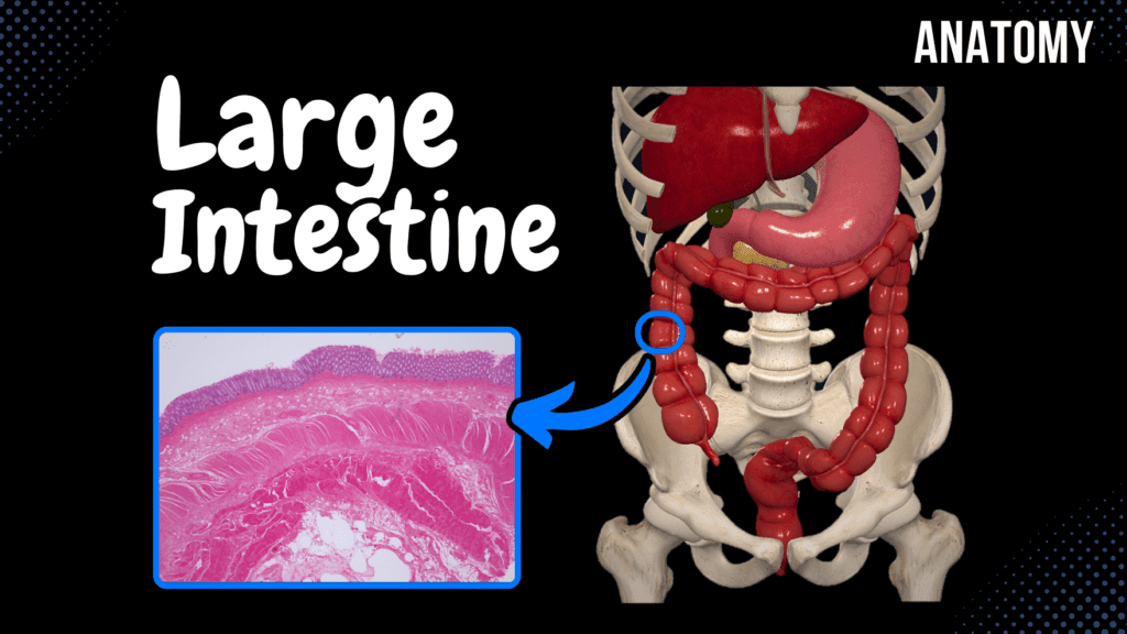
Large Intestine Anatomy (Parts, Topography, Layers) Official Links Instagram Youtube Jki-discord Notes & Illustrations Quizzes Summary & Transcript Notes ☆ Member Only Go to PDF Notes Illustrations ☆ Member Only Go to Illustrations 12345678910 Large Intestine – QUIZ Test your understanding with 10 random multiple-choice questions from the question bank. You're in the preview mode. Note: All elements work correctly on the front end. 1 / 10 What is the primary blood supply to the rectum? A) Inferior Mesenteric Vein B) Superior Rectal Artery C) Middle Rectal Artery D) Inferior Rectal Artery The superior rectal artery, a branch of the inferior mesenteric artery, supplies the rectum. 2 / 10 What is the significance of the teniae coli? A) Neutralize Acid B) Aid Absorption C) Form Segmental Pouches D) Form Circular Folds Teniae coli are three longitudinal muscle bands that contract to form haustra. 3 / 10 What is the term for folds in the rectum that support fecal weight? A) Anal Columns B) Rectal Transverse Folds C) Semilunar Folds D) Haustra The rectal transverse folds (plicae transversales recti) provide structural support. 4 / 10 What is the length of the rectum? A) 10–15 cm B) 20–25 cm C) 15–20 cm D) 5–10 cm The rectum is approximately 15–20 cm long. 5 / 10 What is the function of semilunar folds in the colon? A) Transport Fats B) Store Fecal Material C) Divide Haustra D) Neutralize Acid Semilunar folds divide haustra and assist with segmentation and peristalsis. 6 / 10 What structure prevents fecal matter from re-entering the ileum? A) Rectal Sphincter B) Sigmoid Flexure C) Ileocecal Valve D) Teniae Coli The ileocecal valve prevents backflow of feces into the small intestine. 7 / 10 What structure marks the boundary between the sigmoid colon and the rectum? A) Anal Canal B) Perineal Flexure C) Sacroiliac Joint D) Rectal Ampulla The sacroiliac joint marks this transition. 8 / 10 Which layer of the large intestine contains intestinal crypts? A) Tunica Mucosa B) Tunica Serosa C) Tunica Muscularis D) Tela Submucosa Intestinal crypts are found in the tunica mucosa and house epithelial cells. 9 / 10 What is McBurney’s point associated with? A) Diverticulosis B) Intestinal Obstruction C) Hemorrhoids D) Appendicitis McBurney’s point indicates the surface location of the appendix, often used to diagnose appendicitis. 10 / 10 Which flexure separates the transverse colon from the descending colon? A) Left Colic Flexure B) Sigmoid Flexure C) Hepatic Flexure D) Right Colic Flexure The left colic flexure separates these parts of the colon. Your score is The average score is 0% Description Large Intestine Overview Latin Name: Intestinum Crassum Extends from: End of Ileum to Anus Length: 1-1.65 meters Function: Absorption of water and electrolytes Parts of the Large Intestine: Caecum Appendix Ascending Colon Transverse Colon Descending Colon Sigmoid Colon Rectum Caecum Topography: Right Inguinal Region Size: 6-8 cm long, 7-7.5 cm wide Ileal Orifice: Ostium Ileale Ileocecal Valve: Regulates flow between small and large intestine Appendix Vermiformis Size: 2-4 cm long, 0.5-1 cm wide Orifice: Ostium Appendicis Vermiformis Positions of the Appendix: Caudal Position (40-50%) Medial Position (17-20%) Lateral Position (17-20%) Posterior Position (9-13%) Anterior Position (rare) Appendicitis Inflammation of the appendix May cause peritonitis McBurney’s Point: Common site of pain T10 Dermatome: Early stage pain felt at the umbilical region Ascending Colon (Colon Ascendens) Length: 12-20 cm Topography: Right Lateral Region, Right Hypochondriac Region Syntopy: Anteriorly: Anterior Abdominal Wall Laterally: Lateral Abdominal Wall Posteriorly: Right Kidney, Quadratus Lumborum, Transverse Abdominal Muscle Transverse Colon (Colon Transversum) Length: 30-80 cm Topography: Right Colic Flexure (Flexura Coli Dextra) – Right Hypochondriac Region Umbilical Region Left Colic Flexure (Flexura Coli Sinistra) – Left Hypochondriac Region Skeletopy: L2 – L1 (Left colic flexure is higher, held by the phrenocolic ligament) Syntopy: Transverse Mesocolon, Greater Omentum Descending Colon (Colon Descendens) Length: 15-20 cm Topography: Left Lateral Region, Left Iliac Fossa Syntopy: Small Intestine, Left Kidney, Quadratus Lumborum, Abdominal Wall Ascending and Descending Colon: Mesoperitoneal Viscera (Partly covered by peritoneum) Sigmoid Colon (Colon Sigmoideum) Length: 15-80 cm Extends from: Left Iliac Fossa to the Sacroiliac Joint Continues into: Rectum Held in place by: Root of the Mesentery (Radix Mesenterii) Rectum Length: 15-20 cm Location: Anterior to Sacrum Key Structures: Sacral Flexure (Flexura Sacralis) Perineal Flexure (Flexura Perinealis) Rectal Ampulla (Ampulla Recti) Anal Canal (Canalis Analis) Anus Layers of the Large Intestinal Wall Tunica Mucosa Simple Columnar Epithelium Goblet Cells Intestinal Crypts Lamina Propria Lamina Muscularis Mucosa Semilunar Folds of the Colon (Plicae Semilunares Coli) Solitary and Aggregated Lymph Nodules Transverse Folds in Rectum: Superior Transverse Fold (Plica Transversalis Recti Superior) Middle Transverse Fold (Plica Transversalis Recti Media) Inferior Transverse Fold (Plica Transversalis Recti Inferior) Anal Structures: Anal Columns (Columnae Anales) Anal Sinuses (Sinus Anales) Tela Submucosa Loose Connective Tissue and Blood Vessels Rectal Venous Plexuses (Hemorrhoidal Plexus) – Hemorrhoidal Zone Tunica Muscularis Circular Muscle Fibers: Internal Anal Sphincter External Anal Sphincter Longitudinal Muscle Fibers: Tenia Coli: Tenia Libera (Free Tenia) Tenia Omentalis (Omental Tenia) Tenia Mesocolica (Mesocolic Tenia) Haustra of Colon (Haustrae Coli) Transverse Grooves (Sulci Transversi) Tunica Serosa Intraperitoneal Viscera Mesoperitoneal Viscera Sources: Memorix Anatomy 2nd Edition Biorender University Notes and Lectures Transcript Introduction0:03What’s up. Meditay here, and in this video, we’re gonna go through the anatomy of the0:07Large Intestine. So in the last video, we went through the anatomy of the Small Intestine.0:12Now the step after the Small Intestine is the Large Intestine, as you see here. So in0:18this video, we’re first going to go through the different parts of the large intestine,0:22as well as the topography, basically where the different parts are located in relation0:27to the surrounding structures. Then we’ll go through the layers of the large intestinal wall.Small Intestine Overview0:32So here, I’ve highlighted the large intestine or Intestinum Crassum in Latin.0:37Crassum means dense or thick. It starts at the end of the Ileum0:42and ends at the anus, so the whole colored part is the large intestine.0:47And so, if you’d stretch out the large intestine, you’d see that it would approximately be between0:531-1,5 meters long.
Small Intestine Anatomy
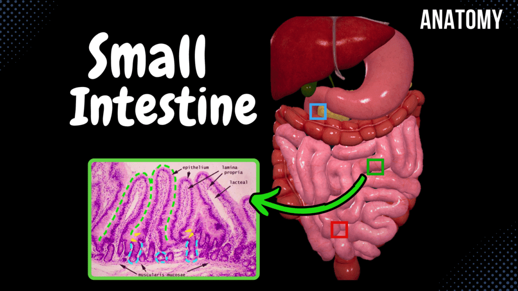
Small Intestine Anatomy (Parts, Topography, Structures, Layers) Official Links Instagram Youtube Jki-discord Notes & Illustrations Quizzes Summary & Transcript Notes ☆ Member Only Go to PDF Notes Illustrations ☆ Member Only Go to Illustrations 12345678910 Small Intestine – QUIZ Test your understanding with 10 random multiple-choice questions from the question bank. You're in the preview mode. Note: All elements work correctly on the front end. 1 / 10 Which layer of the small intestine contains lymphatic vessels called lacteals? A) Tunica Mucosa B) Tela Submucosa C) Tunica Muscularis D) Tunica Serosa Lacteals are present in the villi within the tunica mucosa. 2 / 10 What is the main function of plicae circulares in the small intestine? A) Increase absorption B) Neutralize acids C) Facilitate peristalsis D) Secrete enzymes Plicae circulares increase the surface area for nutrient absorption. 3 / 10 Which region of the small intestine absorbs iron? A) Ileum B) Duodenum C) Caecum D) Jejunum Iron absorption occurs primarily in the duodenum. 4 / 10 What is the landmark where the duodenum transitions into the jejunum? A) Duodenojejunal Flexure B) Ampulla of Vater C) Minor Duodenal Papilla D) Pyloric Orifice The duodenojejunal flexure marks this transition and is supported by the ligament of Treitz. 5 / 10 Which part of the small intestine is more vascularized? A) Ileum B) Jejunum C) Colon D) Duodenum The jejunum is more vascularized, giving it a darker color compared to the ileum. 6 / 10 Which part of the small intestine is primarily intraperitoneal? A) Jejunum and Ileum B) Ileum C) Entire Small Intestine D) Duodenum The jejunum and ileum are intraperitoneal and supported by the mesentery. 7 / 10 What is the function of crypts of Lieberkühn in the small intestine? A) Absorb nutrients B) Absorb fats C) Secrete intestinal juice D) Neutralize acid These glands secrete intestinal juice and renew epithelial cells. 8 / 10 Which part of the duodenum crosses anterior to the inferior vena cava and aorta? A) Superior Part B) Horizontal Part C) Descending Part D) Ascending Part The inferior (horizontal) part of the duodenum crosses these structures. 9 / 10 Which feature of the ileum is specific for immune function? A) Plicae Circulares B) Villi C) Goblet Cells D) Peyer’s Patches Peyer’s patches are clusters of lymphoid nodules in the ileum for immune defense. 10 / 10 What structure allows bile and pancreatic secretions to enter the duodenum? A) Major Duodenal Papilla B) Minor Duodenal Papilla C) Hepatic Duct D) Ampulla of Vater The major duodenal papilla allows bile and pancreatic juices to enter the duodenum. Your score is The average score is 0% Description Anatomy of the Small Intestine Small Intestine Overview: Extends from the Pylorus to the Caecum. Approximately 7 meters long in adults. Divided into three parts: Duodenum Jejunum Ileum Duodenum: Length: 20-30 cm Functions: Iron absorption Chemical digestion Chemical neutralization Processes chyme from the stomach Topography of the Duodenum: Skeletopy: Extends from L1 to L3. Syntopy: Posteriorly: Inferior Vena Cava, Aorta, Right Kidney Superiorly: Liver, Pancreas Anteriorly: Transverse Colon Holotopy: Epigastric and Umbilical Regions Jejunum and Ileum: No clear demarcation between them. Jejunum: 2-3 meters long Primary function: Absorption Ileum: 2-4 meters long Absorbs remaining nutrients and vitamins (especially Vitamin B12). Topography of the Jejunum and Ileum: Skeletopy: L2 to Right Iliac Fossa Syntopy: Surrounded by Caecum, Ascending Colon, Transverse Colon, Descending Colon, and Sigmoid Colon. Fixated posteriorly to the abdominal wall through the Root of the Mesentery (Radix Mesenterii). Covered anteriorly by the Greater Omentum. Holotopy: Ileum: Umbilical and Left Lateral Region Jejunum: Left Lateral Region, Right Inguinal Region, Pubic Region Anatomical Structures: Duodenum: Superior Part: From Pyloric Orifice (Ostium Pyloricum) to Superior Duodenal Flexure (Flexura Duodeni Superior). Descending Part: From Superior Duodenal Flexure to Inferior Duodenal Flexure (Flexura Duodeni Inferior). Inferior Horizontal Part: From Inferior Duodenal Flexure. Ascending Part: Until Duodenojejunal Flexure (Flexura Duodenojejunalis) / Ligament of Treitz. Jejunum: Wider and thicker than the ileum. More vascular. Darker in color. Ileum: Thinner walls. Less vascular. Lighter in color. Layers of the Small Intestinal Wall: Tunica Mucosa: Simple Columnar Epithelium Circular Folds (Plicae Circulare) – Absent in the Duodenal Bulb Intestinal Villi (Villi Intestinales) Microvilli Intestinal Glands (Glandulae Intestinales) Solitary Lymphoid Nodules (Noduli Lymphoidei Solitarii) Aggregated Lymphoid Nodules (Noduli Lymphoidei Aggregati) Specialized Structures in Duodenum: Major Duodenal Papilla (Papilla Duodeni Major) Minor Duodenal Papilla (Papilla Duodeni Minor) Tela Submucosa: Duodenum: Contains Brunner’s Glands. Jejunum: Longer villi. Ileum: More goblet cells and Peyer’s Patches (GALT). Tunica Muscularis: Responsible for Peristaltic Movement Inner Layer: Circular Muscle Layer Hepatopancreatic Sphincter (Sphincter of Oddi) (M. Sphincter Ampullae Hepatopancreatica) Outer Layer: Longitudinal Muscle Layer Tunica Serosa: Intraperitoneal Organs: Covered by Peritoneum. Retroperitoneal Organs (e.g., Duodenum): Have Tunica Adventitia in certain areas. Sources: Memorix Anatomy, 2nd Edition by Hudák Radovan, Kachlík David, and Volný Ondřej Biorender University Notes and Lectures Transcript Introduction0:04What’s up. Meditay here, and in this video, we’re gonna go through the anatomy of the0:08Small Intestine. So in the last video, we went through the anatomy of the Stomach.0:12Now the step after the Stomach is the Small Intestine, as you see here. So in this video,0:18we’re first going to look at the components that make up the small intestine, then we’re going0:23to look at their topography and go through how the small intestine is fixated to the posterior0:28abdominal wall. After that, we’ll look at some important anatomical structures related to the0:34three parts of it. And then, we’ll go through the layers that make up the small intestinal wall.Small Intestine Overview0:40Alright, so here you see the anterior view of the abdominal cavity,0:44the whole small intestine is highlighted in blue here.0:47It reaches from the Stomach all the way to the large intestine, as you see here.0:52To be more specific, it goes from the pylorus of the Stomach to the caecum of the large intestine.0:58Now the length of the small intestine may vary, but in total, if you stretch it out,1:04it’d be approximately 7 meters long. Let’s now look at the parts of the small1:08intestine to get a
Stomach Anatomy
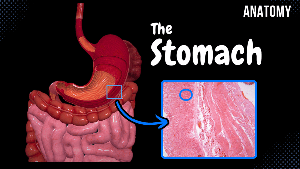
Stomach Anatomy (Topography, External Features, Parts, Layers) Official Links Instagram Youtube Jki-discord Notes & Illustrations Quizzes Summary & Transcript Notes ☆ Member Only Go to PDF Notes Illustrations ☆ Member Only Go to Illustrations 12345678910 Stomach – QUIZ Test your understanding with 10 random multiple-choice questions from the question bank. You're in the preview mode. Note: All elements work correctly on the front end. 1 / 10 Which ligament contains the portal triad? A) Hepatogastric Ligament B) Hepatoduodenal Ligament C) Gastrosplenic Ligament D) Gastrocolic Ligament The hepatoduodenal ligament encloses the portal vein, hepatic artery, and bile duct. 2 / 10 Which part of the stomach transitions into the duodenum? A) Fundus B) Cardiac Part C) Pyloric Part D) Body The pyloric part transitions into the small intestine via the pyloric canal and sphincter. 3 / 10 Which ligament forms part of the greater omentum and attaches the stomach to the diaphragm? A) Gastrophrenic Ligament B) Gastrosplenic Ligament C) Hepatogastric Ligament D) Gastrocolic Ligament The gastrophrenic ligament connects the stomach to the diaphragm. 4 / 10 What is the role of parietal cells in the stomach? A) Mucus B) HCl and Intrinsic Factor C) Hormones D) Pepsinogen Parietal cells secrete hydrochloric acid (HCl) and intrinsic factor. 5 / 10 What is the main venous drainage of the greater curvature of the stomach? A) Gastric Veins B) Splenic Veins C) Gastro-omental Veins D) Short Gastric Veins The gastro-omental veins drain the greater curvature. 6 / 10 What is the primary parasympathetic innervation of the stomach? A) Hypoglossal Nerve B) Sympathetic Chain C) Vagus Nerve D) Splanchnic Nerve The vagus nerve provides parasympathetic innervation to the stomach. 7 / 10 Which stomach wall layer facilitates peristaltic movement? A) Tunica Serosa B) Tela Submucosa C) Tunica Muscularis D) Tunica Mucosa The tunica muscularis, with its three muscle layers, facilitates peristalsis. 8 / 10 What is the term for the narrowing of the pyloric canal due to hypertrophy in infants? A) Achalasia B) Gastritis C) Gastric Ulcer D) Pyloric Stenosis Pyloric stenosis involves hypertrophy of the pyloric muscle, leading to narrowing. 9 / 10 What is the significance of the areae gastricae in the stomach? A) Secretes enzymes B) Protects the mucosa C) Provides blood supply D) Increases surface area Areae gastricae are small elevations of mucosa that increase the stomach’s absorptive surface. 10 / 10 Which ligament anchors the lesser curvature of the stomach to the liver? A) Gastrosplenic Ligament B) Gastrocolic Ligament C) Hepatogastric Ligament D) Gastrophrenic Ligament The hepatogastric ligament is part of the lesser omentum and connects the stomach to the liver. Your score is The average score is 0% Description Anatomy of the Stomach Topography of Gaster: Holotopy: Located in the Epigastric Region and Left Hypochondriac Region (Left Upper Quadrant). Extends from the Cardiac Orifice (Ostium Cardiacum) to the Pyloric Orifice (Ostium Pyloricum). Syntopy (Relations to Neighboring Structures): Anterior Wall: Diaphragm, Liver, Anterior Abdominal Wall Posterior Wall: Left Kidney, Spleen, Pancreas, Transverse Colon External Features of the Stomach: Lesser Curvature (Curvatura Minor) Angular Notch (Incisura Angularis) Greater Curvature (Curvatura Major) Cardiac Notch (Incisura Cardialis) Parts of the Stomach: Cardiac Part (Pars Cardiaca) Fundus (Fundus Gastricus) Body (Corpus Gastricum) Pyloric Part (Pars Pylorica) Pyloric Antrum (Antrum Pyloricum) Pyloric Canal (Canalis Pyloricus) Layers of the Gastric Wall: Tunica Mucosa (2-3 mm thick) Gastric Folds (Plicae Gastricae) Gastric Areas (Areae Gastricae) Simple Columnar Epithelium Gastric Pits (Foveolae Gastricae) Lamina Propria Muscularis Mucosae Tela Submucosa Loose connective tissue Tunica Muscularis (Smooth Muscle Layers) Inner: Oblique Muscle Layer Middle: Circular Muscle Layer Pyloric Sphincter (Musculus Sphincter Pyloricus) Outer: Longitudinal Muscle Layer Tunica Serosa Formed by the peritoneum Sources: Memorix Anatomy, 2nd Edition by Hudák Radovan, Kachlík David, and Volný Ondřej Biorender University Notes and Lectures Transcript Introduction0:00What’s up. Meditay here, and in this video, we’re gonna go through the anatomy of the stomach.0:04So in the last video, we went through the anatomy of the Esophagus. Now the step after0:09the Esophagus is the Stomach, as you see here. So in this video, we’re first going to go through0:14the topography of the stomach, basically where it is related to surrounding structures. After that,0:21we’re going to look at the external features of the stomach and the different parts of it. Then0:26we’ll go through the layers of the gastric wall. Now. Let’s go ahead and start with the topography.Topography of the Stomach0:31So the stomach lies in the epigastric region and the left hypochondriac region, as you see here.0:36Or, if you’re studying using the 4 quadrant division,0:38you’ll find it in the left upper quadrant. If we expose the stomach a little more,0:44you’ll see that it starts from the abdominal part of the Esophagus,0:48and then it curves to the left and then reaches out to the small intestine.0:53And these two openings that go towards the Esophagus and the small intestine has unique names0:59The opening by which the Esophagus communicates with the stomach is known as the cardiac orifice1:06at the level of the 10th to 11th thoracic vertebrae. And the opening by which the1:11stomach communicates with the duodenum of the small intestine is known as the pyloric orifice1:16at the level of the 1st lumbar vertebra So if we look at the stomach from this1:21perspective, laterally, you’ll see this. The stomach has an Anterior wall and a1:27Posterior wall. So from this position, we can look at the1:30Syntopy, meaning the position of the Stomach To other organs. Anteriorly1:36We can find the Diaphragm being very close to the anterior wall; we’ll find the liver,1:41and we’ll also find the stomach facing the anterior abdominal wall aswell. And the1:46Posterior wall of the Stomach is close to the left kidney. You’ll also see the spleen,1:51the pancreas, and the transverse colon facing the posterior surface of the stomach as well.External Features of the Stomach1:56So that was the Syntopy. Now, let’s go back and look at the external structures of the stomach.2:01We can see two distinct curvatures; we have the lesser curvature2:06Here. And since we have a lesser curvature, we also have a greater curvature here. On the2:11lesser curvature,
How to Divide the Abdomen
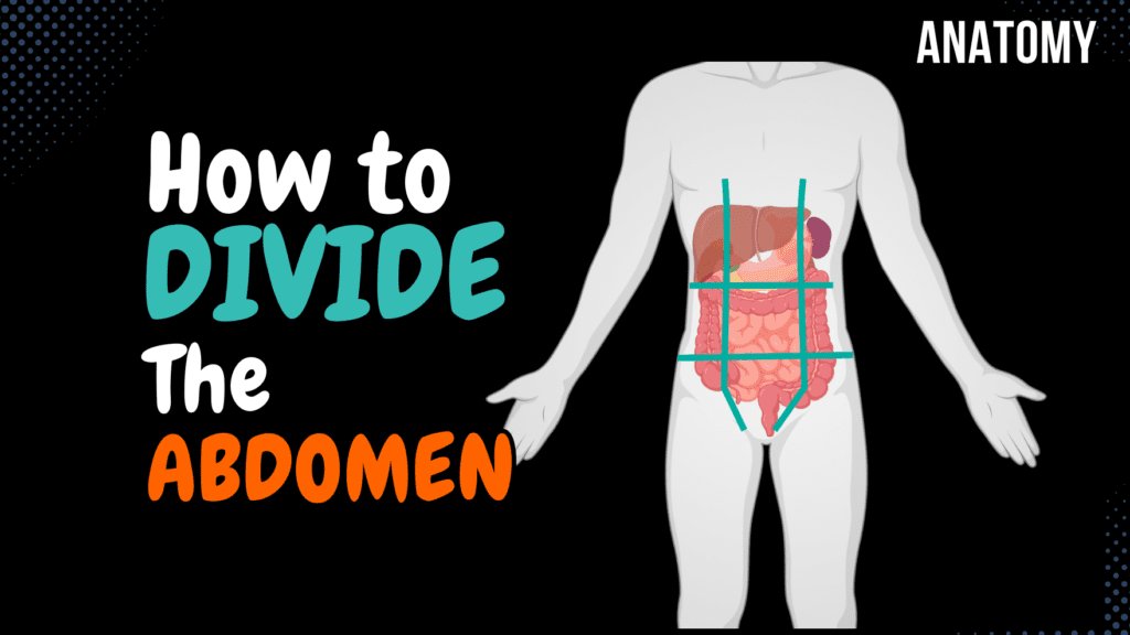
How to Divide the Abdomen (9 Regions) Official Links Instagram Youtube Jki-discord Notes & Illustrations Quizzes Summary & Transcript 📢 Currently, there is no PDF for this video.If you’re interested in having one, feel free to send an inquiry, and I may create it in the future. BUT! There’s a quiz available in the next tab. 12345678910 Dividing the Abdomen – QUIZ Test your understanding with 10 random multiple-choice questions from the question bank. You're in the preview mode. Note: All elements work correctly on the front end. 1 / 10 Which region lies inferior to the epigastric region in the nine-region scheme? A) Hypochondriac Region B) Inguinal Region C) Umbilical Region D) Pubic Region The umbilical region lies directly inferior to the epigastric region. 2 / 10 Which region is located at the center of the nine abdominal regions? A) Right Hypochondriac Region B) Epigastric Region C) Umbilical Region D) Pubic Region The umbilical region is located at the center of the nine abdominal regions. 3 / 10 What region contains most of the small intestine? A) Right Lateral Region B) Pubic Region C) Umbilical Region D) Epigastric Region The umbilical region contains most of the small intestine. 4 / 10 What is the distance between the ribs used for abdominal division called? A) Distantia Costarum B) Costal Angle C) Rib Separation D) Subcostal Width The distance is called distantia costarum. 5 / 10 What is the function of the interspinal plane in abdominal division? A) Identify Organs B) Separate Middle and Lower Levels C) Divide Quadrants D) Aid Vascular Mapping The interspinal plane separates the middle and lower levels of the abdomen. 6 / 10 Which organ is primarily located in the pubic region? A) Small Intestine B) Bladder C) Liver D) Appendix The bladder is the primary organ in the pubic region. 7 / 10 What anatomical landmark is used for the vertical lines in abdominal division? A) Sternum B) Iliac Crest C) Rectus Abdominis and Clavicle D) Umbilicus The vertical lines follow the rectus abdominis muscle and pass through the midpoint of the clavicle. 8 / 10 What is the name of the lower horizontal plane? A) Subcostal Plane B) Interclavicular Plane C) Interspinal Plane D) Suprapubic Plane The interspinal plane is the lower horizontal plane used in abdominal division. 9 / 10 Which vertical lines are used in the nine-region scheme? A) Sagittal Lines B) Intercostal Lines C) Subclavicular Lines D) Midclavicular Lines The vertical lines pass through the midpoint of the clavicle and the pubic tubercle. 10 / 10 What is the anatomical name for the vertical lines in the four-quadrant scheme? A) Transumbilical Line B) Midclavicular Line C) Median Plane D) Sagittal Line The vertical line is called the median plane. Your score is The average score is 0% Description Dividing the Abdominal Region This video covers how the abdominal region is divided using anatomical planes and regions. Holotopy: The relation between an organ and the body as a whole. Horizontal Lines: Upper Horizontal Plane: Subcostal Plane Lower Horizontal Plane: Interspinal Plane Distance between the ribs: Distantia Costarum Divided into: Upper Level Middle Level Lower Level Vertical Lines: Along the Rectus Abdominis Muscle From the Pubic Tubercle (Tuberculum Pubicum) Extends approximately to the middle point of the clavicle 9-Region Scheme: Upper Level: Epigastric Region Right Hypochondriac Region Left Hypochondriac Region Middle Level: Umbilical Region Right Lateral (Lumbar) Region Left Lateral (Lumbar) Region Lower Level: Pubic (Hypogastric) Region Right Inguinal Region Left Inguinal Region 4-Quadrant Scheme: The abdomen can also be divided into four quadrants: Right Upper Quadrant (RUQ) Left Upper Quadrant (LUQ) Right Lower Quadrant (RLQ) Left Lower Quadrant (LLQ) Why Divide the Abdomen? Used in clinical practice to locate pain and pathology. Helps in understanding the placement of abdominal organs. Essential for surgical and diagnostic procedures. Sources: Memorix Anatomy, 2nd Edition by Hudák Radovan, Kachlík David, and Volný Ondřej Biorender University Notes and Lectures Transcript Introduction0:03What’s up, Meditay here, and in this video, we’ll be going through the procedure on how0:08we divide the abdomen into 9 distinct regions. Because as you’re studying the organs of the0:13abdominal cavity, you’ll most probably see the word Holotopy, which means the relationship0:18between the organs and the body as a whole, and by describing the holotopy of an organ, you’ll0:23be stating at which region the organ Is located. So, Let’s start by dividing the abdominal regionHorizontal Lines0:29using two horizontal lines first. We call these the Upper horizontal plane, and the0:34lower horizontal plane. The upper horizontal plane is at the lowest point of the costal arch and is0:40therefore also called the subcostal place. While the lower horizontal plane is between the Spina0:46Iliaca ant. Superior, so the lower horizontal place is sometimes called the interspinal plane.0:53This will divide the abdomen into three levels using this arch0:56Right here that the lower edges of the rib cartilage form, that’s the distance between1:00the ribs – called distantia costarum. Now We get an upper level, above this arch,1:06We get a middle level under the costal arch, until the1:09Interspinous place, and then under the interspinous plane, you’ll find the lower level.Vertical Lines1:15And now, we need to add the vertical lines. And we add them using the rectus1:20Abdominis muscle, as you see here. And the way we add the vertical lines is along this muscle,1:25along the Musculus rectus abdominis. It starts Down here at pubic tubercule1:30Then it goes along the rectus abdominis and then reaches approximately at the1:34middle point of the clavicula up here. So now we get nine distinct regions, right?Nine Regions1:40At the upper level, we get the epigastric region and the Right and Left hypochondriac region on1:45either side beneath the cartilage. And then on the middle level, we get1:48the umbilical region in the middle, since the umbilicus, or the navel, is here in the middle.1:53And then, laterally, we have the right and left lateral region.1:57And then, at the lower region, we got the pubic region and the right and left inguinal2:02region or sometimes also referred to as the right and left Iliac
Esophagus
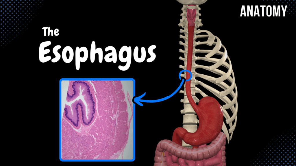
Esophagus (Parts, Curvatures, Constrictions, Layers) Official Links Instagram Youtube Jki-discord Notes & Illustrations Quizzes Summary & Transcript Notes ☆ Member Only Go to PDF Notes Illustrations ☆ Member Only Go to Illustrations 12345678910 Esophagus – QUIZ Test your understanding with 10 random multiple-choice questions from the question bank. You're in the preview mode. Note: All elements work correctly on the front end. 1 / 10 Which condition is associated with esophageal varices? A) Hiatal Hernia B) Achalasia C) GERD D) Portal Hypertension Esophageal varices are dilated veins in the lower esophagus due to portal hypertension, often associated with liver disease. 2 / 10 What is the function of the esophageal cardiac glands? A) Facilitate Digestion B) Produce Enzymes C) Increase Peristalsis D) Protect Against Gastric Reflux Esophageal cardiac glands secrete mucus to protect the esophagus from gastric reflux. 3 / 10 What is the primary function of the esophagus? A) Hormonal Regulation B) Digestion of Proteins C) Transport Food D) Absorption of Nutrients The esophagus transports food and liquids from the pharynx to the stomach via peristaltic contractions. 4 / 10 What type of muscle fibers dominate the middle third of the esophagus? A) Cardiac Muscle B) Skeletal Muscle C) Smooth Muscle D) Mixed Skeletal and Smooth The middle third of the esophagus contains a mix of skeletal and smooth muscle fibers. 5 / 10 What is the histological characteristic of the esophageal submucosa? A) Dense Connective Tissue B) Smooth Muscle C) Elastic Tissue D) Loose Connective Tissue with Glands The esophageal submucosa contains loose connective tissue and esophageal glands. 6 / 10 What is the primary innervation of the esophagus? A) Vagus Nerve B) Accessory Nerve C) Hypoglossal Nerve D) Phrenic Nerve The esophagus is primarily innervated by the esophageal plexus, formed by the vagus nerve and sympathetic trunk. 7 / 10 What is the function of the tunica muscularis in the esophagus? A) Absorbs Nutrients B) Secretes Mucus C) Supports Lamina Mucosa D) Generates Peristalsis The tunica muscularis generates peristaltic waves to propel food into the stomach. 8 / 10 Which nerve primarily controls the peristalsis of the esophagus? A) Vagus Nerve B) Sympathetic Nerve C) Hypoglossal Nerve D) Phrenic Nerve The vagus nerve controls the peristalsis of the esophagus via the esophageal plexus. 9 / 10 What condition is caused by dysfunction of the LES? A) Barrett's Esophagus B) GERD C) Achalasia D) Esophageal Cancer Dysfunction of the LES leads to gastroesophageal reflux disease (GERD). 10 / 10 What is the physiological function of the lower esophageal sphincter (LES)? A) Prevent Backflow B) Absorb Stomach Acid C) Promote Backflow D) Facilitate Peristalsis The LES prevents the backflow of stomach contents into the esophagus, protecting against acid reflux. Your score is The average score is 0% Description Esophagus: Length: ~25 cm Located between the pharynx and the stomach Divided into three parts: Cervical Part (Pars Cervicalis) Thoracic Part (Pars Thoracica) Abdominal Part (Pars Abdominalis) Parts of the Esophagus: Cervical Part: Begins at the Pharyngeal Opening into Esophagus (Ostium Oesophageum) Ends at the Superior Thoracic Aperture (Apertura Thoracica Superior) Thoracic Part: Starts at the Superior Thoracic Aperture (Apertura Thoracica Superior) Ends at the Esophageal Hiatus (Hiatus Oesophageus Diaphragmatis) Abdominal Part: Begins at the Esophageal Hiatus (Hiatus Oesophageus Diaphragmatis) Ends at the Cardiac Orifice (Ostium Cardiacum Gastrici) Curvatures of the Esophagus: Curves to the left at the beginning (Cervical Part) Curves to the right at the middle (Thoracic Part) Curves to the left above the diaphragm Constrictions of the Esophagus: Anatomical Constrictions: Pharyngoesophageal Constriction Bronchoaortic Constriction Diaphragmatic Constriction Physiological Constrictions: At the level of T8-T9 At the level of T11 (Lower Esophageal Sphincter) Layers of the Esophageal Wall: Tunica Mucosa: Stratified Squamous Non-Keratinized Epithelium Contains mucous glands and vasculature Includes Lamina Muscularis Mucosa Tela Submucosa: Loose connective tissue Contains esophageal glands and esophageal cardiac glands Tunica Muscularis: Inner circular muscle fibers Outer longitudinal muscle fibers Muscle composition: Upper 1/3: Skeletal muscle Middle 1/3: Mixed skeletal and smooth muscle Lower 1/3: Smooth muscle Tunica Adventitia & Tunica Serosa: Tunica Adventitia: Covers the esophagus in the thoracic part Tunica Serosa: Present in the abdominal part Sources: Memorix Anatomy, 2nd Edition by Hudák Radovan, Kachlík David, and Volný Ondřej Biorender University Notes and Lectures Transcript Introduction0:00What’s up. Meditay here, and in this video, we’re gonna go through the anatomy of the Esophagus.0:04So in the last video, we went through the anatomy of the Pharynx. Now the step after0:09the Pharynx is the Esophagus, as you see here. So in this video, we’re first going to look at the0:14parts of the Esophagus. After that, look at the curvatures and the constrictions of the esophagus.0:20Then we’re gonna go through the layers of the esophageal wall through a histology slide.0:24Cool, let’s start by looking at an anterior view of the Esophagus. The esophagus lies between theParts of the Esophagus0:31Pharynx, and the stomach as you see here. The length of the esophagus varies a lot, but in0:36average It should be approximately 25 cm long. Now. The esophagus is divided into three parts0:42according to their anatomical location. First we have the cervical part, which lies in the neck.0:48Then we have the Thoracic Part, which lies in the thoracic cavity. And then we have the Abdominal0:52Part, which lies in the abdominal cavity. So these are the three parts of the esophagus,0:58let’s go through where each of them start and end. The cervical part starts from the pharynx.1:03So remember the Pharynx has two openings. There’s the Laryngeal Inlet, which leads into the Larynx.1:08And there’s the Pharyngeal Opening into the Esophagus, or Ostium esophageum, which is1:13going to be the start of the cervical part of the esophagus. And the cervical part ends just1:18before it enters the thoracic cavity through the superior thoracic aperture, or the upper opening1:24of the ribcage. So the cervical part starts at the Pharyngeal opening into he esophagus,1:30and ends at the Superiro thoracic aperture. After the cervical part is the thoracic part.1:36And this one is going to start at the superior thoracic aperture and go all the way
Salivary Glands & Saliva
Salivary Glands & Saliva (Parotid, Submandibular, Sublingual) Official Links Instagram Youtube Jki-discord Notes & Illustrations Quizzes Summary & Transcript Notes ☆ Member Only Go to PDF Notes Illustrations ☆ Member Only Go to Illustrations 12345678910 Salivary Glands – QUIZ Test your understanding with 10 random multiple-choice questions from the question bank. You're in the preview mode. Note: All elements work correctly on the front end. 1 / 10 Which glands are located within the labial mucosa? A) Palatine Glands B) Buccal Glands C) Labial Glands D) Lingual Glands Labial glands are minor salivary glands located in the labial mucosa. 2 / 10 Which glands are located on the tongue? A) Palatine Glands B) Lingual Glands C) Buccal Glands D) Sublingual Glands Lingual glands, such as von Ebner’s glands, are located on the tongue. 3 / 10 Where is the accessory parotid gland located? A) Anterior to the Parotid Duct B) Near the Submandibular Duct C) Near the Sublingual Caruncle D) Posterior to the Parotid Gland The accessory parotid gland is located near the parotid duct. 4 / 10 What is the clinical significance of the parotid duct? A) Drains Submandibular Secretions B) Potential for Obstruction and Infection C) Enhances Mucous Production D) Lymphatic Drainage The parotid duct can be obstructed or infected, leading to sialadenitis or salivary stones. 5 / 10 What type of secretion do labial glands produce? A) Mucous B) Serous C) Enzymatic D) Mixed Labial glands primarily produce mucous secretions. 6 / 10 What is the role of von Ebner’s glands? A) Protect Against Infection B) Aid in Lubrication C) Secrete Mucus D) Cleanse Circumvallate Papillae Von Ebner’s glands secrete serous fluid to cleanse the circumvallate papillae. 7 / 10 What structure surrounds the parotid gland? A) Parotid Fascia B) Buccal Fascia C) Submandibular Fascia D) Sublingual Fascia The parotid gland is enclosed in the parotid fascia. 8 / 10 Which gland contributes the least to total salivary output? A) Sublingual Gland B) Submandibular Gland C) Minor Salivary Glands D) Parotid Gland The sublingual gland contributes the least to overall salivary output. 9 / 10 What are the secretory units of salivary glands called? A) Acini B) Nodes C) Lobules D) Ducts The secretory units are acini, classified as serous, mucous, or mixed. 10 / 10 Where does the parotid duct open in the oral cavity? A) Opposite First Lower Molar B) Opposite Canine Tooth C) Opposite Premolars D) Opposite Second Upper Molar The parotid duct opens into the oral cavity near the second upper molar. Your score is The average score is 0% Description Salivary Glands: Minor Salivary Glands Major Salivary Glands Components of Saliva: Serous Component (Contains Enzymes) Produced by Serous Glands Mucous Component (Mucus) Produced by Mucous Glands Seromucous Glands (Mixed Secretion) Minor Salivary Glands: Labial Glands (Glandulae Labiales) Buccal Glands (Glandulae Buccales) Palatine Glands (Glandulae Palatinae) Lingual Glands (Glandulae Linguales) Major Salivary Glands: Parotid Gland (Glandula Parotidea) Submandibular Gland (Glandula Submandibularis) Sublingual Gland (Glandula Sublingualis) Parotid Gland: Largest Salivary Gland Divided into: Superficial Part (Pars Superficialis) Located near the Zygomatic Arch and Angle of Mandible Deep Part (Pars Profunda) Located in the Retromandibular Fossa Encased by Parotid Fascia Parotid Duct (Ductus Parotideus) / Stensen’s Duct Opens at the Papilla of the Parotid Duct (Papillae Ductus Parotidei) Accessory Parotid Gland (Glandula Parotidea Accessoria) Submandibular Gland: Seromucous Gland Located in the Submandibular Space Submandibular Duct (Ductus Submandibularis) / Wharton’s Duct Opens at the Sublingual Caruncle (Caruncula Sublingualis) Sublingual Gland: Smallest Major Salivary Gland Major Sublingual Duct (Ductus Sublingualis Major) / Duct of Bartholin Opens at the Sublingual Caruncle Minor Sublingual Ducts (Ductus Sublinguales Minores) Located along the Sublingual Folds (Plica Sublingualis) Sources: Memorix Anatomy, 2nd Edition by Hudák Radovan, Kachlík David, and Volný Ondřej Biorender University Notes and Lectures Transcript Introduction0:03What’s up, meditay here and in this video, we’re going to go through the different0:07salivary glands you have around the oral cavity. So the salivary glands are divided based on their0:14size. There are the minor salivary glands, that are scattered throughout the oral cavity,0:18and there’s the major salivary glands, which has ducts that open into the oral cavity0:23to secrete out its saliva. But first, we need to address some words I’m gonna useSaliva0:28throughout this video so that you understand the whole idea regarding salivary glands0:33Our saliva is made up two components. There’s a serous component, and there’s a mucous component.0:40The serous component contains enzymes that help us digest the food we eat.0:45And the mucous component is mucous, that lubricates the inner surfaces of our mouth,0:50as well as lubricating the food we eat so that it passes easily down to the0:54next step of the digestive system. And these two components are produced by two different glands.1:01They’re the Serous gland, and the mucous gland, so let’s go through these a little bit.1:06The serous gland looks like this. It contains a lot of granules that produces watery secretions1:12containing enzymes like alpha amylase. Mucous glands look like this. They stain1:18lighter than the serous gland because they don’t have these granules that the serous gland does,1:23and it mainly produces mucin, that absorbs water to form a lubricating secretion called mucus.1:30Then there’s a combination of those, called seromucous gland, which look like this.1:34That produces both mucous and enzymes. Alright. Now that you have a generalMinor Salivary Glands1:40knowledge of the different glands. Let’s start with the minor salivary glands. The1:44minor salivary glands are scattered throughout the oral cavity. And they produce saliva continuously,1:51without any neuronal stimulation. The majority of those are gonna be seromucous glands.1:57There are minor salivary glands in the lips. Called labial glands.2:01There are glands in the buccal region,2:03called buccal glands. Then there’s the Palatine glands,2:06and lingual glands. These are the main minor salivary glands that we have in the oral cavity.Major Salivary Glands2:12Then we have the Major Salivary Glands, which look like this. They are the Parotid Gland,2:18Submandibular gland, and the sublingual glands. Cool. Let’s start with the parotid gland!Parotid Gland2:23Which is this one. Now there are three things that I want you to remember2:28when it comes to
Oral Cavity Proper
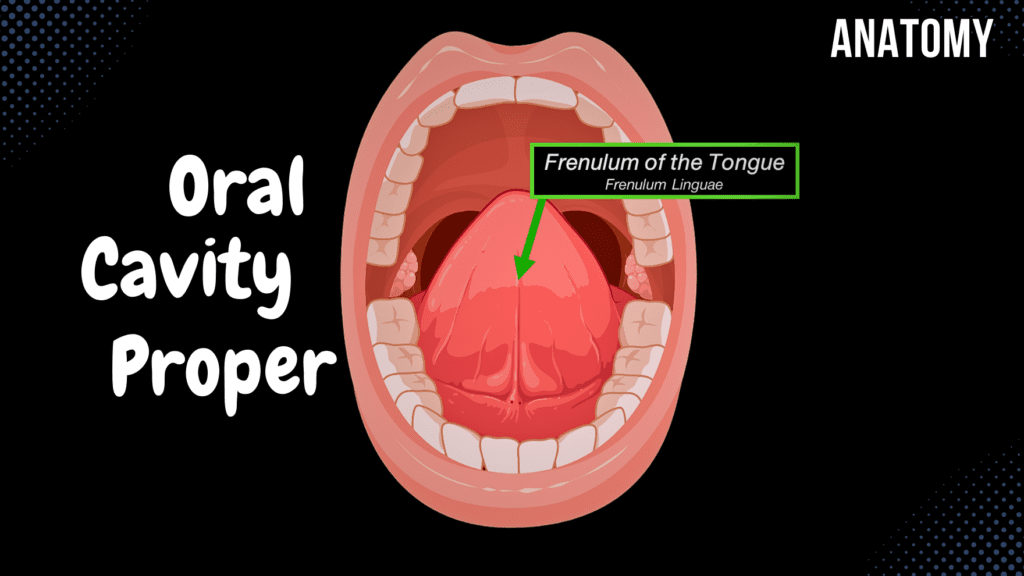
Oral Cavity Proper (Palate & Tongue) Official Links Instagram Youtube Jki-discord Notes & Illustrations Quizzes Summary & Transcript Notes ☆ Member Only Go to PDF Notes Illustrations ☆ Member Only Go to Illustrations 12345678910 Oral Cavity Proper – QUIZ Test your understanding with 10 random multiple-choice questions from the question bank. You're in the preview mode. Note: All elements work correctly on the front end. 1 / 10 Which part of the tongue contains the foramen caecum? A) Root B) Apex C) Body D) Terminal sulcus The terminal sulcus of the tongue contains the foramen caecum. 2 / 10 What is the role of the palatopharyngeus muscle? A) Moves the soft palate B) Elevates the pharynx C) Changes the tongue shape D) Forms the soft palate arch It elevates the pharynx during swallowing. 3 / 10 Which intrinsic muscle of the tongue alters its shape? A) Superior and inferior longitudinal muscles B) Hyoglossus C) Palatoglossus D) Styloglossus The superior and inferior longitudinal muscles alter the shape of the tongue. 4 / 10 What is the structure that divides the tongue into the anterior and posterior regions? A) Terminal sulcus B) Lingual papillae C) Median glossoepiglottic fold D) Frenulum of the tongue The terminal sulcus divides the tongue into anterior and posterior regions. 5 / 10 What structure is located between the palatoglossal and palatopharyngeal arches? A) Terminal sulcus B) Palatine tonsil C) Uvula D) Lingual tonsil The palatine tonsil is located between the palatoglossal and palatopharyngeal arches. 6 / 10 What is the function of the soft palate during swallowing? A) Protects teeth B) Anchors tongue muscles C) Blocks the nasal cavity D) Guides food into the oesophagus The soft palate blocks the nasal cavity during swallowing to prevent food from entering. 7 / 10 Which structure forms the superior border of the soft palate? A) Hard palate B) Incisive papilla C) Palatoglossal arch D) Frenulum The superior border of the soft palate is attached to the posterior part of the hard palate. 8 / 10 Which structures form the superior border of the oral cavity proper? A) Palatine tonsils B) Mylohyoid muscle C) Oropharyngeal isthmus D) Hard and soft palate The hard and soft palate form the superior border. 9 / 10 What is the anterior boundary of the oral cavity proper? A) Soft palate B) Oropharyngeal isthmus C) Mylohyoid muscle D) Teeth and gums The teeth and gums form the anterior boundary of the oral cavity proper. 10 / 10 Which muscle forms the bulk of the tongue? A) Palatoglossus B) Genioglossus C) Styloglossus D) Hyoglossus The genioglossus muscle forms the bulk of the tongue. Your score is The average score is 0% Description Borders of the Oral Cavity Proper: Anterior and Lateral Border: Teeth, Gums Superior Border: Hard Palate, Soft Palate Inferior Border: Mylohyoid, Digastric, Geniohyoid Muscles, Tongue Posterior Border: Oropharyngeal Isthmus Superior Border: Hard Palate (Palatum Durum) Anterior Part: Alveolar Processes of Maxilla Posterior Part: Horizontal Plate of Palatine Bone Covered by Periosteum Covered by Mucous Membrane containing Palatine Glands Key Structures: Incisive Papilla Palatine Raphe Transverse Palatine Folds Soft Palate (Palatum Molle) Velum Palatinum – Free part of the posterior palate Contains muscle tissue, palatine aponeurosis, vasculature, and mucous glands Key Structure: Uvula (Uvula Palatina) Swallowing Process: Tongue Blocks the Oral Cavity Soft Palate Blocks the Nasal Cavity Epiglottis Blocks the Larynx Muscles of the Soft Palate: Muscle of the Uvula (Musculus Uvulae) Levator Veli Palatini (Musculus Levator Veli Palatini) Tensor Veli Palatini (Musculus Tensor Veli Palatini) Palatoglossus Muscle (Musculus Palatoglossus) Palatopharyngeus Muscle (Musculus Palatopharyngeus) Structures Formed by the Palatoglossal and Palatopharyngeal Muscles: Palatoglossal Arch (Arcus Palatoglossus) Palatopharyngeal Arch (Arcus Palatopharyngeus) Palatine Tonsil (Tonsilla Palatina) Inferior Border: Floor of the Oral Cavity Mylohyoid Muscle Anterior Belly of Digastric Muscle Geniohyoid Muscle Structures of the Tongue: Muscle organ composed of several muscles Divided into 3 parts: Apex, Body, and Root of Tongue Key Features: Medial Sulcus (Sulcus Medianus) Terminal Sulcus (Sulcus Terminalis) Foramen Caecum of the Tongue (Foramen Linguae) Lingual Tonsil (Tonsilla Lingualis) Glossoepiglottic Folds: Right/Left Glossoepiglottic Folds (Plica Glossoepiglottica Dextra et Sinistra) Median Glossoepiglottic Fold (Plica Glossoepiglottica Mediana) Epiglottic Vallecula (Valleculae Epiglotticae) Frenulum of the Tongue (Frenulum Linguae) Fimbriated Folds (Plica Fimbriata) Sublingual Folds (Plica Sublingualis) Sublingual Caruncle (Caruncula Sublingualis) Muscles of the Tongue: Extrinsic (Extraglossal) Muscles: Genioglossus Muscle (Musculus Genioglossus) Styloglossus Muscle (Musculus Styloglossus) Hyoglossus Muscle (Musculus Hyoglossus) Palatoglossus Muscle (Musculus Palatoglossus) Intrinsic (Intraglossal) Muscles: Superior and Inferior Longitudinal Muscles Vertical Muscles of the Tongue Transverse Muscles of the Tongue Lingual Papillae: Filiform Papilla Fungiform Papilla Vallate Papilla Foliate Papilla Posterior Border: Oropharynx Isthmus of Fauces (Isthmus Faucium) Sources: Memorix Anatomy, 2nd Edition by Hudák Radovan, Kachlík David, and Volný Ondřej. Biorender University Notes and Lectures Transcript Introduction0:00What’s up, Meditay here. Let’s talk about the digestive system anatomy. In our last video,0:01we covered the anatomical structures associated with the Oral Vestibule. Which remember consists0:06of the external borders with the Lips and Cheeks, and the Inter borders with the Teeth and Gums.0:12Now let’s wrap up the anatomy of the oral cavity, by going through the anatomical structures0:17associated with the Oral Cavity Proper. And to do that, we need to go through the0:21borders of the oral cavity proper. First, we have the Anterior and Lateral border,0:27which are the alveolar processes and the gums, as well as the teeth, we already covered themBorders of the Oral Cavity Proper0:31in the last video, since the teeth and gums are also the internal borders of the oral vestibule.0:36Superiorly however, you’ll find the hard palate, and the soft palate.0:40The inferior border consist of the floor of the oral cavity, which is made up by0:45the mylohyoid muscle, anterior belly of digastric muscle and the geniohyoid muscle. And you’ll find0:51the tongue here in the inferior border. Posteriorly, the oral cavity proper will0:56continue into the pharynx through the oropharyngeal isthmus.1:00So our goal in this video is to go through all the structures associated with these borders here.1:06We talked through the anterior and lateral border in our last video. Now, let’s start with the1:11superior