
Muscles of the Shoulder
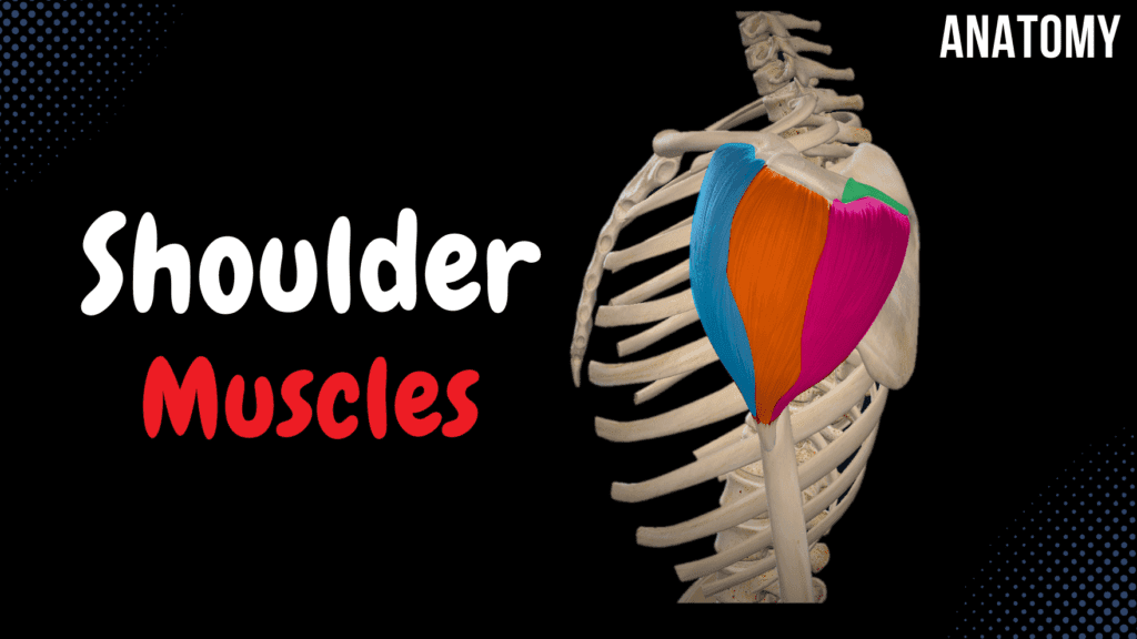
Muscles of the Shoulder (Division, Origin, Insertion, Function) Official Links Instagram Youtube Jki-discord Notes & Illustrations Quizzes Summary & Transcript Notes ☆ Member Only Go to PDF Notes Illustrations ☆ Member Only Go to Illustrations 12345678910 Muscles of the Shoulder – QUIZ Test your understanding with 10 random multiple-choice questions from the question bank. You're in the preview mode. Note: All elements work correctly on the front end. 1 / 10 Which part of the deltoid muscle is responsible for flexion of the shoulder? A) Clavicular part B) Acromial part C) All parts D) Spinal part The clavicular part of the deltoid muscle flexes the shoulder. 2 / 10 What is the primary function of the deltoid’s acromial part? A) Internal rotation B) Extension C) Flexion D) Abduction The acromial part of the deltoid abducts the arm. 3 / 10 Which muscle inserts into the lesser tubercle of the humerus? A) Subscapularis B) Infraspinatus C) Teres minor D) Supraspinatus The subscapularis muscle inserts into the lesser tubercle of the humerus. 4 / 10 What is the function of the teres major muscle? A) Abduction B) External rotation C) Adduction, extension, and internal rotation D) Flexion The teres major performs adduction, extension, and internal rotation of the arm. 5 / 10 What is the primary role of the rotator cuff muscles? A) Abduction of arm B) External rotation C) Flexion of shoulder D) Stabilization of shoulder joint The rotator cuff muscles stabilize the shoulder joint during movement. 6 / 10 What is the function of the teres major muscle? A) Abduction B) Internal rotation C) Elevation D) External rotation The teres major is responsible for adduction, extension, and internal rotation of the arm. 7 / 10 What is the insertion of the teres major muscle? A) Deltoid tuberosity B) Greater tubercle C) Crest of the lesser tubercle D) Radial tuberosity The teres major muscle inserts into the crest of the lesser tubercle of the humerus. 8 / 10 What is the main action of the deltoid’s spinal part? A) Adduction B) Flexion C) Extension and external rotation D) Abduction The spinal part of the deltoid extends and externally rotates the arm. 9 / 10 What is the insertion point of the infraspinatus muscle? A) Deltoid tuberosity B) Lesser tubercle C) Acromion D) Greater tubercle The infraspinatus inserts into the greater tubercle of the humerus. 10 / 10 Which muscle inserts into the crest of the greater tubercle? A) Subscapularis B) Infraspinatus C) Supraspinatus D) Pectoralis major The pectoralis major inserts into the crest of the greater tubercle of the humerus. Your score is The average score is 0% Description This video covers the muscles of the shoulder joint, including their origins, insertions, functions, and clinical significance. Muscles of the Shoulder Joint Deltoid Subscapularis Supraspinatus Infraspinatus Teres Minor Teres Major Deltoid (Musculus Deltoideus) Spinal Part Origin: Spine of Scapula (Spina Scapulae) Acromial Part Origin: Acromion of Scapula Clavicular Part Origin: Clavicle Insertion: Deltoid Tuberosity of Humerus (Tuberositas Deltoidei Humeri) Function: A: Abduction (Abductio Brachii) C: Internal Rotation (Rotatio Brachii Interna) S: External Rotation (Rotatio Brachii Externa) S+C: Adduction A+C: Flexion A+S: Extension Subscapularis (Musculus Subscapularis) Origin: Subscapular Fossa (Fossa Subscapularis) Insertion: Lesser Tubercle + Crest of Humerus (Tuberculum Minus Humeri et Crista Tuberculi Minoris) Function: Adduction, Internal Rotation Supraspinatus (Musculus Supraspinatus) Origin: Supraspinous Fossa (Fossa Supraspinata) Insertion: Greater Tubercle of Humerus (Tuberculum Majus Humeri) Function: Abduction Infraspinatus (Musculus Infraspinatus) Origin: Infraspinous Fossa (Fossa Infraspinata) Insertion: Greater Tubercle of Humerus (Tuberculum Majus Humeri) Function: External Rotation Teres Minor (Musculus Teres Minor) Origin: Lateral Margin of Scapula (Margo Lateralis Scapulae) Insertion: Greater Tubercle of Humerus (Tuberculum Majus Humeri) Function: External Rotation, Extension Teres Major (Musculus Teres Major) Origin: Inferior Angle of Scapula (Angulus Inferior Scapulae) Insertion: Crest of Lesser Tubercle of Humerus (Crista Tuberculi Minoris Humeri) Function: Adduction Extension Internal Rotation Rotator Cuff Muscles Subscapularis (Musculus Subscapularis) Supraspinatus (Musculus Supraspinatus) Infraspinatus (Musculus Infraspinatus) Teres Minor (Musculus Teres Minor) Function: Stabilizes the Shoulder Joint Clinical Importance ⚠ Rotator Cuff Tear Tendinitis Bursitis Transcript Introduction0:03What’s up. Meditay here and in this video, we’ll be looking at the muscles of the shoulders,0:08which are a part of the upper limb. Alright. So, the muscles of the upper limb are divided into 40:13parts according to their anatomical location. The first group are muscles of the shoulder joint.0:19Then we have the muscles of the arm, muscles of the forearm and then the muscles of the hand.0:24So again, muscles of the shoulder joint are what we’re gonna focus on today. These areDivision of the Shoulder Muscles0:28the Deltoid Muscle, Subscapularis, Supraspinatus, Infraspinatus, Teres Minor and Teres Major. All0:36of these muscles surround the shoulder joint. But the supraspinatus, infraspinatus, teres minor and0:42subscapularis form the rotator cuff muscles, which allows rotation of the humerus at the shoulder0:48joint, they provide stability to the shoulder and prevents dislocation.0:52These muscles are clinically important because any rotator cuff injury can cause a dull pain in0:58the shoulder, which often worsens when you elevate the arm in a certain way.1:02Alright. So let’s talk about these muscles. We’ll add a skeleton to use it as a landmark.Deltoid1:07First we have the deltoid muscle. Which is this large muscle on the shoulder that1:11is used as an injection site in the shoulder. This muscle is divided into 3 parts according1:17to their place of origin. It’s divided into the Spinal part, Acromial Part,1:22and the Clavicular part. So the spinal part originates from the Spine of Scapula, as you1:27see here. Acromial part from the acromion of the scapula, and clavicular part from the…. Clavicle.1:35Then they all unite and insert at a common region, which is the Deltoid tuberosity of the Humerus.1:41The function of the deltoid muscle depends on which muscle fibers contract. For instance,1:46if Acromial part contract alone, it abducts the arm. If clavicular part contracts alone, it1:53rotates the arm internally, and if the Spinal part contracts alone, it rotates the arm externally. If2:00the Spinal part and the Clavicular part contract, they adduct the arm. If the Acromial part and the2:06clavicular part contract,
Fascia of the Thorax
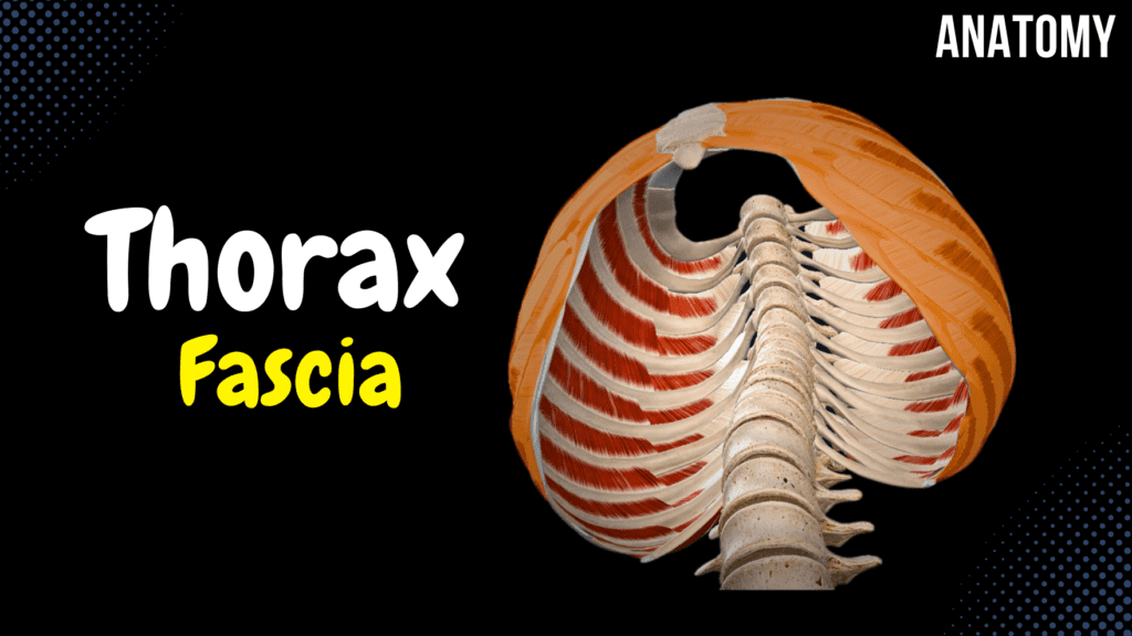
Fascia of the Thorax (Endothoracic, Thoracic, Clavipectoral, Pectoral Fascia) Official Links Instagram Youtube Jki-discord Notes & Illustrations Quizzes Summary & Transcript Notes ☆ Member Only Go to PDF Notes Illustrations ☆ Member Only Go to Illustrations 12345678910 Fascia of the Thorax – QUIZ Test your understanding with 10 random multiple-choice questions from the question bank. You're in the preview mode. Note: All elements work correctly on the front end. 1 / 10 Which fascia encloses the sternocleidomastoid and trapezius muscles? A) Clavipectoral fascia B) Pectoral fascia C) Cervical fascia D) Thoracic fascia The cervical fascia encloses these muscles and lies superiorly. 2 / 10 Which fascia is stretched between the clavicle and cartilaginous ribs? A) Clavipectoral fascia B) Endothoracic fascia C) Thoracic fascia D) Pectoral fascia The clavipectoral fascia stretches between the clavicle and ribs. 3 / 10 Which fascia connects the clavicle to the cartilaginous ribs? A) Pectoral fascia B) Endothoracic fascia C) Clavipectoral fascia D) Thoracic fascia The clavipectoral fascia connects the clavicle to the cartilaginous ribs. 4 / 10 Which fascia connects the clavicle to the coracoid process and cartilaginous ribs? A) Pectoral fascia B) Thoracic fascia C) Clavipectoral fascia D) Endothoracic fascia The clavipectoral fascia connects these structures in the thorax. 5 / 10 What fascia is considered the deep fascia of the thorax, covering the ribs and intercostal spaces? A) Pectoral fascia B) Clavipectoral fascia C) Endothoracic fascia D) Thoracic fascia The thoracic fascia covers the ribs and intercostal spaces. 6 / 10 What structure passes through the clavipectoral fascia at the infraclavicular region? A) Axillary artery B) Cephalic vein C) Internal thoracic vein D) Subclavian nerve The cephalic vein passes through the clavipectoral fascia in the infraclavicular region. 7 / 10 The fascia overlying the internal surface of the thoracic cavity is known as? A) Pectoral fascia B) Endothoracic fascia C) Thoracic fascia D) Clavipectoral fascia The endothoracic fascia overlies the internal thoracic cavity surface. 8 / 10 What structure is covered by the clavipectoral fascia? A) Serratus anterior B) External intercostal muscles C) Pectoralis major D) Subclavius and pectoralis minor The clavipectoral fascia covers the subclavius and pectoralis minor muscles. 9 / 10 Which fascia lies superficial to the thoracic fascia? A) Pectoral fascia B) Aponeurosis C) Endothoracic fascia D) Clavipectoral fascia The clavipectoral fascia lies superficial to the thoracic fascia. 10 / 10 What is the main function of the thoracic fascia? A) Provides fat storage B) Forms the pleural cavity lining C) Covers the lungs D) Covers ribs and intercostal spaces The thoracic fascia stabilizes and separates the ribs and intercostal muscles. Your score is The average score is 0% Description This video covers the fascia of the thorax, including its superficial and deep layers. Superficial Fascia of the Thorax Pectoral Fascia (Fascia Pectoralis) Covers the Pectoralis Major Clavipectoral Fascia (Fascia Clavipectoralis) Covers the Subclavius and Pectoralis Minor Stretched between the: Clavicle Coracoid Process Cartilaginous Ends of the Ribs Deep Fascia of the Thorax Thoracic Fascia (Fascia Thoracica) A fascia over the surface of the ribs and intercostal spaces Endothoracic Fascia (Fascia Endothoracica) Covers the internal surface of the thoracic cavity Includes: Diaphragmatic Fascia Suprapleural Membrane Transcript Introduction0:03What’s up. Meditay here and in this video, we’ll be going through the0:06Fascia you’ll find in the thoracic region. Aight. So, here’s the plan anterior view0:12of the thorax. And Here, we’ll add all the main muscles associated with thoracic region.Content0:17So in this video, we’re mainly gonna cover the Pectoral Fascia. The Clavipectoral Fascia.0:22Thoracic Fascia and Endothoracic Fascia. So these are the fascias we’re gonna go through, and we’llPectoral Fascia and Clavipectoral Fascia0:28start with the Pectoral and Clavipectoral Fascia. These two fascias are pretty simple. The Pectoral0:34Fascia covers the pectoralis Major, as you see here in blue.0:38And the Clavipectoral Fascia covers the subclavius muscle and the pectoralis minor, which are both0:43located underneath the pectoralis major. The Clavipectoral fascia is stretched0:47between the clavicle, coracoid process and the cartilaginous ends of the ribs, as you see here.0:53These two fascias you see here, are considered as the Superficial Fascia of the Thorax,0:58so when we remove them, we’ll see the actual anterior wall of the thorax.Thoracic Fascia1:03The whole external surface of the thorax is covered by a fascia called Thoracic Fascia.1:08As you see here. So again, it’s a fascia that lies over the surface of the ribs and intercostal1:14spaces. The other fascia is associated with the internal surface of the thoracic wall,1:20so let’s look at the thorax from this perspective.1:23And here still we have the thoracic fascia in orange.Endothoracic Fascia1:26The inner surface of the thoracic cavity is covered by a fascia called Endothoracic Fascia.1:31As you see here in red. But there are a couple of things that we need to know1:34about the endothoracic fascia. SO let’s add the diaphragm and look at the thorax from an anterior1:40view. And then cut the thorax like this. And look at it from this perspective, we’ll see1:44this. So again. Here’s the Endothoracic Fascia. Now the Endothoracic fascia is going to go covert1:50eh diaphragm as well, and get the name diaphragmatic fascia, which is just a fancy name1:55for the diaphragmatic part of the endothoracic fascia. Another thing the endothoracic fascia is2:00going to form is the suprapleural membrane, which is a thickened part of the endothoracic fascia2:06on the pleural cupula. Now if you’re unsure about what the pleura is. It’s basically a2:12covering around the actual lungs. And the upper apex of the pleura is what we call pleural cupula.2:18So, the endothoracic fascia is going to cover the pleural cupula and form a membrane.2:23So that was all the main fascia of the thorax, and I hope that was helpful. Notes & Illustrations Quizzes Summary & Transcript Notes ☆ Member Only Go to PDF Notes Illustrations ☆ Member Only Go to Illustrations Fascia of the Thorax – QUIZ Test your understanding with 10 random multiple-choice questions from the question bank. Start Become a Member You have to become a member before you can access the Notes and the Quizzes. Membership Plans Description This video covers
Diaphragm
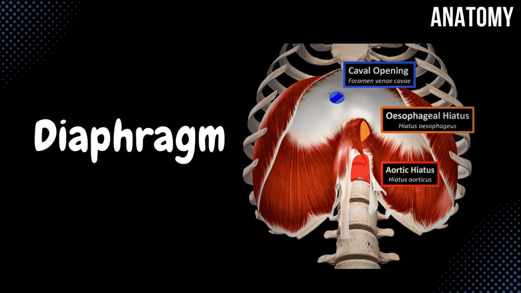
Diaphragm – Origin Points, Openings and Coverings Official Links Instagram Youtube Jki-discord Notes & Illustrations Quizzes Summary & Transcript Notes ☆ Member Only Go to PDF Notes Illustrations ☆ Member Only Go to Illustrations 12345678910 Diaphragm – QUIZ Test your understanding with 10 random multiple-choice questions from the question bank. You're in the preview mode. Note: All elements work correctly on the front end. 1 / 10 What structure passes through the oesophageal hiatus of the diaphragm? A) Internal thoracic artery B) Esophagus C) Inferior vena cava D) Aorta The oesophageal hiatus, located at T10, allows passage of the esophagus and vagus nerves. 2 / 10 Which opening in the diaphragm allows passage of the inferior vena cava? A) Aortic hiatus B) Sternocostal triangle C) Oesophageal hiatus D) Caval opening The caval opening, located at T8, allows the inferior vena cava and right phrenic nerve to pass through the diaphragm. 3 / 10 What structure passes through the aortic hiatus of the diaphragm? A) Aorta B) Phrenic nerve C) Inferior vena cava D) Esophagus The aortic hiatus, located at T12, allows passage of the aorta and thoracic duct. 4 / 10 What is the anatomical landmark for the caval opening? A) Central tendon at T8 B) Costal part at T10 C) Lumbar part at T12 D) Sternal origin The caval opening is located in the central tendon at the T8 vertebral level. 5 / 10 Which nerves pass through the caval opening? A) Right phrenic nerve B) Sympathetic nerve trunk C) Left vagus nerve D) Left phrenic nerve The right phrenic nerve passes through the caval opening alongside the inferior vena cava. 6 / 10 From which vertebral levels do the right and left crura of the diaphragm arise? A) L2-L4 B) L1-L4 C) T11-T12 D) L1-L3 (right), L1-L2 (left) The right crus arises from L1–L3 vertebral bodies, while the left crus arises from L1–L2 vertebral bodies. 7 / 10 What ligament of the diaphragm is associated with the quadratus lumborum muscle? A) Median arcuate ligament B) Crural ligament C) Medial arcuate ligament D) Lateral arcuate ligament The lateral arcuate ligament arches over the quadratus lumborum muscle, providing structural support to this muscle. 8 / 10 Which diaphragm part allows the passage of the thoracic duct? A) Sternocostal triangle B) Lumbocostal triangle C) Aortic hiatus D) Caval opening The aortic hiatus transmits the thoracic duct alongside the aorta. 9 / 10 Which muscle is associated with the lateral arcuate ligament of the diaphragm? A) Psoas major B) Rectus abdominis C) Quadratus lumborum D) Internal oblique The lateral arcuate ligament arches over the quadratus lumborum muscle. 10 / 10 What is the function of the sternocostal triangle (trigonum sternocostale)? A) Sympathetic trunk B) Splanchnic nerves C) Thoracic duct D) Internal thoracic artery and vein The sternocostal triangle allows passage of the internal thoracic artery and vein through the diaphragm. Your score is The average score is 0% Description This video covers the diaphragm, including its parts, origin points, openings, and coverings. Diaphragm Parts Central Tendinous Part Muscular Part Main Muscle of Inspiration Origin Points Lumbar Part Right Crus: Crus dextrum Left Crus: Crus sinistrum Suspensory Muscle of Duodenum: (Muscle of Treitz) Lateral Arcuate Ligament (Quadratus Lumborum Muscle) Median Arcuate Ligament (Aorta) Medial Arcuate Ligament (Psoas Major Muscle) Costal Part Sternal Part Xiphoid Process of Sternum Openings of the Diaphragm Caval Opening (Foramen Venae Cavae) Structures Passing Through: Inferior Vena Cava Right Phrenic Nerve Oesophageal Hiatus (Hiatus Oesophageus) Structures Passing Through: Esophagus Vagus Nerve (N. Vagus) Aortic Hiatus (Hiatus Aorticus) Structures Passing Through: Aorta Thoracic Duct Sternocostal Triangle (Trigonum Sternocostale) Structures Passing Through: Internal Thoracic Vein Internal Thoracic Artery Lumbocostal Triangle (Trigonum Lumbocostale) Also known as “Bochdalek’s Foramen“ Lumbar Part (Pars Lumborum) Structures Passing Through: Sympathetic Trunk Splanchnic Nerves Azygos Vein Coverings of the Diaphragm Endothoracic Fascia (Fascia Endothoracica) Diaphragmatic Part of Parietal Pleura Endoabdominal Fascia (Fascia Endoabdominalis) Peritoneum Transcript Introduction0:03What’s up. Meditay here and in this video, we’ll be covering the whole anatomy of the Diaphragm.0:08Aight, so here you see the anterior view of the thorax and the abdomen. The Lungs, as you seeDiaphragm Function0:13here, are located within the thoracic cavity. While organs like your liver, and intestines,0:18are a part of the abdominal cavity. Now between the Thoracic cavity and the abdominal cavity,0:24there’s gonna be a large muscle called the Diaphragm, which is our muscle for today.0:26Just by looking at the diaphragm, you can see that it consists of two main parts. There’s a0:31central tendinous part, and a muscular part around the central tendinous part. And the main reason0:37why we need to diaphragm, is because when the muscle fibers contract, they’ll pull the central0:42tendinous part down. That’ll increase the volume of the thoracic cavity, and decrease the pressure,0:48so that air can easily come in into the lungs. So the diaphragm is our main muscle of0:52inspiration. One that e can’t live without. So let’s go ahead and isolate the diaphragmContent0:57and understand the actual anatomy of it. So in this video, we’ll first cover the1:02three origin points. Which are the Lumbar part, the costal part and the sternal part.1:06After that, we’re going to look at the openings of the diaphragm, basically which1:10structures go through it. and then we’ll see what type of coverings it has from both sides.1:15So, let’s start with the origin points, and do the lumbar part first.Lumbar Origin1:19Now the lumbar attachment of the diaphragm is from this area so let’s go ahead and focus on that.1:24The main thing you can see looking at the lumbar part of the diaphragm1:28are two muscular fibers going down along the vertebral column, called the Right crus,1:33and the Left Crus. The right crus being longer than the left crus as you see here.1:38Don’t get confused by this long structure here, it’s called suspensory muscle of the duodenum. It1:43just goes down and grabs the duodenum to keep a certain part of it flexed. It’s not a part1:49of the origin points and it’s not a part of the lumbar part, so don’t mind this structure for
Muscles of the Thorax
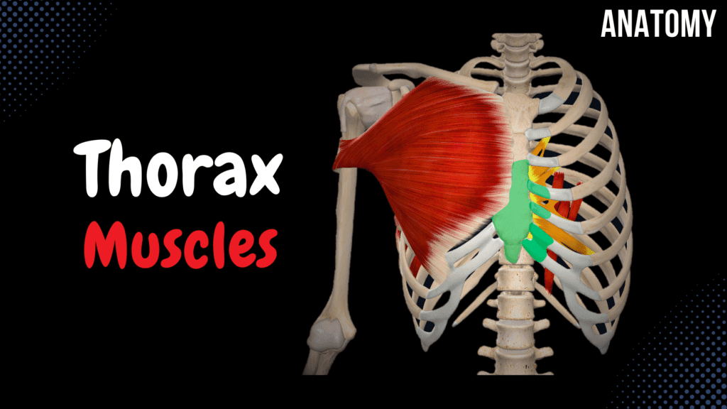
Muscles of the Thorax (Origin, Insertion, Function) Official Links Instagram Youtube Jki-discord Notes & Illustrations Quizzes Summary & Transcript Notes ☆ Member Only Go to PDF Notes Illustrations ☆ Member Only Go to Illustrations 12345678910 Muscles of the Thorax – QUIZ Test your understanding with 10 random multiple-choice questions from the question bank. You're in the preview mode. Note: All elements work correctly on the front end. 1 / 10 Which layer of the thoracic fascia forms the suprapleural membrane? A) Endothoracic fascia B) Pectoral fascia C) Clavipectoral fascia D) Thoracic fascia The endothoracic fascia forms the suprapleural membrane and stabilizes the pleural apex. 2 / 10 Which muscle originates from the transverse processes of T7–T10? A) Subcostal muscles B) External intercostals C) Subclavius D) Levatores costarum longi The levatores costarum longi originates from these transverse processes and elevates the ribs. 3 / 10 Which thoracic muscle is referred to as the “kissing muscle”? A) Orbicularis oris B) External intercostals C) Subclavius D) Transverse thoracis The orbicularis oris is involved in closing the lips tightly and is colloquially referred to as the “kissing muscle.” 4 / 10 What is the insertion of the subclavius muscle? A) Subclavian groove on clavicle B) Ribs 1–3 C) Medial scapular border D) Crest of greater tubercle The subclavius inserts into the subclavian groove on the clavicle. 5 / 10 What is the origin of the transverse thoracis muscle? A) Costal cartilage ribs 4–7 B) Transverse processes of thoracic vertebrae C) Internal surface of sternum D) Superior surface of rib below The transverse thoracis originates from the internal surface of the sternum and xiphoid process. 6 / 10 Which thoracic muscle inserts into the crest of the greater tubercle of the humerus? A) Internal intercostals B) Transverse thoracis C) Pectoralis major D) Subcostal muscles The pectoralis major muscle inserts into the humerus and contributes to arm adduction and internal rotation. 7 / 10 Which muscle inserts into the medial border of the scapula? A) Transverse thoracis B) Pectoralis minor C) Subclavius D) Serratus anterior The serratus anterior inserts into the medial border of the scapula, protracting and stabilizing it against the thoracic wall. 8 / 10 Which muscle originates from the internal surface of the sternum and xiphoid process? A) Transverse thoracis B) Internal intercostals C) Levatores costarum D) Subcostal muscles The transverse thoracis muscle helps in depressing the ribs during forced expiration. 9 / 10 Which thoracic fascia is continuous with the superficial abdominal fascia? A) Endothoracic fascia B) Clavipectoral fascia C) Pectoral fascia D) Thoracic fascia The pectoral fascia is continuous with the superficial abdominal fascia. 10 / 10 Which thoracic muscle is innervated by the subclavian nerve? A) Serratus anterior B) Levatores costarum C) Pectoralis minor D) Subclavius The subclavius muscle is innervated by the subclavian nerve and stabilizes the clavicle. Your score is The average score is 0% Description This video covers the muscles of the thorax, including their origins, insertions, and classifications. Superficial Muscles Thoracohumeral Muscles Subclavius Serratus Anterior Pectoralis Minor Pectoralis Major Deep Muscles Proper Muscles of the Thorax External Intercostal Muscles Internal Intercostal Muscles Subcostal Muscles Transverse Thoracis Levatores Costarum Superficial Muscles Subclavius (Musculus Subclavius) Origin: First Rib Insertion: Lower surface of Clavicle Serratus Anterior (Musculus Serratus Anterior) Origin: External Surface of Ribs 1-9 Insertion: Medial Border of Scapula Pectoralis Minor (Musculus Pectoralis Minor) Origin: External Surface of Ribs 3-5 Insertion: Coracoid Process of Scapula (Processus coracoideus scapulae) Pectoralis Major (Musculus Pectoralis Major) Clavicular Part: Pars Clavicularis Sternocostal Part: Pars Sternocostalis Abdominal Part: Pars Abdominalis Insertion: Crest of the Greater Tubercle (Crista tuberculi majoris humeri) Deep Muscles Subcostal Muscles (Musculi Subcostales) Origin: Lower Rib Insertion: Upper Rib Transverse Thoracis (Musculus Transversus Thoracis) Origin: Internal Surface of Sternum Xiphoid Process Cartilage of 4th-7th Rib Insertion: Cartilage of 2nd-6th Rib Internal Intercostal Muscles (Musculi Intercostales Interni) Origin: Lower Rib Insertion: Upper Rib External Intercostal Muscles (Musculi Intercostales Externi) Origin: Upper Rib Insertion: Lower Rib Levatores Costarum (Musculi Levatores Costarum) Levatores Costarum Breves Origin: Transverse Process C7-T11 Insertion: One Rib Below Levatores Costarum Longi Origin: Transverse Process T8-T11 Insertion: Two Ribs Below Transcript Introduction0:03Hey what’s up. Meditay here and in this video,0:06we’re gonna talk about the superficial and the deep muscles of the thorax. Alright, dependingDivision of the Thoracic Muscles0:11on the source you’re studying from, muscles of the thorax may be divided into 3 groups.0:15The first group are the superficial muscles of the thorax, or also called the thoracohumeral muscles0:21because they act on the upper limb movement as well. These are the Subclavius, Serratus Anterior,0:26Pectoralis Minor and Pectoralis Major. Then we have the Deep muscles of the Thorax,0:32which are also called the Proper muscles of the thorax because they both originate0:36and insert in the thorax. These are the External Intercostal muscle. Internal Intercostal muscles.0:42Subcostal muscles, transverse thoracis and then the levatores Costarum.0:47The 3rd group is the Diaphragm. It’s also considered as a muscle of the Thorax. We’re0:52not gonna talk about the diaphragm in this video, there’s gonna be a separate video on that one.0:57So in this video, we’re first going to talk about the 4 superficial muscles, and then do1:02the 5 deep muscles in the thorax. Awesome. So, the subclavius muscle is located here.Subclavius Muscle1:08It’s sub-clavius so it’s under the clavicle. It originates at the first rib as you see here,1:15and it inserts at the lower surface of the clavicle. So, when this muscle contracts, it1:20stabilized the clavicle by pulling it downwards. Next, we have the Serratus Anterior muscle. TheseSerratus Anterior1:26are large muscle fiber groups located on the lateral aspect of the 1-9th rib.1:31So it originates from the external surface of the ribs 1-9; and it inserts1:37at the medial border of the scapula, back here. Just to visualize it better, here you see the1:42superior view of it. You see it goes back and attaches to the medial margin of the scapula.1:48And when they contract, they pull the medial margin of the scapula forward,1:53which help flex the arm. So that’s the serratus anterior. Next is the Pectoralis Minor.Pectoralis Minor2:00The pec minor
Fascia of the Head and Neck
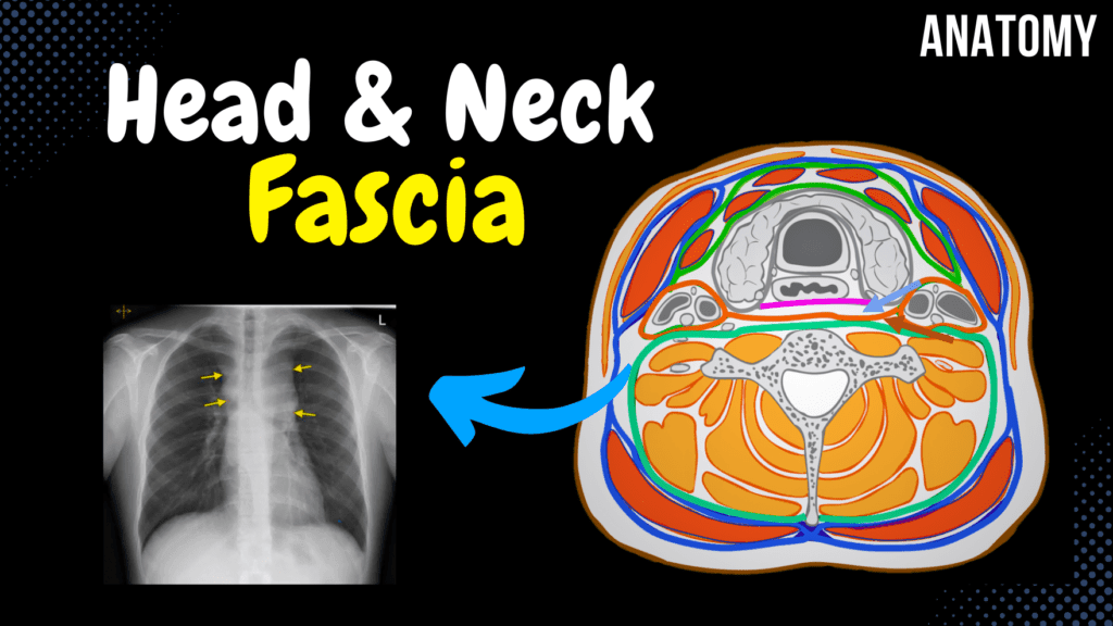
Fascia of the Head and Neck (Groups, Attachment Points, Arrangement) Official Links Instagram Youtube Jki-discord Notes & Illustrations Quizzes Summary & Transcript Notes ☆ Member Only Go to PDF Notes Illustrations ☆ Member Only Go to Illustrations 12345678910 Fascia of Head and Neck – QUIZ Test your understanding with 10 random multiple-choice questions from the question bank. You're in the preview mode. Note: All elements work correctly on the front end. 1 / 10 Which fascia covers the temporal muscle and attaches to the superior temporal line? A) Masseteric fascia B) Buccopharyngeal fascia C) Pretracheal fascia D) Temporal fascia The temporal fascia covers the temporal muscle and attaches to the superior temporal line. 2 / 10 What is the structural role of the superficial cervical fascia? A) Covers neurovascular structures B) Connects with thoracic fascia C) Separates skin and platysma D) Supports deep muscles The superficial cervical fascia separates skin and platysma from underlying structures. 3 / 10 Which cervical fascia layer encloses the infrahyoid muscles? A) Prevertebral fascia B) Temporal fascia C) Buccopharyngeal fascia D) Pretracheal fascia The pretracheal layer of cervical fascia encloses the infrahyoid muscles. 4 / 10 Where does the temporal fascia begin? A) Coronoid process B) Inferior temporal line C) Superior temporal line D) Zygomatic arch The temporal fascia begins at the superior temporal line. 5 / 10 Which muscle lies superficial to the superficial cervical fascia? A) Platysma B) Sternocleidomastoid C) Trapezius D) Omohyoid The platysma lies superficial to the superficial cervical fascia. 6 / 10 What is the function of the cervical fascia? A) Muscle elevation B) Stabilization and separation C) Oxygen exchange D) Synovial fluid transfer The cervical fascia stabilizes and separates neck muscles and organs, forming compartments. 7 / 10 Which fascia serves as a pathway for lymphatic drainage in the neck? A) Buccopharyngeal fascia B) Superficial cervical fascia C) Deep cervical fascia D) Temporal fascia The deep cervical fascia, including the pretracheal and prevertebral layers, aids in lymphatic drainage. 8 / 10 Which fascia covers the deep cervical muscles? A) Prevertebral layer B) Temporal fascia C) Masseteric fascia D) Superficial cervical fascia The prevertebral layer of cervical fascia covers the deep cervical muscles. 9 / 10 Which fascia of the neck contributes to the compartmentalization of neck structures? A) Buccopharyngeal fascia B) Pretracheal fascia C) Temporal fascia D) Masseteric fascia The pretracheal fascia contributes to the compartmentalization of the neck structures. 10 / 10 Which fascia of the neck covers the deep cervical muscles? A) Temporal fascia B) Superficial cervical fascia C) Prevertebral fascia D) Buccopharyngeal fascia The prevertebral layer of cervical fascia covers the deep cervical muscles. Your score is The average score is 0% Description This video covers the fascia of the head and neck, including its functions, types, and anatomical distribution. Fascia Functions Stabilizes and separates muscles from other internal organs Forms compartments Passage for nerves, blood vessels, and lymph Storage medium for fat and water Three Types of Fascia Superficial Fascia Deep Fascia Visceral Fascia Fascia of the Head Temporal Fascia Starts at the Superior Temporal Line Splits into two layers: Deep Layer: Lamina Profunda Superficial Layer: Lamina Superficialis Parotid Fascia Covers the Parotid Gland Masseteric Fascia Covers the Masseter Muscle Buccopharyngeal Fascia Covers the Buccinator Muscle Extends behind the pharynx Fuses with the Pterygomandibular Raphe Fascia of the Neck Cervical Fascia Superficial Layer Covers the surface of the neck Envelops the Sternocleidomastoid and Trapezius Platysma is superficial to this fascia Pretracheal Layer Envelops the Infrahyoid Muscles Forms the Carotid Sheath (surrounds the neurovascular bundle) Prevertebral Layer Covers the Deep Cervical Muscles Transcript Introduction0:03Hey what’s up. Meditay here and in this video, we’re gonna look at the main fascia covering the0:08head and neck. But first, If I would ask you, what s a fascia? And how do we categorize them, wouldWhat is a Fascia?0:14you be able to answer? Because these ae important things to understand before you actually learn0:19about the different fascia we have in the body. So a fascia is just a connective tissue0:24surrounding structures within the body. So here is a muscle, just a raw muscle within our body.0:30And here is a fascia. It surrounds the muscle. Now why do we need them?0:34Well one thing is that fascia stabilizes and separates muscles from other internal organs.0:40Fascia form compartments. Specially in clinics if you get patients with edema0:48within the compartment that the fascia forms, we’ll get the so called compartment syndrome,0:53which could be very dangerous as blood supply may get cut off due to the pressure.0:57Fascia also forms a passage for nerves, blood vessels and lymph. And this is also important1:03to keep in mind. Specially in people with chronic neck pain who are on constant pain medications. It1:09doesn’t necessarily have to be your muscle that’s still, it could also be the fascia. So massage and1:13stretching exercises are important factors which can stretch the fascia and help loosening it up.1:19Fascia also function as a storage medium for fat and water. And lastly. There are1:25three types of fasciae that you need to know. These are Superficial fascia,1:30Deep fascia, and Visceral Fascia. Ok. So here is the skin without removing1:35any layers. If you remove just the layers of the skin, you’ll see a superficial fascia,1:41located right underneath the skin. And then when you remove the superficial fascia, you’ll see1:46the deep fascia. The deep fascia is actually the that can surround individual muscles and groups of1:52muscles to separate into compartments. And when we talk about fascia within the body, it’s most1:58often the deep fascia we’re talking about. SO when you remove the deep fascia,2:03and enough muscle and bone to see an organ, we’ll see the visceral fascia,2:08that surrounds the organs within our body. Here we see the fascia covering the lungs, called pleura.2:14So that is the three types of fasciae we have And if we go back here. This fascia I showed you2:20earlier, was a deep fascia. Alright. So finally- In this video, we’re first going to look at theContent2:26fascia of the head, which consist of the Temporal
Muscles of the Neck
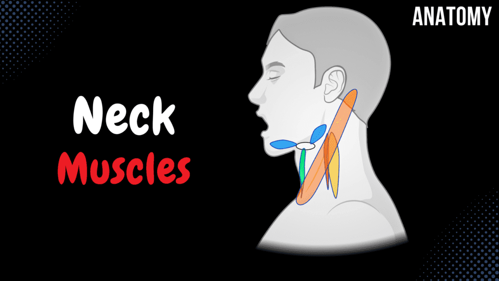
Muscles of the Neck (Groups, Origin, Insertion, Function) Official Links Instagram Youtube Jki-discord Notes & Illustrations Quizzes Summary & Transcript Notes ☆ Member Only Go to PDF Notes Illustrations ☆ Member Only Go to Illustrations 12345678910 Muscles of the Neck – QUIZ Test your understanding with 10 random multiple-choice questions from the question bank. You're in the preview mode. Note: All elements work correctly on the front end. 1 / 10 Where does the scalenus posterior muscle insert? A) Transverse processes of C6 B) First rib C) Lateral clavicle D) Second rib The scalenus posterior muscle inserts on the second rib. 2 / 10 Which muscle inserts at the basilar part of the occipital bone and originates from the atlas? A) Scalenus medius B) Rectus capitis anterior C) Longus capitis D) Rectus capitis lateralis The rectus capitis anterior muscle inserts at the basilar part of the occipital bone and originates from the lateral mass of the atlas (C1). 3 / 10 What is the function of the sternothyroid muscle? A) Elevates the thyroid cartilage B) Depresses the mandible C) Depresses the thyroid cartilage D) Rotates the thyroid cartilage The sternothyroid depresses the thyroid cartilage. 4 / 10 Which muscle has both superior and inferior bellies and originates from the scapula? A) Platysma B) Sternohyoid C) Thyrohyoid D) Omohyoid The omohyoid muscle has superior and inferior bellies, with the inferior belly originating from the scapula. 5 / 10 Where does the scalenus medius muscle originate? A) Superior surface of first rib B) First rib C) Transverse processes of C1/C2-C7 D) Lateral border of clavicle The scalenus medius originates from the transverse processes of C1/C2 to C7. 6 / 10 What is the insertion of the sternocleidomastoid muscle? A) Clavicle B) Mastoid process and superior nuchal line C) External occipital protuberance D) Manubrium of sternum The sternocleidomastoid muscle inserts on the mastoid process and superior nuchal line. 7 / 10 What is the function of the scalenus anterior muscle? A) Elevates the second rib B) Depresses the clavicle C) Elevates the first rib D) Rotates the head laterally The scalenus anterior aids in elevating the first rib during inspiration and laterally flexes the neck. 8 / 10 Where does the platysma muscle originate? A) Pectoral and deltoid fascia B) Clavicle C) Sternum D) Base of mandible The platysma muscle originates from the pectoral and deltoid fascia. 9 / 10 What is the origin of the longus capitis muscle? A) Lateral mass of atlas B) Transverse processes of C5-C7 C) Transverse processes of C1-C4 D) Transverse processes of C3-C6 The longus capitis originates from the transverse processes of C3-C6. 10 / 10 What is the insertion of the rectus capitis anterior muscle? A) Lateral part of occipital bone B) Inferior border of mandible C) Basilar part of occipital bone D) Atlas (C1) transverse process The rectus capitis anterior inserts on the basilar part of the occipital bone. Your score is The average score is 0% Description This video covers the Division of the Neck Muscles, including their origins, insertions, and classifications. Division of the Neck Muscles Deep Muscles of the Neck Lateral Muscles of the Neck Suprahyoid Muscles Infrahyoid Muscles Craniothoracal Muscles Deep Muscles of the Neck Longus Capitis (Musculus Longus Capitis) Origin: Transverse Processes of C3-C6 (Proc. transversus vertebrae cervicalis III – VI) Insertion: Basilar part of Occipital Bone (Pars basilaris ossis occipitalis) Longus Colli (Musculus Longus Colli) Superior Oblique Part Origin: Transverse Processes of C3-C5 (Proc. transversus vertebrae cervicalis III – V) Insertion: Anterior Tubercle of Atlas C1 (Tuberculum anterius atlantis) Vertical Part Origin: C5-T3 Body Insertion: C2-C4 Body Inferior Oblique Part Origin: T1-T3 Body Insertion: C5-C6 Transverse Process Rectus Capitis Anterior (Musculus Rectus Capitis Anterior) Origin: Lateral Mass of Atlas C1 (Massa lateralis atlantis) Insertion: Basilar Part of Occipital Bone (Pars basilaris ossis occipitalis) Rectus Capitis Lateralis (Musculus Rectus Capitis Lateralis) Origin: Transverse Process of Atlas C1 (Processus transversus atlantis) Insertion: Lateral Part of Occipital Bone (Pars lateralis ossis occipitalis) Lateral Muscles of the Neck Scalenus Anterior (Musculus Scalenus Anterior) Origin: Transverse Process of C3-C6 (Proc. transversus vertebrae cervicalis III – VI) Insertion: Scalenus Tubercle on 1st Rib (Tuberculum m. scaleni costa primae) Scalenus Medius (Musculus Scalenus Medius) Origin: Transverse Process of C1/C2-C7 (Proc. transversus vertebrae cervicalis I/II – VII) Insertion: Surface of 1st Rib (behind scalenus anterior) (Facies superior costae prima) Scalenus Posterior (Musculus Scalenus Posterior) Origin: Transverse Process of C5-C7 (Proc. transversus vertebrae cervicalis V – VII) Insertion: Surface of 2nd Rib (Costae II) Suprahyoid Muscles Digastricus (Musculus Digastricus) Posterior Belly: Origin: Mastoid Notch of Temporal Bone (Incisura mastoidea) Insertion: Tendon inserting at hyoid bone Anterior Belly: Origin: Tendon inserting at hyoid bone Insertion: Digastric Fossa of Mandible (Fossa digastrica) Infrahyoid Muscles Sternohyoid (Musculus Sternohyoideus) Origin: Sternum and Clavicle Insertion: Hyoid Bone Craniothoracal Muscles Sternocleidomastoid (Musculus Sternocleidomastoideus) Origin: Sternum + Clavicle Insertion: Mastoid Process + Superior Nuchal Line Trapezius (Musculus Trapezius) Superior Part Origin: Superior Nuchal Line, External Occipital Protuberance, Nuchal Ligament Insertion: Acromial End of Clavicle, Acromion of Scapula Middle Part Origin: Spinous Process of C7-T3/T4 Insertion: Spine and Acromion of Scapula Inferior Part Origin: Spinous Process of T4-T12 Insertion: Spine of Scapula Platysma Origin: Pectoral and Deltoid Fascia Insertion: Base of Mandible, Lower Lip, Skin around Mouth Transcript Introduction0:00Hey what’s up.0:04Meditay here and in this video, we’re gonna cover all the muscles of the neck.0:08Alright so The muscles of the neck can be divided into0:105 groups based on their anatomical location.0:13And keep in mind the classification of the neck muscles may vary depending on the source0:18you’re studying from, but all the muscles are the same.0:21So first are the deep muscles of the neck.0:24Those are the deepest close to the vertebra.0:26Then we have the Lateral Muscles of the neck.0:29The other two groups are related with the hyoid bone.0:32The suprahyoid muscles are located above the hyoid bone, and the infrahyoid muscles are0:36located below the hyoid bone.0:38And notice that both groups attach to the hyoid bone.0:42Then we have the cardiothoracic muscles, and they go from the cranium to
Muscles of Facial Expression
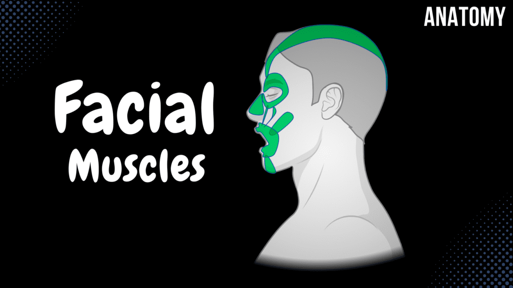
Muscles of Facial Expression (Parts, Origin, Insertion, Function) Official Links Instagram Youtube Jki-discord Notes & Illustrations Quizzes Summary & Transcript Notes ☆ Member Only Go to PDF Notes Illustrations ☆ Member Only Go to Illustrations 12345678910 Muscles of Facial Expression – QUIZ Test your understanding with 10 random multiple-choice questions from the question bank. You're in the preview mode. Note: All elements work correctly on the front end. 1 / 10 Which muscle originates from the temporal fascia and inserts into the auricle? A) Occipitofrontalis B) Auricularis posterior C) Buccinator D) Auricularis anterior The auricularis anterior muscle originates from the temporal fascia and inserts into the auricle. 2 / 10 Which muscle inserts into the auricle and originates from the temporal bone? A) Auricularis posterior B) Auricularis anterior C) Orbicularis oris D) Corrugator supercilii The auricularis posterior muscle originates from the temporal bone and inserts into the auricle. 3 / 10 What is the origin of the occipital belly of the occipitofrontalis muscle? A) Temporal fascia B) Epicranial aponeurosis C) Zygomatic arch D) Highest nuchal line The occipital belly of the occipitofrontalis originates from the highest nuchal line of the occipital bone. 4 / 10 Which muscle originates from the maxilla and inserts onto the skin of the upper lip and nasal wing? A) Zygomaticus minor B) Levator labii superioris alaeque nasi C) Buccinator D) Nasalis The levator labii superioris alaeque nasi originates from the maxilla and inserts onto the skin of the upper lip and nasal wing. 5 / 10 What is the primary function of the buccinator muscle? A) Depresses the lower lip B) Raises the eyebrows C) Compresses the cheeks D) Elevates the corners of the mouth The buccinator muscle compresses the cheeks, aiding in chewing and preventing food from collecting in the oral vestibule. 6 / 10 Which muscle elevates the upper lip and nasal wing, aiding in opening the nostrils? A) Orbicularis oris B) Levator labii superioris alaeque nasi C) Zygomaticus major D) Nasalis The levator labii superioris alaeque nasi elevates the upper lip and nasal wing, assisting in opening the nostrils. 7 / 10 What is the origin of the orbicularis oculi’s orbital part? A) Zygomatic bone B) Lacrimal bone C) Infraorbital margin of maxilla D) Medial palpebral ligament The orbital part of the orbicularis oculi originates from the medial palpebral ligament. 8 / 10 Which muscle forms the majority of the cheek structure? A) Buccinator B) Orbicularis oris C) Risorius D) Zygomaticus major The buccinator forms the majority of the cheek and assists in mastication and speech. 9 / 10 Which muscle has two parts, the transverse and alar parts, and controls the nasal opening? A) Levator labii superioris alaeque nasi B) Procerus C) Nasalis D) Zygomaticus major The nasalis muscle consists of the transverse and alar parts, functioning to control the nasal opening. 10 / 10 What is the primary function of the occipitofrontalis muscle’s occipital belly? A) Tilts the head backward B) Puckers the lips C) Retracts the scalp D) Elevates the eyebrows The occipital belly of the occipitofrontalis retracts the scalp. Your score is The average score is 0% Description This video covers the Division of Facial Muscles, their origins, insertions, and functions. Division of Facial Muscles Muscles of the Scalp Muscles around the Eye Opening Muscles around the Oral Opening Muscles of the Nasal Opening Muscles around the Ear Opening All insert into the skin All are innervated by the facial nerve All originate from the 2nd Pharyngeal Arch during development Muscles of the Scalp Occipitofrontal Muscle (Musculus Occipitofrontalis) Occipital Belly (Venter Occipitalis) Origin: Highest Nuchal Line (Linea Nuchalis Superior) Insertion: Epicranial Aponeurosis (Galea Aponeurosis) Frontal Belly (Venter Frontalis) Origin: Epicranial Aponeurosis (Galea Aponeurosis) Insertion: Skin of Eyebrows Muscles around the Eye Opening Orbicularis Oculi (Musculus Orbicularis Oculi) Orbital Part Origin/Insertion: Medial Palpebral Ligament Palpebral Part Origin: Medial Palpebral Ligament Insertion: Lateral Palpebral Ligament Lacrimal Part Origin: Lacrimal Bone Insertion: Lacrimal Sac Procerus (Musculus Procerus) Origin: Bridge of Nose Insertion: Skin of Forehead Corrugator Supercilii (Musculus Corrugator Supercilii) Origin: Glabella, Supraorbital Margin Insertion: Skin of Eyebrow Muscles around the Oral Opening Sides (“Smile”) Zygomaticus Major (Musculus Zygomaticus Major) Origin: Zygomatic Bone Zygomaticus Minor (Musculus Zygomaticus Minor) Origin: Zygomatic Bone Risorius (Musculus Risorius) Origin: Masseteric Fascia Insertion: Skin at the Angle of the Mouth Angle of the Mouth Levator Anguli Oris (Musculus Levator Anguli Oris) Origin: Anterior Surface of Maxilla Insertion: Skin at the Angle of the Mouth Levator Labii Superioris (Musculus Levator Labii Superioris) Origin: Infraorbital Margin of the Maxilla Insertion: Skin of the Upper Lip Depresses the Angle of the Mouth Depressor Anguli Oris (Musculus Depressor Anguli Oris) Depressor Labii Inferioris (Musculus Depressor Labii Inferiores) Lateral Wall of the Oral Cavity (“Satisfaction”) Buccinator (Musculus Buccinator) Origin: Alveolar Processes of Maxilla and Mandibula, Pterygomandibular Raphe Insertion: Skin at the Angle of the Mouth Lips (“Kissing Muscle”) Orbicularis Oris (Musculus Orbicularis Oris) Marginal Part Labial Part Chin (“Muscle of Doubt”) Mentalis (Musculus Mentalis) Origin: Alveolar Processes of Mandibula Insertion: Skin of the Chin Muscles of the Nasal Opening Nasalis (Musculus Nasalis) Transverse Part: Origin: Anterior Surface of Maxilla, Insertion: Dorsal Cartilage of the Nose Alar Part: Origin: Anterior Surface of Maxilla, Insertion: Dorsal Cartilage of the Nose Levator Labii Superioris Alaeque Nasi (Musculus Levator Labii Superioris Alaeque Nasi) Origin: Frontal Process of Maxilla Insertion: Skin of the Upper Lip, Skin of the Nasal Wing Muscles around the Ear Opening Extrinsic Muscles of the Ear Auricularis Anterior: Origin: Temporal Fascia, Insertion: Auricle Auricularis Superior: Origin: Epicranial Aponeurosis, Insertion: Auricle Auricularis Posterior: Origin: Temporal Bone, Insertion: Auricle Transcript Introduction0:03What’s up. Meditay here and in this video, we’re gonna cover all the muscles0:07of facial expression. Which are a part of the muscles of the head. Alright so0:12All muscles of the head are divided into two groups. The first group is the muscles0:16of mastication. Mastication means to chew, so those are the muscles responsible for chewing0:21when you’re eating. And the second group are gonna be fascial muscles or the muscles that0:26are gonna be
Connective Tissue
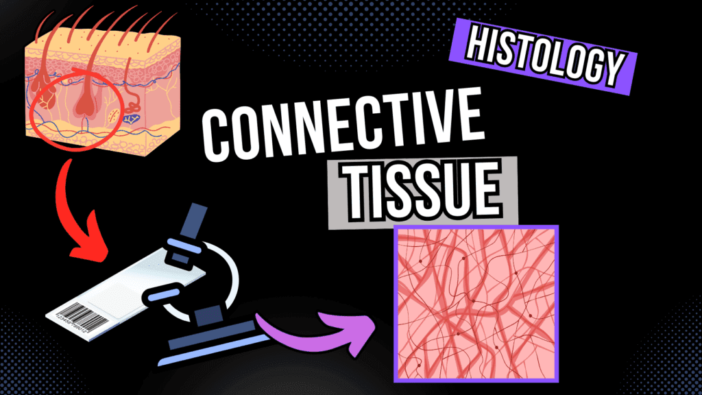
Connective Tissue Under the Microscope (Development and Structure) Official Links Instagram Youtube Jki-discord Notes & Illustrations Quizzes Summary & Transcript 📢 Currently, there is no PDF for this video.If you’re interested in having one, feel free to send an inquiry, and I may create it in the future. BUT! There’s a quiz available in the next tab. 12345678910 Connective Tissue – QUIZ Test your understanding with 10 random multiple-choice questions from the question bank. You're in the preview mode. Note: All elements work correctly on the front end. 1 / 10 Which connective tissue cell is derived from monocytes and involved in immune defense? A) Mast cells B) Fibroblasts C) Macrophages D) Plasma cells Macrophages are derived from monocytes and play a role in immune defense and phagocytosis. 2 / 10 Which component of the extracellular matrix provides tensile strength? A) Ground substance B) Elastic fibers C) Collagen fibers D) Reticular fibers Collagen fibers provide tensile strength in connective tissue. 3 / 10 What is the primary structural unit of collagen fibers? A) Tropocollagen B) Reticulin C) Elastin D) Proteoglycans Tropocollagen is the primary structural unit of collagen fibers. 4 / 10 Which type of collagen is found in the basal lamina? A) Type III collagen B) Type IV collagen C) Type II collagen D) Type I collagen Type IV collagen supports the basal lamina in epithelial tissues. 5 / 10 What distinguishes mesenchyme in histology? A) Hematopoietic tissue B) Embryonic tissue C) Vascular tissue D) Dense fibrous tissue Mesenchyme has nuclei and cell processes forming a syncytium in embryonic tissue. 6 / 10 What is the primary function of type V collagen fibers? A) Resists pressure B) Supports lymph nodes C) Provide tensile strength D) Connects basal lamina Type V collagen provides tensile strength and is found alongside type I collagen. 7 / 10 What is the role of Matrix Metalloproteinase in connective tissue? A) Synthesizes collagen B) Produces elastin C) Degrades ECM proteins D) Lubricates tissues Matrix Metalloproteinase degrades extracellular matrix proteins for remodeling. 8 / 10 Which connective tissue type provides structural support in tendons? A) Reticular CT B) Dense regular CT C) Dense irregular CT D) Loose CT Dense regular connective tissue provides structural support in tendons due to its parallel collagen fibers. 9 / 10 What is the origin of connective tissue? A) Mesenchyme B) Ectoderm C) Epidermis D) Endoderm Connective tissue originates from the mesoderm through mesenchyme. 10 / 10 Which cells in connective tissue are involved in wound healing? A) Adipocytes B) Myofibroblasts C) Macrophages D) Mast cells Myofibroblasts are specialized fibroblasts involved in wound healing. Your score is The average score is 0% Description This video is about the Connective Tissue 🔹 Development of Connective Tissue (Mesenchyme) Embryonic Development: Zygote → Blastula → Gastrula Germ Layers: Ectoderm → Skin and Nervous System Endoderm → GI Tract, Glands, Respiratory Tract Mesoderm → Connective Tissue (CT), Bones, Cartilage, Hematopoietic Cells Mesenchyme is an embryonic tissue, found near the neural tube during development. 🔹 Classification of Connective Tissue Common Origin: Mesenchyme Connective Tissue Proper: Loose CT, Dense CT CT with Special Properties: Adipose Tissue, Hematopoietic Cells, Elastin, Mucous Tissue Supportive CT: Cartilage, Bone 🔹 Composition of Connective Tissue Located under the basement membrane Consists of: Cells (Fibroblasts, Reticulocytes, Myofibroblasts) Extracellular Matrix: Fibers: Collagen, Elastin, Reticular Ground Substance: Glycosaminoglycans, Proteoglycans, Multi-Adhesive Glycoproteins Extracellular Fluid 🔹 Dense Connective Tissue Regular Dense CT: Found in tendons, collagen fibers aligned. Irregular Dense CT: Found in deep dermis, collagen fibers arranged randomly. 🔹 Collagen Fiber Types Type 1: Resistant to tension. Type 2: Resistant to pressure. Type 3: Maintenance in organs. Type 4: Supports basal lamina. Type 5: Resistant to tension (works with Type 1). Type 7: Connects basal lamina with reticular lamina. Type 9 & 10: Found in cartilage and bone. 🔹 Types of Connective Tissue Fibers Elastic Fibers: Oxytalan (Strong fibrillin fibers) Elaunin Fibers (Elastin + Fibrillin) Proper Elastic Fibers (Elastin + Fibrillin) Reticular Fibers: Consist of Type 3 Collagen. Produced by Reticulocytes. Histology: Found in lymph nodes. 🔹 Ground Substance Located between cells and fibers. Acts as a lubricant and barrier against invaders. Contains Glycosaminoglycans, Proteoglycans, Multi-adhesive Glycoproteins. 🔹 Histology of Connective Tissue Cells Macrophage: From bone marrow, bean-shaped nucleus. Mast Cell: Large, contains granules with histamine and heparin. Plasma Cell: Derived from B-Lymphocytes, “cartwheel” nucleus. White Adipocyte: Energy storage, nucleus at the periphery. Brown Adipocyte: Found in newborns, rich in mitochondria. 🔹 Challenge at the End of the Video! Transcript Introduction0:00hello and welcome to another video in0:02this video I’m gonna cover the basics of0:04connected tissue in terms of histology0:06so as you probably know we have four0:08main types of tissue in the body we have0:11epithelial tissue we have nervous tissue0:13we have muscle tissue and connective0:15tissue so in this video I’m going to0:17mainly focus on the connective tissue0:19all right so the first thing we start0:21with is the development of connective0:23tissue CT stand for connective tissue0:25what I’m gonna mention the mesenchyme0:27and I’m also going to talk about the0:29classification of connective tissue and0:31then I’m going to talk about the0:32extracellular matrix which include the0:35fibroblast types of dense connective0:36tissue reticular tissue and ground0:39substance and I’m also going to talk0:40about the cells in connective tissue0:42which include the macrophage must cell0:45plasma cell and the two different types0:48of adipocyte we have or a so let’s startDevelopment of Connective Tissue0:51with the development of connective0:52tissue so it all starts when sperm0:55fertilizes an egg and becomes what is0:57called a zygote and then the zygote is1:00going to divide a lot and become a ball1:02of cells called a blastula and then the1:05blaster is going to keep dividing and1:07become what is called a gastrula and1:10already here you can see that the cells1:12are starting to differentiate you can1:14see that you can have an outer layer in1:16blue and inner layer in orange and then1:19a middle layer in green all right so the1:22outer layer in blue right here we call1:24this one the ectoderm1:25and the ectoderm is eventually going to1:28form things like the skin and nervous1:30system alright and the inner layer in1:33orange we call
What Is Epithelial Tissue?

What Is Epithelial Tissue? Official Links Instagram Youtube Jki-discord Notes & Illustrations Quizzes Summary & Transcript 📢 Currently, there is no PDF for this video.If you’re interested in having one, feel free to send an inquiry, and I may create it in the future. BUT! There’s a quiz available in the next tab. 12345678910 Epithelial Tissue – QUIZ Test your understanding with 10 random multiple-choice questions from the question bank. You're in the preview mode. Note: All elements work correctly on the front end. 1 / 10 Which structure anchors epithelial cells to one another? A) Tight junctions B) Gap junctions C) Desmosomes D) Hemidesmosomes Desmosomes anchor epithelial cells together to provide structural integrity. 2 / 10 What is the primary function of simple cuboidal epithelium? A) Filtration B) Secretion and absorption C) Diffusion D) Protection Simple cuboidal epithelium functions in secretion and absorption, often in glands and renal tubules. 3 / 10 Where is transitional epithelium most commonly found? A) Lungs B) Skin C) Stomach D) Urinary bladder Transitional epithelium is found in the urinary system, including the bladder and ureters. 4 / 10 Which epithelium specializes in sensory perception? A) Transitional epithelium B) Simple squamous C) Neuroepithelium D) Stratified squamous Neuroepithelium is specialized for sensory perception, found in taste buds and olfactory epithelium. 5 / 10 Which epithelium forms the outermost layer of the skin? A) Stratified squamous keratinized B) Simple squamous C) Simple cuboidal D) Transitional epithelium Stratified squamous keratinized epithelium forms the outermost layer of the skin. 6 / 10 What type of epithelial tissue is found in alveoli? A) Stratified squamous B) Transitional epithelium C) Simple squamous D) Simple cuboidal Simple squamous epithelium facilitates gas exchange in the alveoli. 7 / 10 What is the role of basal lamina in epithelial tissue? A) Produces mucus B) Facilitates blood flow C) Forms cell junctions D) Provides structural support Basal lamina provides structural support and acts as a filter for molecules. 8 / 10 What is the primary location of simple squamous epithelium in the kidney? A) Loop of Henle B) Bowman's capsule C) Collecting duct D) Proximal tubule Simple squamous epithelium lines the Bowman’s capsule in the kidney. 9 / 10 Which epithelial cell type has a brush border of microvilli? A) Transitional epithelium B) Stratified squamous C) Simple cuboidal D) Simple columnar Simple columnar epithelium with microvilli forms the brush border, aiding absorption in intestines. 10 / 10 Which type of epithelium is specialized for rapid diffusion and filtration? A) Simple squamous epithelium B) Transitional epithelium C) Simple columnar D) Stratified squamous Simple squamous epithelium is specialized for diffusion and filtration. Your score is The average score is 0% Description This video covers the topic “What Is Epithelial Tissue?” 🔹 Introduction to Epithelial Tissue The body consists of four main tissue types: epithelial, nervous, muscle, and connective. Epithelial tissue is divided into: Covering epithelium: Lines surfaces. Glandular epithelium: Secretes substances. 🔹 Development & Structure Forms early from a zygote, developing through stages like the blastula and gastrula before differentiating into specialized tissues. Key surfaces: Apical: Free surface. Lateral: Contains cell junctions. Basal: Attached to the basement membrane. 🔹 Cell Junctions & Basement Membrane Tight junctions: Prevent leakage. Gap junctions: Allow communication & ion transport. Adherens & desmosomes: Provide structural support. Basement membrane: Anchors epithelium & aids nutrient diffusion. 🔹 Classification of Epithelial Tissue Layers: Simple (one layer) vs. Stratified (multiple layers). Cell shapes: Squamous (flat), Cuboidal (cube-like), Columnar (tall). 🔹 Types of Epithelium & Locations Simple Squamous: Found in alveoli, aids gas exchange. Simple Cuboidal: Kidney tubules & glands, functions in secretion & absorption. Simple Columnar: Intestinal lining, specialized for absorption (often with microvilli). Stratified Squamous: Found in skin & mouth; can be keratinized (dry) or non-keratinized (moist). Transitional Epithelium: Adaptable, found in the urinary bladder, allowing stretching. Glandular Epithelium: Includes: Endocrine glands: Hormone secretion. Exocrine glands: Secretion to surfaces. 🔹 Neuroepithelium Specialized epithelial cells involved in sensory functions, linked to the nervous system. 🔹 Histological Perspectives Epithelial tissue is best understood through microscopic slides, helping in the identification of different types. Transcript Introduction 0:00 hello and welcome to another video in this video I want to talk about the epithelial tissue so as you probably 0:05 know the body consists of four type of tissues we have epithelial tissue nervous and muscle and connective tissue 0:11 and each of these tissue types have under group so in this video we’re going to cover the epithelial tissue look at 0:18 their under group and how they are divided into the body and we’re also gonna see the development of epithelial 0:24 tissue and at the end of this video I’m gonna put some random at the feel of the tissue so you can try to pause the video 0:30 and try to guess which cell types you’re looking at so talking about epithelial tissue you’re gonna find two main types 0:36 we have the covering epithelia and we have glandular epithelium and then I’m gonna talk a lot about glandular fever 0:42 as most of the times when we look at epithelial tissue you can think of covering epithelia but I’m gonna mention 0:49 the most important structures you’ll find the glandular epithelium pourtant to know so covering epithelia is called 0:57 covering because this covers the structures you have in the body you know the skin cells gonna composed of the 1:02 epithelial tissue you have the outside surfaces of organs gonna be epithelial 1:09 tissue and also going to line the internal surfaces of organs so as an example of that if you look at this 1:14 intestines right here you can see that you can see if a fatty tissue covering the external surfaces of the the 1:23 intestines but it’s also going to line the internal surfaces of it you can have different types of epithelial tissue 1:29 covering and lining but they’re all gonna be a Patil tissue now when you talk about glandular epithelium you can 1:36 you
What is HISTOLOGY?
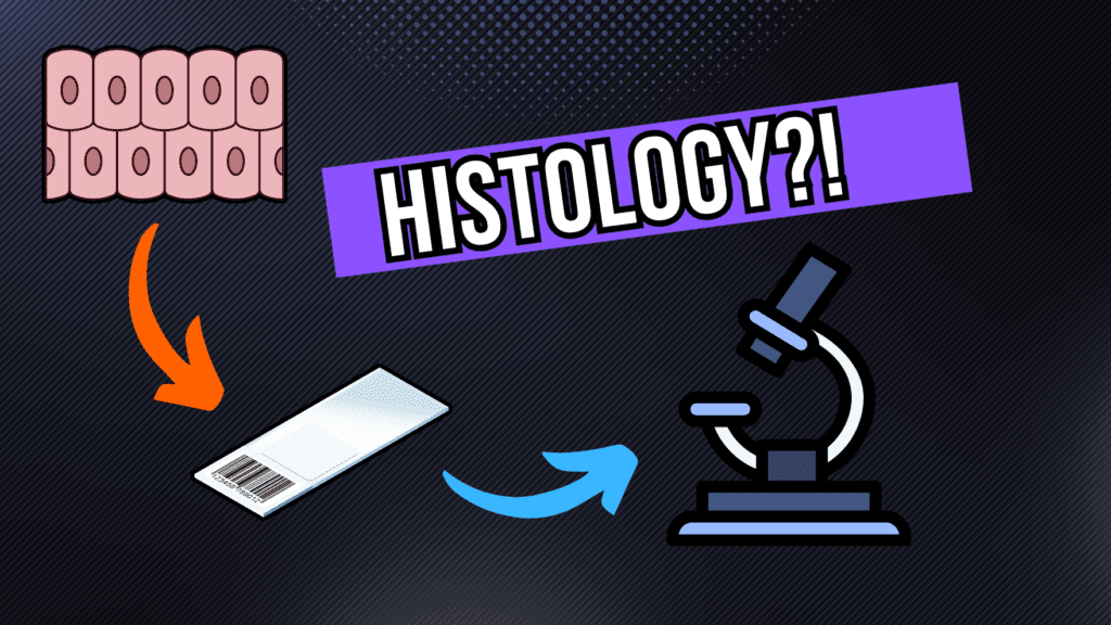
What is HISTOLOGY? A quick TOUR Official Links Instagram Youtube Jki-discord Notes & Illustrations Quizzes Summary & Transcript 📢 Currently, there is no PDF for this video.If you’re interested in having one, feel free to send an inquiry, and I may create it in the future. BUT! There’s a quiz available in the next tab. 12345678910 What is HISTOLOGY? – QUIZ Test your understanding with 10 random multiple-choice questions from the question bank. You're in the preview mode. Note: All elements work correctly on the front end. 1 / 10 Who first used the term “cell” in the context of biology? A) Rudolf Virchow B) Robert Hooke C) Louis Pasteur D) Antonie van Leeuwenhoek Robert Hooke coined the term “cell” when observing cork tissue under a microscope. 2 / 10 What is the main component of the microtome used in histology? A) Objective lens B) Stage C) Condenser lens D) Blade A microtome is used to cut tissue into ultra-thin sections. 3 / 10 What substance is commonly used for fixation in histology? A) Xylene B) Ethanol C) Paraffin D) Formalin Formalin is commonly used for fixation. 4 / 10 What type of fixation is most commonly used for preserving tissue samples? A) Ethanol B) Formalin C) Paraffin D) Xylene Formalin fixation is widely used for preserving tissue samples. 5 / 10 Which cellular component is specifically highlighted by eosin staining? A) Golgi apparatus B) Cytoplasm C) Ribosomes D) Nucleus Eosin stains the cytoplasm and extracellular matrix pink. 6 / 10 Which microscope provides the highest magnification and resolution? A) Phase-contrast microscope B) Scanning electron microscope C) Transmission electron microscope D) Brightfield microscope A transmission electron microscope provides the highest magnification and resolution. 7 / 10 What is the primary difference between prokaryotic and eukaryotic cells? A) Prokaryotes have organelles B) Both lack nuclei C) Eukaryotes have nuclei D) Both have nuclei Eukaryotic cells have a nucleus, while prokaryotic cells do not. 8 / 10 Which microscope is best for observing intracellular organelles at a high resolution? A) Transmission electron microscope B) Fluorescence microscope C) Phase-contrast microscope D) Light microscope Transmission electron microscopes are ideal for observing intracellular organelles at high resolution. 9 / 10 Which type of microscopy is suitable for observing live cells in real time? A) Brightfield microscopy B) Electron microscopy C) Fluorescence microscopy D) Phase-contrast microscopy Phase-contrast microscopy is ideal for observing live cells without staining. 10 / 10 What is the primary purpose of histological staining? A) Dehydrate tissues B) Enhance visualization C) Embed tissues D) Fix tissues Staining enhances the visualization of cellular and tissue structures. Your score is The average score is 0% Description This video is about histology, its history, and how histological slides are prepared. In this video, I cover: Short history of histology: First light microscope and early discoveries. Basic cell properties: Differences between prokaryotic and eukaryotic cells. Microscopy techniques: Light Microscope: Brightfield Phase-Contrast Darkfield Fluorescence Microscope Transmission Electron Microscope (TEM) Scanning Electron Microscope (SEM) Histological slide preparation and staining: Biopsy Fixation Dehydration Paraffin Infiltration Staining (Hematoxylin & Eosin – H&E) Quick look into how to differentiate cells under a microscope (Muscle Tissue). Enjoy! Transcript Introduction0:00hello welcome to a video in this video0:01I’m going to talk about the basics of0:03cell biology where I’m going to talk0:04about the properties of the you credit0:06cell I’m also going to talk about the0:07differences between the eukaryotic cell0:09and appropriate Excel and I also got0:12attacked by the basics of histology0:13where I’m gonna take it through the0:15journey of how tissue in the human body0:17ends up looking like this so over many0:20years we have been looking at cells0:22through simple light microscopes just0:25like the one you see right here so if weHistory of Histology0:28take a quick look into the history of0:30histology there has been a lot of people0:31that have contributed to the development0:33of microscope but it’s all we really0:36started with Robert Hooke and Marshall0:38OMA Pinckney who in year 1700 were the0:42first people who used the term cell they0:45use this simple light microscope you see0:48right here and how do they work well0:51they used candle just like this one this0:54is they used this as a source of light0:56because they didn’t have any electricity0:57at that time and they use this glass1:00bulb filled with water to focus the1:03light into a specimen something they1:05wanted to observe and then that light1:07would then reflect it into this1:09microscope where they had different1:12lenses to magnify this image and with1:16those lenses they could actually see the1:19the cells of the specimen they’re1:22looking at so here’s what they looked at1:24this is a bark tissue from a tree called1:26cork oak so this is a plant you Kratt1:29Excel and each black spot we’re gonna1:32present one eukaryotic cell so in theDifferent Cellular Characteristics1:35astrology in order to understand the1:37differences between tissues in order to1:39understand the differences between1:40nervous and muscle tissues for example1:42you need to understand the different1:44cellular characteristics so I’m not1:46gonna go too much in details under this1:48because that’s gonna be for other videos1:49but mainly the first thing you want to1:52look at when you look at tissues like1:53that is what cells are you looking at1:55what are their functions here I’ve used1:58three muscle tissues as an example here2:01you see the skeletal muscles and the2:03cardiac muscles and small muscles so2:06what cells are looking at what are their2:08functions will they know that muscle2:09tissues I need to apply force so they’re2:12movable2:13so how do you differentiate between them2:14even though they have the same function2:16well you know that skeletal muscles are2:18going to be long cylindrical just like2:19you see here2:20cardiac muscles are going to be short2:22and branched some of these muscles for2:25examples for me like that2:26well this muscle is cells from a dead2:30while swarm us also I’m going to be very2:33narrow and elongated so that we’re gonna2:38be very close to each other so you need2:41to look at the shape of the cells but2:44you also need to look at the cell2:45nucleus so you know that the skeletal2:47muscles are gonna be multi nuclide and2:49they’re gonna have many nuclei it’s like2:51you see here while cardiac muscles are2:54gonna have one nucleus not be the same2:56goes