
CN 4: Trochlear Nerve

CN 4: Trochlear Nerve Official Links Instagram Youtube Jki-discord Notes & Illustrations Quizzes Summary & Transcript Notes ☆ Members Only Go to PDF Notes Illustrations ☆ Members Only Go to Illustrations 12345678910 Trochlear Nerve – QUIZ Test your understanding with 10 random multiple-choice questions from the question bank. You're in the preview mode. Note: All elements work correctly on the front end. 1 / 10 Which cranial nerve palsy leads to an upwardly rotated eye? A) Abducens Nerve B) Optic Nerve C) Oculomotor Nerve D) Trochlear Nerve Trochlear nerve palsy leads to an upwardly rotated eye due to unopposed inferior oblique and superior rectus activity. 2 / 10 Which artery is most closely associated with trochlear nerve injury? A) Basilar Artery B) Superior Cerebellar Artery C) Posterior Cerebral Artery D) Middle Cerebral Artery The superior cerebellar artery is closely associated with the trochlear nerve. 3 / 10 Which condition results from trochlear nerve damage? A) Mydriasis B) Ptosis C) Horizontal Diplopia D) Vertical Diplopia Trochlear nerve damage causes vertical diplopia and difficulty looking downward. 4 / 10 Which cranial nerve has the longest intracranial course? A) Trochlear Nerve B) Oculomotor Nerve C) Abducens Nerve D) Optic Nerve The trochlear nerve has the longest intracranial course of all cranial nerves. 5 / 10 What is the function of the superior oblique muscle? A) Dilates the Pupil B) Adducts the Eye C) Depresses the Eye D) Elevates the Eye The superior oblique depresses, abducts, and internally rotates the eye. 6 / 10 What is unique about the trochlear nerve? A) Exits Dorsally B) Innervates Two Muscles C) Originates from the Pons D) Contains Parasympathetic Fibers It is the only cranial nerve that exits dorsally from the brainstem and crosses to innervate the contralateral side. 7 / 10 What is the origin of the superior oblique muscle? A) Sphenoid Bone B) Orbit C) Frontal Bone D) Maxilla The superior oblique muscle originates from the body of the sphenoid bone. 8 / 10 Which cranial nerve is the longest intracranially? A) Trochlear Nerve B) Trigeminal Nerve C) Oculomotor Nerve D) Abducens Nerve The trochlear nerve is the longest intracranial cranial nerve. 9 / 10 Which action is NOT performed by the superior oblique muscle? A) Elevation B) Intorsion C) Depression D) Abduction The superior oblique does not perform elevation of the eye. 10 / 10 What does the superior oblique tendon pass through before inserting? A) Orbit B) Trochlea C) Common Tendinous Ring D) Tendinous Ring The superior oblique tendon passes through the trochlea, acting as a pulley. Your score is The average score is 0% Description Trochlear Nerve Scheme / Overview Trochlea is Latin for “Pulley,” referring to the function of the superior oblique muscle. Nucleus of the trochlear nerve is located in the midbrain at the level of the inferior colliculi. Emerges from the posterior surface of the midbrain and turns anteriorly. Enters and runs on the lateral wall of the cavernous sinus. Enters the orbit via the superior orbital fissure to innervate the superior oblique muscle. Exclusively a somatomotor nerve. Course of the Trochlear Nerve Nucleus of the Trochlear Nerve (nuclei nervi trochlearis) is located in the midbrain at the level of the inferior colliculus (colliculi inferiores). Nucleus is in the same area as the oculomotor nerve nuclei, just a level lower. The trochlear nerve crosses to the contralateral side, exits from the posterior surface of the midbrain, and turns anteriorly. Pierces the dura mater and runs through the lateral wall of the cavernous sinus. Goes through the superior orbital fissure to innervate the superior oblique muscle. Functions of the Superior Oblique Muscle Origin: Body of the sphenoid bone, medial to the common tendinous ring. Runs anteriorly, with its tendon hooking around the trochlea of the superior oblique. Then takes a sharp posterior-lateral turn. Insertion: Posterior superolateral aspect of the eyeball (deep to rectus superior, via trochlea orbitae). Causes: Abduction Depression Internal rotation of the eyeball Clinical Relevance Damage to the trochlear nerve can be either congenital or acquired. When damaged, the eye naturally shifts upwards and externally rotates, leading to diplopia (double vision). To compensate and reduce diplopia, patients often adopt a head tilt and chin tuck. Congenital Causes: Malformations of the nucleus or nerve. Acquired Causes: Trauma or stroke in the midbrain. Sources Singh, I. (2017). Human Neuroanatomy (10th ed.). Helwany M, Bordoni B. Neuroanatomy, Cranial Nerve 1 (Olfactory) [Updated 2022 Aug 8]. In: StatPearls [Internet]. Treasure Island (FL): StatPearls Publishing; 2022 Jan-. Kozlowski, T. (2017). Memorix Anatomy: The Complete Study Guide. 2nd ed. Thieme Medical Publishers. Pictures and Visuals Complete Anatomy Biorender PowerPoint Camtasia 2021 Transcript Introduction0:04What’s up, Taim Talks Med here. Let’s continue our Cranial nerve series.0:11Cranial nerves are twelve pairs of nerves that exit the brain and the brainstem, and in this0:16segment, we’ll talk detailed about the fourth cranial nerve, which is the Trochlear nerve.0:21And we’ll do that by first making a quick scheme of the trochlear pathway to get an overview of it.0:28Then we’ll cover the course of the trochlear nerve and go detailed into its pathway and which0:33structures the oculomotor nerve goes through, and while doing so we’ll talk through the function0:39of the muscle the trochlear nerve innervates the superior oblique muscle. Then at the end,0:45we’ll talk a little bit about the clinical relevance, and pathologies0:49related to the fourth cranial nerve pathway. So, the trochlear nerve is the fourth cranialTrochlear Nerve Scheme0:55nerve, and it gets its name from the Latin word pulley, “trochleae.” Now a pulley is a device1:02that lifts an object, right? In each eye, the superior oblique muscle functions as the trochlea,1:10or a pulley. The trochlear nerve innervates the superior oblique muscle to lift the eyes so you1:17can look down. So, the trochlear nerve innervates the superior oblique muscle to move the eye in a1:24down-and-out position, and intort the eye. Let’s see how it innervates it.1:29The nerve starts from a nucleus called the nucleus of the trochlear nerve, located in1:34the midbrain at level of the inferior colliculus. Form the nucleus of
CN 3: Oculomotor Nerve
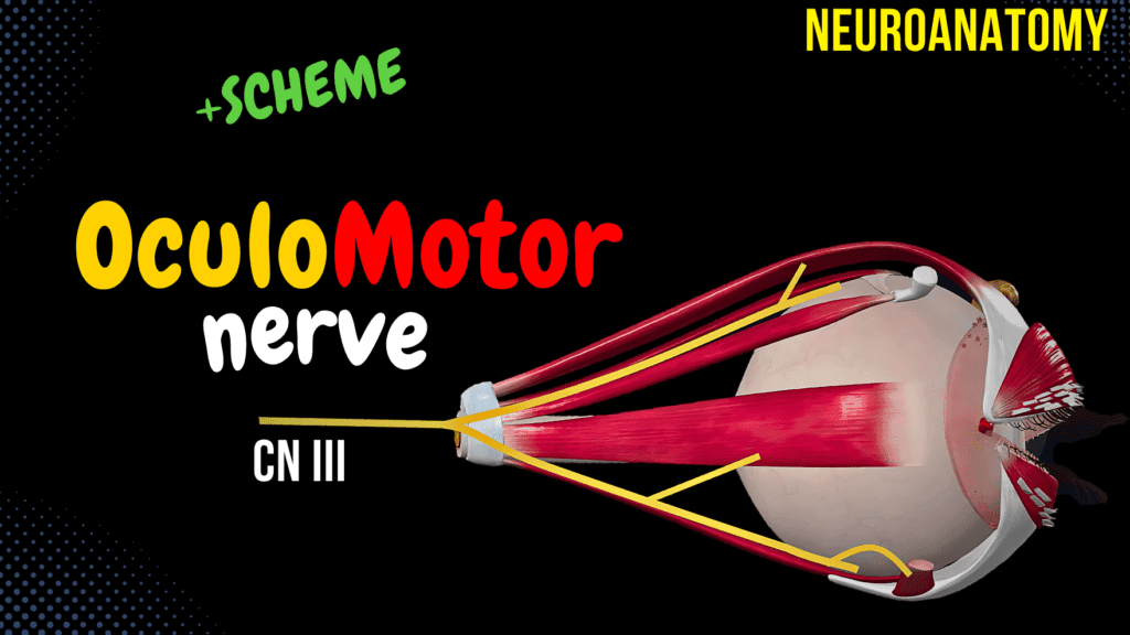
CN 3: Oculomotor Nerve Official Links Instagram Youtube Jki-discord Notes & Illustrations Quizzes Summary & Transcript Notes ☆ Members Only Go to PDF Notes Illustrations ☆ Members Only Go to Illustrations 12345678910 Oculomotor Nerve – QUIZ Test your understanding with 10 random multiple-choice questions from the question bank. You're in the preview mode. Note: All elements work correctly on the front end. 1 / 10 What causes diplopia in oculomotor nerve palsy? A) Mydriasis B) Constricted Pupil C) Misalignment of the Eyes D) Ptosis Diplopia is caused by misalignment of the eyes due to impaired eye muscle function. 2 / 10 What causes mydriasis in oculomotor nerve palsy? A) Overactive Sympathetic Response B) Loss of Parasympathetic Control C) Damage to Medial Rectus D) Inferior Oblique Weakness Mydriasis occurs due to loss of parasympathetic innervation to the sphincter pupillae. 3 / 10 Which muscle is responsible for elevation and lateral rotation of the eye? A) Superior Rectus B) Medial Rectus C) Inferior Rectus D) Inferior Oblique The inferior oblique is responsible for elevation and lateral rotation of the eye. 4 / 10 What is the function of the inferior oblique muscle? A) Elevation and Medial Rotation B) Depression and Medial Rotation C) Elevation and Lateral Rotation D) Depression and Lateral Rotation The inferior oblique muscle is responsible for elevation and lateral rotation of the eye. 5 / 10 From which part of the brain does the oculomotor nerve originate? A) Midbrain B) Pons C) Medulla D) Spinal Cord The oculomotor nerve originates in the midbrain at the level of the superior colliculi. 6 / 10 What is the role of the superior rectus muscle? A) Elevates the Eye B) Abducts the Eye C) Depresses the Eye D) Adducts the Eye The superior rectus muscle elevates the eye. 7 / 10 What is the role of the ciliary muscle? A) Contraction of Retina B) Pupil Constriction C) Elevation of Eyelid D) Lens Accommodation The ciliary muscle adjusts the lens for near vision (accommodation). 8 / 10 Which nerve innervates the lateral rectus muscle? A) Trigeminal Nerve B) Trochlear Nerve C) Abducens Nerve D) Oculomotor Nerve The lateral rectus muscle is innervated by the abducens nerve (CN VI). 9 / 10 What is the role of short ciliary nerves in the visual pathway? A) Relay Visual Signals B) Adjust Lens Shape C) Control Lateral Eye Movement D) Carry Parasympathetic Fibers Short ciliary nerves carry parasympathetic fibers to the sphincter pupillae and ciliary muscles. 10 / 10 What structure does the oculomotor nerve pass through within the cavernous sinus? A) Anterior Border B) Lateral Wall of Cavernous Sinus C) Center of Cavernous Sinus D) Inferior Wall The oculomotor nerve runs along the lateral wall of the cavernous sinus. Your score is The average score is 0% Description Oculomotor Pathway Scheme / Overview Oculomotor nerve (nervus oculomotorius) allows movement of the eye muscles, constriction of the pupil, and the position of the upper eyelid. Starts with the nucleus of the oculomotor nerve and the accessory oculomotor nucleus in the midbrain. Runs together and exits through the oculomotor sulcus in the interpeduncular fossa. Passes through the lateral wall of the cavernous sinus, superior orbital fissure, and common tendinous ring. Divides into superior and inferior branches (somatomotor fibers). Parasympathetic fibers from the Edinger-Westphal nucleus run with the inferior branch to the ciliary ganglion (pre-ganglionic fibers). Short ciliary nerves exit towards the ciliary muscle and sphincter pupillae. Midbrain Anatomy Posterior View: Cerebral Peduncles (pedunculus cerebri) Tectal Plate: Superior Colliculi (colliculi superiores) Brachium of the Superior Colliculus (brachium colliculi superioris) Inferior Colliculi (colliculi inferiores) Brachium of the Inferior Colliculus (brachium colliculi inferior) Anterior View: Cerebral Peduncles Interpeduncular Fossa (fossa interpeduncularis) Oculomotor Sulcus of the Mesencephalon (sulcus nervi oculomotorii) Internal View: Aqueduct of Midbrain Substantia Nigra Superior Colliculi Periaqueductal Grey Substance (substantia grisea centralis) Reticular Formation (formatio reticularis) Red Nucleus (nucleus ruber) Nucleus of the Oculomotor Nerve (nucleus nervi oculomotorii) Edinger-Westphal Nucleus / Accessory Nucleus of Oculomotor Nerve (nucleus accessorii nervi oculomotorii) Course of the Oculomotor Nerve Starts at the midbrain (level of superior colliculi) Somatic fibers originate from the nucleus of the oculomotor nerve Preganglionic parasympathetic fibers from the Edinger-Westphal nucleus Leaves through the oculomotor sulcus of the mesencephalon on the anterior surface Passes through the dura mater → lateral border of the cavernous sinus → superior orbital fissure → common tendinous ring Splits into: Superior Branch: Innervates the superior rectus and levator palpebrae superioris Inferior Branch: Innervates the medial rectus, inferior rectus, and inferior oblique Extraocular Muscles Medial Rectus Lateral Rectus Inferior Rectus Superior Rectus Superior Oblique Inferior Oblique Levator Palpebrae Superioris Superior Branch Superior Rectus: Elevates the eye Levator Palpebrae Superioris: Elevates the eyelid Inferior Branch Medial Rectus: Adducts the eye Inferior Rectus: Depresses the eye Inferior Oblique: Causes superior and lateral rotation Autonomic Innervation Pre-ganglionic parasympathetic fibers from the Edinger-Westphal nucleus Runs with the inferior branch and goes to the ciliary ganglion Ciliary ganglia give off post-ganglionic parasympathetic motor neurons as short ciliary nerves Functions: Pupillary Sphincter → Pupillary Constriction Ciliary Muscles → Accommodation Reflex Clinical Relevance Stroke/bleeding of the midbrain can cause damage Increased pressure in cavernous sinus Posterior communicating artery aneurysms Meningitis Paranasal sinus infection Trauma Uncontrolled diabetes Chronic hypertension Manifestation of Oculomotor Nerve Damage Gaze stuck in downward-out position Video Illustration: Left-sided oculomotor nerve palsy Neurological examination: Eye with oculomotor nerve palsy cannot rotate medially Condition causes: Diplopia (Double Vision) Ptosis (Drooping Eyelid) Dilated Pupil Sources Singh, I. (2017). Human Neuroanatomy (10th ed.). Helwany M, Bordoni B. Neuroanatomy, Cranial Nerve 1 (Olfactory) [Updated 2022 Aug 8]. In: StatPearls [Internet]. Treasure Island (FL): StatPearls Publishing; 2022 Jan- Kozlowski, T. (2017). Memorix Anatomy: The Complete Study Guide. 2nd ed. Thieme Medical Publishers. Oculomotor nerve palsy illustration video Transcript Introduction0:06What’s up, Taim Talks Med here.0:08Let’s continue our Cranial nerve series.0:11Cranial nerves are twelve pairs of nerves that exit the brain and the brainstem, and0:16in this segment, we’ll talk detailed about the third cranial nerve, which is the Oculomotor0:21nerve.0:22And we’ll do that by first making a
CN 2: Optic Nerve
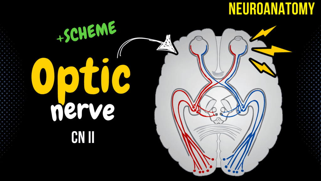
CN 2: Optic Nerve Official Links Instagram Youtube Jki-discord Notes & Illustrations Quizzes Summary & Transcript Notes ☆ Members Only Go to PDF Notes Illustrations ☆ Members Only Go to Illustrations 12345678910 Optic Nerve – QUIZ Test your understanding with 10 random multiple-choice questions from the question bank. You're in the preview mode. Note: All elements work correctly on the front end. 1 / 10 What is the location of the primary visual cortex? A) Occipital Lobe B) Parietal Lobe C) Frontal Lobe D) Temporal Lobe The primary visual cortex is located in the occipital lobe. 2 / 10 Which retinal fibers remain ipsilateral? A) Superior Fibers B) Inferior Fibers C) Temporal Fibers D) Nasal Fibers Temporal fibers remain ipsilateral and do not cross at the optic chiasma. 3 / 10 What is the blind spot caused by? A) Macula Lutea B) Optic Disc C) Peripheral Retina D) Fovea Centralis The optic disc causes the physiological blind spot due to the absence of photoreceptors. 4 / 10 What visual defect is caused by damage to the lateral geniculate body? A) Bitemporal Hemianopia B) Homonymous Hemianopia C) Quadrantanopia D) Total Blindness Damage to the lateral geniculate body causes homonymous hemianopia. 5 / 10 Which retinal region is specialized for sharp central vision? A) Peripheral Retina B) Fovea Centralis C) Optic Disc D) Parafovea The fovea centralis is specialized for sharp central vision. 6 / 10 What structure integrates reflexive eye movements? A) Pretectal Nucleus B) Superior Colliculus C) Lateral Geniculate Body D) Hypothalamus The superior colliculus integrates reflexive eye movements in response to visual stimuli. 7 / 10 What is the role of the suprachiasmatic nucleus in the visual pathway? A) Circadian Rhythm Regulation B) Reflex Integration C) Saccadic Eye Movement D) Depth Perception The suprachiasmatic nucleus regulates circadian rhythms based on visual input. 8 / 10 Which brain structure regulates circadian rhythms via visual input? A) Hypothalamus B) Superior Colliculus C) Thalamus D) Pretectal Nucleus The hypothalamus regulates circadian rhythms through the suprachiasmatic nucleus. 9 / 10 What is the function of the pretectal nucleus in the visual pathway? A) Motion Detection B) Visual Acuity C) Color Perception D) Pupillary Reflex The pretectal nucleus is involved in the pupillary light reflex. 10 / 10 What type of neurons are ganglion cells? A) Second-Order Neurons B) Fourth-Order Neurons C) Third-Order Neurons D) First-Order Neurons Ganglion cells are third-order neurons in the visual pathway. Your score is The average score is 0% Description Visual Pathway Scheme / Overview Visual Stimuli → 1st, 2nd, and 3rd Order Neurons in Retina → Optic Nerve → Half Fibers Cross the Optic Chiasma → Optic Tract → Lateral Geniculate Body → Primary Visual Cortex Eye Anatomy Cornea Anterior Chamber Pupil Iris Lens Posterior Chamber Ciliary Muscles Sclera Choroid Retina Optic Disc Vitreous Humor Hyaloid Canal Retina 1st Order Neurons: Rods and Cones 2nd Order Neurons: Bipolar Cells 3rd Order Neurons: Ganglion Cells Visual Fields Fovea: Highest Acuity of Vision Retina is Split into Temporal Half and the Retinal Half Medial Fibers Go to Contralateral Side Lateral Fibers Go to Ipsilateral Side Course of the Visual Pathway 1st Neuron: Rods and Cones 2nd Neurons: Bipolar Cells 3rd Neurons: Ganglion Cells Optic Nerve (Nervus Opticus) Optic Chiasma (Chiasma Opticum) Optic Tract (Tractus Opticus) 4th Order Neuron: Lateral Geniculate Bodies Optic Radiations (Radiato Optica) and Meyer’s Loop Primary Visual Cortex (Area 17) Visual Pathway Collaterals Accommodation Reflex Direct and Consensual Light Reflex Saccadic Eye Movement Tectospinal Tract (For Reflex Movements Due to Unexpected Visual Irritation) Medial Longitudinal Fasciculus Clinical Relevance Lesion in Optic Nerve: Complete Blindness of Affected Side Lesion in Optic Chiasma: Visual Field Bitemporal Hemianopia Lesion in Optic Tract and Geniculate Body: Homonymous Hemianopia Lesion in Optic Radiation: Quadrantanopia Lesion in the Visual Cortex: Cortical Blindness → Homonymous Hemianopias with or without Macular Involvement Sources Singh, I. (2017). Human Neuroanatomy (10th ed.). Helwany M, Bordoni B. Neuroanatomy, Cranial Nerve 1 (Olfactory) [Updated 2022 Aug 8]. In: StatPearls [Internet]. Treasure Island (FL): StatPearls Publishing; 2022 Jan- Kozlowski, T. (2017). Memorix Anatomy: The Complete Study Guide. 2nd ed. Thieme Medical Publishers. Transcript Introduction0:06What’s up!0:08Let’s continue our Cranial nerve series.0:11Cranial nerves are twelve pairs of nerves that exit the brain and the brainstem, and0:16in this segment, we’ll talk detailed about the second cranial nerve, which is the Optic0:21nerve.0:22And we’ll do that by first making a quick scheme of the optic pathway to get an overview0:26of it.0:28Then we’ll cover the very basic of eye anatomy, and talk a little bit about the visual field.0:33Afterwards we’ll go through the course of the visual pathway, and talk about the all0:38the steps that the optic nerve go through in order to make us perceive and understand0:43what we see.0:44We’re also going to shortly go through the visual pathway collaterals.0:49The optic nerve gives off collateral branches that aid with he pupillary light reflex, and0:54the pathway of convergance.0:56At the end we’ll talk a little bit about the clinical relevance around pathologies1:01related with the optic pathway.Optic Nerve Scheme1:03So, I wanna start by saying that the olfactory and the optic nerves are not true nerves.1:10They are just extensions of the diencephalon.1:14They take the meninges with them as they extend towards the eyes.1:18They are not covered by Schwann cells like the peripheral nervous system are.1:23It’s covered by oligodendrocytes, like the Central nervous system is.1:27Now.1:28Another thing I want you know know about the optic nerve is that the optic nerve carries1:32visual information to the brain via the optic tract.1:36Let’s visualize this sentence.1:38Our vision starts at the eye, specifically the retina.1:42The visual pathway is what we call a four-neurone tract, meaning it consists of 4 different1:48neurons.1:50The first, second and third order neurons are located in the retina of the eyeball,1:54I’ll show you a scheme later when we get to it.1:58But it all starts with a visual stimuli from the three neurons in the retina.2:02The visual stimuli is then sent through the optic tract, which goes through the optic2:07canal of the spenoid bone, behind the eyes.2:10The optic nerve then crosses with the
CN 1: Olfactory Nerve
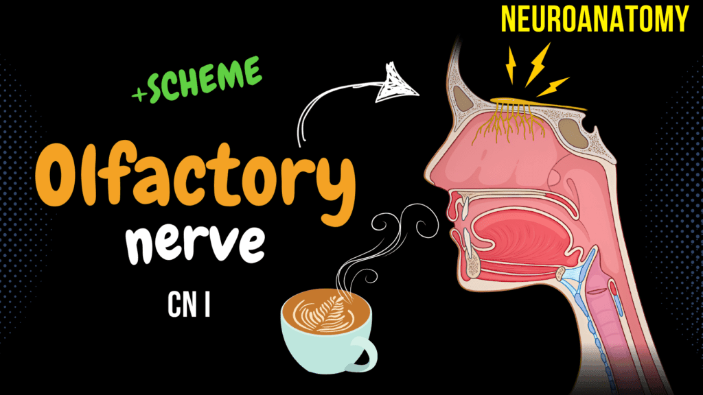
CN 1: Olfactory Nerve Official Links Instagram Youtube Jki-discord Notes & Illustrations Quizzes Summary & Transcript Notes ☆ Members Only Go to PDF Notes Illustrations ☆ Members Only Go to Illustrations 12345678910 Olfactory Nerve – QUIZ Test your understanding with 10 random multiple-choice questions from the question bank. You're in the preview mode. Note: All elements work correctly on the front end. 1 / 10 What is unique about the olfactory pathway compared to other sensory pathways? A) Bypasses the Thalamus B) Requires Interneuron Relay C) Always Relays in Thalamus D) No Cortical Projection The olfactory pathway bypasses the thalamus in its primary projection to the cortex. 2 / 10 Which cranial fossa contains the olfactory bulb? A) Middle Cranial Fossa B) Anterior Cranial Fossa C) Ethmoidal Fossa D) Posterior Cranial Fossa The anterior cranial fossa contains the olfactory bulb. 3 / 10 What is the primary role of mitral cells in the olfactory bulb? A) Inhibit Signals B) Support Other Cells C) Modulate Synapses D) Principal Output Neurons Mitral cells are the principal output neurons of the olfactory bulb. 4 / 10 Which part of the olfactory system is part of the limbic system? A) Mitral Cells B) Subcallosal Gyrus C) Olfactory Glomeruli D) Amygdala The amygdala is part of the limbic system and integrates smell with emotions. 5 / 10 Where does the lateral olfactory stria terminate? A) Hippocampus B) Thalamus C) Amygdala D) Primary Olfactory Cortex The lateral olfactory stria terminates in the primary olfactory cortex. 6 / 10 Which clinical condition involves smelling odors that are not present? A) Phantosmia B) Parosmia C) Anosmia D) Dysosmia Phantosmia involves detecting smells that are not actually present. 7 / 10 What is the primary neurotransmitter used by granule cells? A) Dopamine B) GABA C) Glutamate D) Serotonin GABA is the primary neurotransmitter used by granule cells for lateral inhibition. 8 / 10 Which olfactory stria projects to the primary olfactory cortex? A) Medial Olfactory Stria B) Intermediate Olfactory Stria C) Lateral Olfactory Stria D) Contralateral Olfactory Bulb The lateral olfactory stria projects to the primary olfactory cortex. 9 / 10 What is the main neurotransmitter used in olfactory signal transduction? A) Serotonin B) GABA C) Dopamine D) Glutamate Glutamate is the main neurotransmitter used in the olfactory bulb for signal transduction. 10 / 10 What is the function of tufted cells in the olfactory bulb? A) Detect Odorants B) Transmit Olfactory Information C) Modulate Interneurons D) Produce Mucus Tufted cells transmit olfactory information from the olfactory bulb to the olfactory cortex. Your score is The average score is 0% Description Olfactory Scheme / Overview Smell → Olfactory Nerves → Olfactory Bulb → Olfactory Tract → Olfactory Trigone → Lateral Stria, Medial Stria, and Intermediate Stria Olfactory Epithelium Pseudostratified Columnar Epithelium (Supporting Cells) Consists of: Basal Cells Sustentacular Cells Olfactory Gland Cells (Bowman’s Glands) Olfactory Receptor Neurons Olfactory Neurons Are Bipolar Neurons How We Detect Smell Odorants Enter the Nasal Cavity Through Either the Nasal Opening or Nasopharynx Olfactory Neuron Dendrites Detect Smell Through a G-Protein Coupled Membrane Receptor Signals Pass Through the Cell Body and Axon Axons Form Nerve Bundles (Fila Olfactoria) Olfactory Bulb (Bulbus Olfactorius) Mitral Cells Olfactory Glomerulus (Glomeruli) Tufted Cells Amacrine Granular Cells Periglomerular Cells Olfactory Pathway Olfactory Bulb → Olfactory Tract When the Tract Gets Closer to the Olfactory Tract, It Widens as the Olfactory Trigone (Trigonum Olfactorium) and Divides Into: Lateral Olfactory Stria Medial Olfactory Stria Intermediate Olfactory Stria Lateral Olfactory Stria (Stria Olfactoria Lateralis) Towards the Primary Olfactory Cortex, Which Sends Information to the Secondary Olfactory Cortex Medial Olfactory Stria (Stria Olfactoria Medialis) Goes Towards the Subcallosal Gyrus (Part of the Limbic System) Intermediate Olfactory Stria (Stria Olfactoria Intemedialis) Goes Towards the Olfactory Tubercle (Part of the Limbic System and the Rewards System) Clinical Relevance Anosmia: Temporary or Permanent Loss of the Sense of Smell Dysosmia: Distortion of the Perception of Smell Parosmia: Normal Smell Perceived as Unpleasant/Disgusting Phantosmia: Detecting Smells That Aren’t Really Present in the Environment Sources Singh, I. (2017). Human Neuroanatomy (10th ed.). Olfaction: The Sense of Smell (biology-pages.info) Physiology, Olfactory – StatPearls – NCBI Bookshelf (nih.gov) Olfactory Nerve | Radiology Key Frontiers | Effects of Odor on Emotion, with Implications (frontiersin.org) Other Sources Used Helwany M, Bordoni B. Neuroanatomy, Cranial Nerve 1 (Olfactory) [Updated 2023 Aug 14]. In: StatPearls [Internet]. Treasure Island (FL): StatPearls Publishing; 2024 Jan-. Available from: NCBI Bookshelf Kozlowski, T. (2017). Memorix Anatomy: The Complete Study Guide. 2nd ed. Thieme Medical Publishers. Transcript Introduction0:06What’s up! Taim Talks Med here, let’s start with our Cranial nerve series.0:10Cranial nerves are twelve pairs of nerves that exit the brain and the brainstem,0:15and in this segment, we’ll talk detailed about the first cranial nerve, which is the Olfactory nerve.0:20And we’ll do that by first making a quick scheme of the olfactory pathway to get an overview of it.0:27Then we’ll talk detailed about the Olfactory epithelium and nerve. Basically which striuctures0:32are involved in sensing particles of smell. Then we’ll follow the olfactory pathway and0:38look at which structures in the brain are involved when you smell something.0:42And after that, we’ll do some clinical relevance and examples of conditions that can alter the0:48normal functioning of this nerve. So, the olfactory nerve conductsOlfactory Nerve Scheme0:53the sensation of smell to the brain via the olfactory bulb and tract.0:57Let’s visualize this sentence. Here we got a funky smell of some sort, let’s say someone opened a box1:04of tuna next to you, or maybe someone farted. These gaseous odorants will eventually reach1:10your olfactory nerves. Axons of these olfactory nerves are gonne come together, and run through1:16the cribriform plate, of the ethmoidal bone. So we call the olfactory nerve a 1st order neuron1:23because this is where smell is first registered. Once they go through the cribriform plate,1:29they will synapse with neurons in the olfactory bulb. The olfactory bulb is an elongated structure1:36that lies just above the cribriform plate. It continues posteriorly with the olfactory tract.1:42As the olfactory tract approaches the anterior surface of the midbrain, it
Thoracic Nerves

Thoracic Nerves (Intercostal + Subcostal) Official Links Instagram Youtube Jki-discord Notes & Illustrations Quizzes Summary & Transcript Notes ☆ Members Only Go to PDF Notes Illustrations ☆ Members Only Go to Illustrations 12345678910 Thoracic Nerves – QUIZ Test your understanding with 10 random multiple-choice questions from the question bank. You're in the preview mode. Note: All elements work correctly on the front end. 1 / 10 What is the primary function of the white rami communicantes? A) Preganglionic sympathetic fibers B) Sensory to thoracic skin C) Postganglionic parasympathetic fibers D) Motor to intercostal muscles White rami communicantes carry preganglionic sympathetic fibers from the spinal nerves to the sympathetic trunk. 2 / 10 Which intercostal nerve provides sensory innervation to the skin of the anterior thoracic wall? A) Collateral branch B) Anterior cutaneous branch C) Gray ramus communicans D) Lateral cutaneous branch The anterior cutaneous branch of intercostal nerves provides sensory innervation to the skin of the anterior thoracic wall. 3 / 10 Which thoracic nerve segment extends to form thoracoabdominal nerves? A) T2 B) T6 C) T5 D) T7–T11 The T7–T11 intercostal nerves are referred to as thoracoabdominal nerves, extending beyond the thoracic wall to innervate the abdominal wall. 4 / 10 Which thoracic nerve is referred to as the “subcostal nerve”? A) T11 B) T10 C) T9 D) T12 The T12 spinal nerve is referred to as the subcostal nerve because it lies below the 12th rib. 5 / 10 The intercostobrachial nerve is a branch of which intercostal nerve? A) T2 B) T1 C) T12 D) T3 The intercostobrachial nerve is a branch of the second intercostal nerve and supplies the axilla and medial arm. 6 / 10 What is the anatomical location of the neurovascular bundle in the intercostal space? A) Between the pleura and intercostal muscles B) Superior border of the rib below C) Costal groove D) Posterior border of the rib The neurovascular bundle is located in the costal groove along the inferior border of each rib. 7 / 10 The collateral branch of an intercostal nerve is located in which anatomical position relative to the rib? A) Superior border of the rib below B) Inferior border of the rib below C) Inferior surface of the rib above D) Middle of the intercostal space The collateral branch runs parallel to the main nerve along the superior border of the rib below. 8 / 10 The intercostobrachial nerve is derived from which intercostal nerve? A) T4 B) T1 C) T3 D) T2 The intercostobrachial nerve is derived from the second intercostal nerve and provides sensory innervation to the axilla and medial arm. 9 / 10 What is the primary sensory role of the anterior cutaneous branches of the intercostal nerves? A) Lateral thoracic skin B) Anterior thoracic and abdominal skin C) Axilla skin D) Posterior thoracic skin The anterior cutaneous branches provide sensory innervation to the anterior skin of the thorax and abdomen. 10 / 10 What structure lies superior to the intercostal nerve within the neurovascular bundle? A) Intercostal vein B) Intercostal artery C) Internal intercostal muscle D) Parietal pleura The intercostal vein is the superiormost structure in the neurovascular bundle. Your score is The average score is 0% Description Intro Intercostal Spaces (Spatium Intercostale) Intercostal Nerves (Nervi Intercostales) Typical Intercostal Nerve Muscular Branches (Rami Musculares) Somatosensory Branches Lateral Cutaneous Branches (Rami Cutanei Laterales) Anterior Cutaneous Branches (Rami Cutanei Anteriores) Collateral Branch Atypical Intercostal Nerve 1st Nerve: Superior Branch Joins Brachial Plexus Inferior Branch Continues as Intercostal Nerve 2nd Intercostal Nerve: Has Intercostobrachial Nerve (Nervi Intercostobrachiales) 7th, 8th, 9th, 10th, 11th Intercostal Nerves: Also Called Thoracoabdominal Nerves Innervate Abdominal Wall Run in a Neurovascular Bundle Subcostal Nerve (Nervus Subcostalis) Has the Same Branches as the Intercostal Nerves Gives Off a Communicating Branch That Helps Form Iliohypogastric Nerve of the Lumbar Plexus Transcript Introduction0:06What’s up!0:07Let’s go ahead and continue with our Peripheral Nervous System.0:10Previously we’ve covered the cervical plexus, the brachial, lumbar and sacral plexuses.0:15The next thing that we’re going to talk about now are nerves that’re located within0:19the thoracic wall.0:21The thoracic nerves we call them.0:23So here we see a plan anterior view of the thorax.0:27We see the costal bones, sternum, and the vertebra.0:31The Intercostal space is the space between the ribs, as you see here.0:36There are 11 spaces on each side and they are numbered according to the rib which is0:40the superior border of the space.0:431st intercostal space under the 1st rib, 2nd intercostal space under the 2nd rib, and so0:49on.0:50It goes all the way down to the 11, so again we got 11 intercostal spaces.0:56Now the Throacic nerves are distributed Now.0:57The thoracic spine has 12 nerve roots.1:00So from T1 all the way down to T12, there are nerves that go out from the spine on each1:05side, similar to the rest of the spine.1:08The anterior root of the nerves in the thoracic region branch off and control both the motor1:14and the sensory signals in the thoracic and the abdominal wall.1:18These nerves leave the thoracic spine, and are therefore referred to as the thoracic1:23nerves.1:24So we got one nerve that run underneath each rib, and are numbered according to each nerve1:30that they run underneath.1:32As you see here.1:33If these thoracic nerves run within an intercostal space, they’ll get the name intercostal1:38nerves.1:39So we got 11 intercostal nerves, since we got 11 intercostal spaces.1:44The last nerve that run underneath the 12th rib, is referred to as the subcostal nerve.Content1:50So in this video, we’re first going to go detailed into the intercostal nerves.1:55We’ll talk about their course, and branches.1:59And a traditional division of them called typical and atypical intercostal nerves.2:04Then we’ll talk a little bit about the subcostal nerve so that we cover all the thoracic nerves2:08that’re relevant to know regarding the peripheral nervous system.2:12Awesome.Intercostal Nerve Course2:13So.2:14Course.2:15How do the intercostal nerves go?2:18Here you see a plan anterior view of the thorax along with the intercostal muscles.2:23If we now cross over the thorax like this, right over the rib.2:28Then take away the upper part, and schematically draw what we’ll see
Sacral Plexus
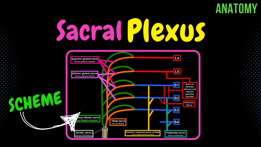
Sacral Plexus (Scheme + Quiz) Official Links Instagram Youtube Jki-discord Notes & Illustrations Quizzes Summary & Transcript Notes ☆ Members Only Go to PDF Notes Illustrations ☆ Members Only Go to Illustrations 12345678910 Sacral Plexus – QUIZ Test your understanding with 10 random multiple-choice questions from the question bank. You're in the preview mode. Note: All elements work correctly on the front end. 1 / 10 The inferior gluteal nerve originates from which spinal levels? A) L2-S1 B) L5-S2 C) L4-S1 D) L3-L5 It arises from the posterior divisions of L5, S1, and S2 spinal nerves. 2 / 10 Which spinal nerves form the superior gluteal nerve? A) L4-S1 B) L3-S1 C) L5-S2 D) S1-S3 The superior gluteal nerve is derived from L4-S1. 3 / 10 What is the largest nerve arising from the sacral plexus? A) Inferior gluteal nerve B) Sciatic nerve C) Tibial nerve D) Pudendal nerve The sciatic nerve is the largest nerve, innervating the posterior thigh, leg, and foot. 4 / 10 Which nerve provides motor supply to the quadratus femoris? A) Pudendal nerve B) Nerve to quadratus femoris C) Inferior gluteal nerve D) Sciatic nerve The nerve to quadratus femoris (L4-S1) supplies this muscle along with inferior gemellus. 5 / 10 What is the primary function of the pudendal nerve? A) Motor and sensory to the perineum B) Sensory to posterior thigh C) Motor to gluteus maximus D) Sensory to pelvic viscera The pudendal nerve provides motor and sensory innervation to the perineum and external genitalia. 6 / 10 The sciatic nerve exits the pelvis through which structure? A) Greater sciatic foramen B) Lesser sciatic foramen C) Obturator canal D) Suprapiriform foramen The sciatic nerve exits the pelvis via the greater sciatic foramen. 7 / 10 Which nerve innervates the gluteus medius and minimus muscles? A) Superior gluteal nerve B) Pudendal nerve C) Sciatic nerve D) Inferior gluteal nerve The superior gluteal nerve innervates both the gluteus medius and gluteus minimus muscles. 8 / 10 Which muscle is innervated by the inferior gluteal nerve? A) Gluteus medius B) Quadratus femoris C) Tensor fasciae latae D) Gluteus maximus The inferior gluteal nerve supplies the gluteus maximus muscle. 9 / 10 Which muscles are innervated by the tibial branch of the sciatic nerve? A) Posterior thigh and leg muscles B) Piriformis C) Gluteus minimus D) Quadratus femoris The tibial nerve innervates the posterior thigh muscles (except short head of biceps femoris), leg, and foot muscles. 10 / 10 The sciatic nerve is composed of fibers from which spinal nerve roots? A) L3–S2 B) L5–S2 C) S1–S4 D) L4–S3 The sciatic nerve originates from the anterior and posterior divisions of L4–S3. Your score is The average score is 0% Description A minor mistake in the video: Superior and Inferior gluteal nerves come from the posterior divisions of L4-S1, not the anterior as shown in the scheme (apologies for this mistake). Topography Anterior branch of spinal nerve From spinal nerve T12, L1, L2, L3, L4 General Outline of Lumbar Plexus Long Branches Sciatic Nerve (Nervus Ischiadicus) Posterior Cutaneous Nerve of the Thigh (Nervus Cutaneus Femoris Posterior) Short Branches Pudendal Nerve (Nervus Pudendus) Nerve to Obturator Internus and Superior Gemellus Muscles Piriformis Nerve Superior Gluteal Nerve (Nervus Gluteus Superior) Inferior Gluteal Nerve (Nervus Gluteus Inferior) Transcript Introduction0:06alright so in this video we’re going to0:07talk about the sacral plexus we already0:10talked about the cervical plexus we0:11talked about the brachial plexus we0:13talked about the lumbar plexus the last0:16large plexus we have now is the sacral0:18plexus so let’s go ahead and get started0:20so first we’re going to go through the0:22topography just talk a little bit about0:24which nerves feed into the sacral plexus0:27then after that we’re going to make a0:29scheme of the sacral plexus and to make0:32this as easy as possible to understand0:34and remember we’re going to draw the0:36general outline first0:38then we’re going to go ahead and divide0:40the nerves into long branches and short0:43branches0:44and go through each nerve and a little0:46bit of clinical importance related to0:49those nerves0:50and at the end there will be a little0:51quizTopography of Sacral Plexus0:52so let’s go ahead and get started with0:54the topography so here we see the spinal0:57nerve within the vertebral canal and1:00here we see the spinal nerve leaving the1:02intervertebral foramen1:04so there will be one spinal nerve1:06leaving on each side they’re paired1:09to be specific though we’re not really1:11talking about the whole spinal nerve1:13right we’re actually talking about the1:15anterior branch of the spinal nerve1:18because the anterior branch is what1:20forms all the plexuses that we’ve talked1:22about in this series of videos1:25alright so the sacral plexus is formed1:27from the anterior branches of the spinal1:30nerves l41:31l51:33s1 s2 s3 and s41:37alright1:38so this is a pretty big plexus one of1:40the main nerves that are coming from the1:42sacral plexus is the sciatic nerve and1:45we’ll talk a little bit about the1:46clinical correlation with that so let’sScheme of the Sacral Plexus1:49go ahead and simplify this drawing what1:52i want you to do is to grab a piece of a1:54paper and a pen and i want you to draw1:57the scheme with me and once you do that1:59i promise you will remember this much2:01much easier2:03alright2:04first off we can start by drawing a line2:06from l4 all the way down to s3 like you2:09see here2:11what happens isSciatic Nerve2:12off from l42:14there’s going to be a division and2:16anterior division that are going to2:18traverse this way2:20on its way down it will pick up a branch2:23from l5 it will pick up a branch from s12:26s2 and s32:29another thing that’s going to happen is2:31that from l4 same thing another branch2:34is going to emerge but this one is going2:36to be more posteriorly and same thing it2:40will pick up a poster of your branch2:42from l5 s1 s2 and s32:46these two nerves are called the tibial2:49nerve and the common feebler nerve so2:52the tibial nerve comes from the anterior2:55division of branches coming from l4 to2:58s32:59from the sacral plexus and the common3:01feebler nerve come from the posterior3:04division of branches coming from l4 to3:07s3 from the sacral plexus3:10however those two nerves run together in3:13a sheath so they go together forming3:16this chaotic nerve it’s
Lumbar plexus
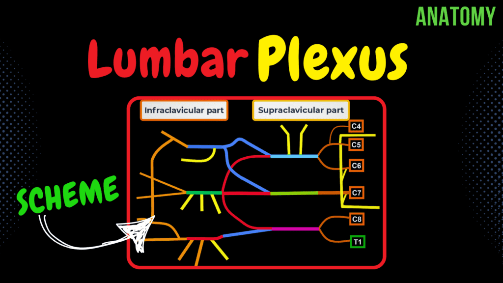
Lumbar plexus (Scheme + Quiz) Official Links Instagram Youtube Jki-discord Notes & Illustrations Quizzes Summary & Transcript Notes ☆ Members Only Go to PDF Notes Illustrations ☆ Members Only Go to Illustrations 12345678910 Lumbar Plexus – QUIZ Test your understanding with 10 random multiple-choice questions from the question bank. You're in the preview mode. Note: All elements work correctly on the front end. 1 / 10 Which branch of the lumbar plexus is located medially to the psoas major? A) Femoral nerve B) Obturator nerve C) Lateral femoral cutaneous nerve D) Ilioinguinal nerve The obturator nerve runs medially to the psoas major. 2 / 10 Which nerve is commonly affected in meralgia paresthetica? A) Femoral nerve B) Obturator nerve C) Ilioinguinal nerve D) Lateral femoral cutaneous nerve The lateral femoral cutaneous nerve is affected, causing pain or numbness in the lateral thigh. 3 / 10 The femoral nerve originates from which spinal segments? A) L1-L3 B) L2-L4 C) L3-L4 D) L2-L5 The femoral nerve is formed by the posterior divisions of L2-L4 and provides motor and sensory innervation to the anterior thigh. 4 / 10 The iliohypogastric and ilioinguinal nerves are branches of which spinal nerve? A) L2-L3 B) L3-L4 C) T12-L1 D) L1-L2 Both iliohypogastric and ilioinguinal nerves originate from L1, with contributions from T12. 5 / 10 What structure does the obturator nerve pass through to reach the thigh? A) Femoral triangle B) Obturator canal C) Obturator foramen D) Inguinal ligament The obturator nerve passes through the obturator foramen to reach the medial compartment of the thigh. 6 / 10 Which nerve passes under the inguinal ligament near the ASIS? A) Obturator nerve B) Genitofemoral nerve C) Femoral nerve D) Lateral femoral cutaneous nerve The lateral femoral cutaneous nerve passes under the inguinal ligament near the anterior superior iliac spine (ASIS). 7 / 10 What spinal levels contribute to the iliohypogastric nerve? A) T11-T12 B) L1-L2 C) L2-L3 D) T12-L1 The iliohypogastric nerve arises from T12 and L1 and supplies motor and sensory functions to the abdominal wall. 8 / 10 Which branch of the lumbar plexus enters the inguinal canal? A) Obturator nerve B) Iliohypogastric nerve C) Femoral nerve D) Genitofemoral nerve The genital branch of the genitofemoral nerve enters the inguinal canal and supplies the cremaster muscle in males. 9 / 10 What is the mnemonic for remembering the branches of the lumbar plexus? A) Interested In Getting Lunch On Friday B) Intelligent Ideas Go Far C) Fridays Always Count D) Love On Friday The mnemonic “Interested In Getting Lunch On Friday” stands for Iliohypogastric, Ilioinguinal, Genitofemoral, Lateral femoral cutaneous, Obturator, and Femoral nerves. 10 / 10 What is the spinal level origin of the genitofemoral nerve? A) T12-L1 B) L2-L3 C) L1-L2 D) L3-L4 The genitofemoral nerve arises from L1 and L2. Your score is The average score is 0% Description Topography Anterior branch of spinal nerve From spinal nerve T12, L1, L2, L3, L4 General Outline of Lumbar Plexus Ilioinguinal Nerve (Nervus Ilioinguinalis) – L1 Subcostal Nerve gives off a branch, which joins L1 branch to form Iliohypogastric Nerve (Nervus Iliohypogastricus) Genitofemoral Nerve (Nervus Genitofemoralis) – L1 and L2 Lateral Femoral Cutaneous Nerve (Nervus Cutaneus Femoris Lateralis) – L2 and L3 Femoral Nerve (Nervus Femoralis) – L2, L3, and L4 Obturator Nerve (Nervus Obturatorius) – L2, L3, and L4 Taim Talks Med Transcript Introduction0:06Alright so in this video we’re going to talk specifically about the Lumbar Plexus.0:10Ok so the lumbar plexus is a really important and we’ll ad a little clinical correlation0:14to it aswell.0:15So first we’re gonna go through the topography.0:18Just talk a little bit about which nerves feed into the lumbar plexus.0:23Then after that we’re gonna make a scheme of the lumbar plexus.0:27And to make this as easy as possible to understand and remember, we’re going to draw the general0:32outline first.0:34Then we’re gonna go ahead and go through each nerve and a little bit of clinical importance0:39related to those nerves.0:41And at the end, there’ll be a little quiz.Topography of Lumbar Plexus0:44So let’s go ahead and get started with the topography.0:47So here we see the spinal cord within the vertebral canal.0:50And here we see the spinal nerve leaving the intervertebral foramen, so there’ll be one0:56spinal nerve leaving on each side.0:59They’re paired.1:00To be specific, though, we’re not really talking about the whole spinal nerve, right?1:05We’re actually talking about the anterior branch of the spinal nerve.1:09Because the anterior branch is what forms all the plexuses that we’re going to talk1:13about in this series of videos.1:15So, lumbar plexus, formed by the spinal nerves.1:19Which branch of the spinal nerves?1:21The anterior branch.1:23More specifically.1:24The lumbar plexus is formed from the anterior branches of spinal nerves L1–L4 and a contributing1:35branch from T12.1:38The plexus is located within the psoas major, lateral to the lumbar vertebrae1:43Let’s now go ahead and simplify this drawing so that we can actually make a scheme outScheme of the Lumbar Plexus1:47of this.1:49Now what I want you to do, is to grab a piece of paper and a pen, and I want you to draw1:54this scheme with me.1:56Once you do that, I promise you’ll remember this much much easier.2:00Aright.2:02First off, L1 is going to give off a branch called the Ilioinguinal nerve.2:07So the primary nerve that the L1 give off is the Ilioinguinal nerve.2:13The nerve above T12 is the subcostal nerve, right?2:17It’s not actually a part of the lumbar plexus, but the Subcostal nerve is, it’s a mixed2:23nerve.2:24It has a sensory and a motor function.2:27Sensory for the skin on the lower abdomen and inguinal regions, and motor for the abdominal2:33wall muscles.2:34Now the subcostal nerve again it’s not a part fot he lumbar plexus, but the reason2:40why I mention it here is because it’s going to give off a branch that’s going to go2:45with the branch of L1 to form the iliohypogastric nerve.2:50We’ll mention all of those in detail in a minute but let’s actually finish off this2:55scheme first.2:57Now.2:58L2 will traverse this way.3:00L1 and L2 will both give off branches that go together to form the so called
Brachial Plexus
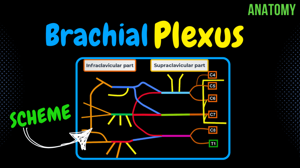
Brachial Plexus (Scheme + Quiz) Official Links Instagram Youtube Jki-discord Notes & Illustrations Quizzes Summary & Transcript Notes ☆ Members Only Go to PDF Notes Illustrations ☆ Members Only Go to Illustrations 12345678910 Brachial Plexus – QUIZ Test your understanding with 10 random multiple-choice questions from the question bank. You're in the preview mode. Note: All elements work correctly on the front end. 1 / 10 The lateral cord gives rise to which nerve? A) Radial nerve B) Ulnar nerve C) Median nerve D) Musculocutaneous nerve The lateral cord gives rise to the musculocutaneous nerve. 2 / 10 Where is the brachial plexus located in relation to the scalene muscles? A) Anterior to both B) Between anterior and middle C) Posterior to both D) Medial to both The roots of the brachial plexus pass between the anterior and middle scalene muscles. 3 / 10 Which nerve runs along the medial side of the arm and passes through Guyon’s canal in the wrist? A) Radial nerve B) Ulnar nerve C) Median nerve D) Axillary nerve The ulnar nerve runs medially along the arm and enters the wrist through Guyon’s canal. 4 / 10 Which nerve originates from the C5 root of the brachial plexus? A) Axillary nerve B) Median nerve C) Dorsal scapular nerve D) Suprascapular nerve The dorsal scapular nerve arises from the C5 root and innervates rhomboid muscles and levator scapulae. 5 / 10 Which artery is closely associated with the brachial plexus at its roots? A) Brachial artery B) Subclavian artery C) Profunda brachii artery D) Axillary artery The subclavian artery is closely associated with the roots of the brachial plexus. 6 / 10 What is the primary function of the posterior division of the brachial plexus? A) Flexor innervation B) Extensor innervation C) Sensory innervation D) Reflex coordination The posterior division of the brachial plexus innervates the extensor compartments of the upper limb. 7 / 10 What is the sensory distribution of the musculocutaneous nerve? A) Medial arm B) Posterior forearm C) Lateral forearm D) Lateral hand The musculocutaneous nerve provides sensation to the lateral forearm via the lateral cutaneous nerve. 8 / 10 Which nerve innervates the teres major muscle? A) Suprascapular nerve B) Radial nerve C) Axillary nerve D) Lower subscapular nerve The lower subscapular nerve innervates the teres major muscle. 9 / 10 Which nerve is most likely affected in a winged scapula? A) Radial nerve B) Ulnar nerve C) Long thoracic nerve D) Axillary nerve Winged scapula occurs when the long thoracic nerve is damaged, impairing serratus anterior function. 10 / 10 Which nerve pierces the middle scalene muscle? A) Median nerve B) Suprascapular nerve C) Long thoracic nerve D) Dorsal scapular nerve The dorsal scapular nerve pierces the middle scalene muscle to reach the rhomboid muscles. Your score is The average score is 0% Description Topography Anterior branch of spinal nerve From spinal nerve C4, C5, C6, C7, C8, T1 General Outline of Brachial Plexus Landmarks mnemonic: Really Tired, Drink Coffee Now Root, Trunk, Division, Cord, Nerves Superior Trunk (Truncus Superior) Middle Trunk (Truncus Medius) Inferior Trunk (Truncus Inferior) Anterior and Posterior Divisions Posterior Cord (Funiculus Posterior) Lateral Cord (Fasciculus Lateralis) Medial Cord (Fasciculus Medialis) Supraclavicular Branches of Brachial Plexus Dorsal Scapular Nerve (Nervus Dorsalis Scapulae) Long Thoracic Nerve (Nervus Thoracicus Longus) Suprascapular Nerve (Nervus Suprascapularis) Subclavian Nerve (Nervus Subclavius) Lateral Pectoral Nerve (Nervus Pectoralis Lateralis) Upper Subscapular Nerve (Nervi Subscapularis) Lower Subscapular Nerve Thoracodorsal Nerve (Nervus Thoracodorsalis) Medial Pectoral Nerve (Nervus Pectoralis Medialis) Infraclavicular Branches of the Brachial Plexus Medial Cutaneous Nerve of Forearm (Nervus Cutaneus Antebrachii Medialis) Medial Cutaneous Nerve of Arm (Nervus Cutaneus Brachii Medialis) Musculocutaneous Nerve (Nervus Musculocutaneus) Axillary Nerve (Nervus Axillaris) Radial Nerve (Nervus Radialis) Median Nerve (Nervi Mediani) Ulnar Nerve (Nervus Ulnaris) Quiz! Transcript Introduction0:06Alright so in this video we’re going to talk about the Brachial Plexus.0:09It’s a super important plexus, so let’s go ahead and get started0:13So first we’re gonna go through the topography.0:14Just talk a little bit about which nerves feed into the brachial plexus.0:19Then after that we’re gonna make a scheme of the brachial plexus.0:20And to make this as easy as possible to understand and remember, we’re going to draw the general0:26outline first.0:27Then we’ll go ahead and go through the supraclavicular parts of the brachial plexus, and then the0:32infraclavicular.0:34And at the end, there’ll be a little quiz.0:36So let’s go ahead and get started with the topography.Topography of Brachial Plexus0:39So here we see the spinal cord within the vertebral canal.0:43And here we see the spinal nerve leaving the intervertebral foramen, so there’ll be one0:48spinal nerve leaving on each side.0:50They’re paired.0:51To be specific, though, we’re not really talking about the whole spinal nerve, right?0:56We’re actually talking about the anterior branch of the spinal nerve.1:00Because the anterior branch is what forms all the plexuses that we’re going to talk1:04about in this series of videos.1:06So, brachial plexus, formed by the spinal nerves.1:09Which branch of the spinal nerves?1:11The anterior branch.1:13More specifically.1:14The brachial plexus is formed by spinal nerve number C5, C6, C7, C8, it’ll also have the1:22T1 join in for the brachial plexus.1:25So we say that it generally starts at C5, it might get some branches from the C4 but1:31for the most part it starts at C5.1:33But we’ll add the C4 too so that we cover all the variations.1:37Alright.Scheme of the Brachial Plexus1:38Let’s now go ahead and simplify this drawing so that we can actually make a scheme out1:42of this.1:43Now what I want you to do, is to grab a piece of paper and a pen, and I want you to draw1:49this scheme with me.1:50Once you do that, I promise you’ll remember this much much easier.1:54Aright.1:55Now the first thing we can do, is lay down the main topographical areas here so that2:00it helps us memorize the scheme.2:02Coming from the spinal segments are nerve roots.2:05They form Trunks, which form divisions.2:09The divisions go together forming cords, which then form the main nerves that supply different2:15structures of the upper extremities.2:17So Root Trunk, Divisions, Chord, Nerves.2:21You’ll find many mnemonics on
Cervical Plexus

Cervical Plexus (Scheme) Official Links Instagram Youtube Jki-discord Notes & Illustrations Quizzes Summary & Transcript Notes ☆ Members Only Go to PDF Notes Illustrations ☆ Members Only Go to Illustrations 12345678910 Cervical Plexus – QUIZ Test your understanding with 10 random multiple-choice questions from the question bank. You're in the preview mode. Note: All elements work correctly on the front end. 1 / 10 Which nerve provides motor innervation to the diaphragm? A) Hypoglossal nerve B) Vagus nerve C) Phrenic nerve D) Accessory nerve The phrenic nerve (nervus phrenicus) from C3–C5 is the primary motor nerve of the diaphragm. 2 / 10 What is the spinal root origin of the supraclavicular nerves? A) C4–C5 B) C2–C3 C) C1–C2 D) C3–C4 The supraclavicular nerves originate from C3 and C4 and innervate the skin over the clavicle. 3 / 10 What type of fibers does the phrenic nerve contain? A) Motor only B) Sympathetic fibers only C) Sensory only D) Mixed fibers The phrenic nerve is a mixed nerve, containing motor, sensory, and sympathetic fibers. 4 / 10 Which sensory nerve from the cervical plexus supplies the occipital region? A) Lesser occipital nerve B) Transverse cervical nerve C) Supraclavicular nerve D) Greater auricular nerve The lesser occipital nerve (nervus occipitalis minor) from C2 and C3 innervates the skin of the occipital region. 5 / 10 What is the anatomical role of the phrenic nerve’s pleural branches? A) Sensory to mediastinal pleura B) Motor to intercostal muscles C) Motor to diaphragm D) Parasympathetic to heart The pleural branches of the phrenic nerve provide sensory innervation to the mediastinal pleura. 6 / 10 Which spinal nerves contribute to the formation of the cervical plexus? A) C2–C5 B) C1–C4 C) C1–C3 D) C3–C6 The cervical plexus arises from the anterior branches of spinal nerves C1–C4. 7 / 10 Which plexus nerve provides sensation to the parotid region? A) Transverse cervical nerve B) Supraclavicular nerves C) Greater auricular nerve D) Lesser occipital nerve The greater auricular nerve provides sensory innervation to the parotid region. 8 / 10 Which muscle is directly innervated by the muscular branches of the cervical plexus? A) Scalene muscles B) Sternocleidomastoid C) Infrahyoid muscles D) Platysma Muscular branches innervate the infrahyoid muscles and other deep neck muscles. 9 / 10 Which nerve supplies the skin of the occiput? A) Transverse cervical nerve B) Lesser occipital nerve C) Greater auricular nerve D) Supraclavicular nerves The lesser occipital nerve innervates the skin of the occiput and the posterior neck region. 10 / 10 Which structure is directly innervated by the phrenic nerve? A) Scalene muscles B) Sternocleidomastoid C) Diaphragm D) Intercostal muscles The phrenic nerve provides motor innervation to the diaphragm and sensory fibers to the pericardium and pleura. Your score is The average score is 0% Description Topography Anterior branch of spinal nerve From spinal nerve C1, C2, C3, C4 Somatosensory Branches (Mechanoreceptors, Exteroreceptors, Proprioceptors, Thermoreceptors) Transverse Cervical Nerve (Nervus Transversus Colli) from C2 and C3 Divides into inferior branch for the skin of the infrahyoid region, and superior branch for the skin of the suprahyoid region. Superior branch forms superficial ansa cervicalis for platysma Greater Auricular Nerve (Nervus Auricularis Magnus) from C2 and C3 Innervates the skin over the ear and parotid glands. Anterior branch for anterior part of auricle and parotid Posterior branch for the posterior parts Lesser Occipital Nerve (Nervus Occipitalis Minor) from C2 and C3 Supplies skin of occiput, the posterior neck, and lateral back of neck Supraclavicular Nerves (Nervi Supraclaviculares) from C3 and C4 Motor Branches Muscular Branches (Rami Musculares) from C1-C4 Deep Cervical Loop (Ansa Cervicalis Profunda) for the infrahyoid muscles Formed by Superior Root (Radix Superior) – C1 and C2 Inferior Root (Radix Inferior) – C2 to C3 Mixed Branches Phrenic Nerve (Nervus Phrenicus) – C3 to C5 Phrenic Branches (Rami Phrenici) Pericardial Branches (Rami Pericardiaci) Pleural Branches (Rami Pleurales) Phrenicoabdominal Branches (Rami Phrenicoabdominales) Sensory Branches for the thymus Accessory Nerve (Spinal Part) Transcript Introduction0:07What’s up! Taim Talks Med here. Let’s talk about the Cervical Plexus!0:11The cervical plexus is extremely important because it’s gonna supply0:15many structures in the head in the neck, and even some other muscles that we’ll talk about.0:20Now we’re gonna talk about in a series of videos, we’re gonna talk about the cervical, the brachial0:25plexus, the lumbar, the sacral. And then we’re gonna talk about the intercostal nerves.0:30So the way I wanna explain this plexus to you all is go through from the very basic,0:36to innervation and then some clinical relevance. So first we’re gonna go through the topography.0:42Just talk a little bit about where these nerves come from specifically.0:46Then we’re gonna make a scheme of the cervical plexus.0:49And to make this scheme, we’ll first go through the somatosensory branches of the cervical plexus.0:55We’re gonna go through the motor branches and talk a little bit about the mixed branch1:00that the cervical plexus contributes to. Now don’t get mad at me for simplifyingTopography of Cervical Plexus1:05it too much now. But here you see the very basic vertebral bodies and the spinous processes of the1:11vertebrae, forming the vertebral canal. And within the vertebral canal, you’ll find the spinal cord.1:18And here we see the spinal cord divided into the cervical segments in orange, then the thoracic1:24segments, the lumbar segments, sacral segments. And then a tiny end the conus medullaris,1:31from where the coccygeal nerve goes out from. Each spinal cord segment give off two spinal1:37nerves. One on each side. And they leave the vertebral cala through the intervertebral1:43foramen. Here they’re super simplified, I know it looks like they’re leaving from between the1:49vertebral bodies, but they’re not. They leave through the intervertebral foramen on each side.1:55Once the spinal nerve leaves the vertebral canal, it starts dividing into a posterior branch,2:02which innervates the back. It divides into an anterior branch, which innervates2:06the anterior part of the trunk and limbs. There’s a meningeal branch that goes back into the2:12vertebral canal to innervate the spinal meninges. And then there’re the rami communicans,2:18connected to the sympathetic chain ganglia. Or the
Internal Cerebrum
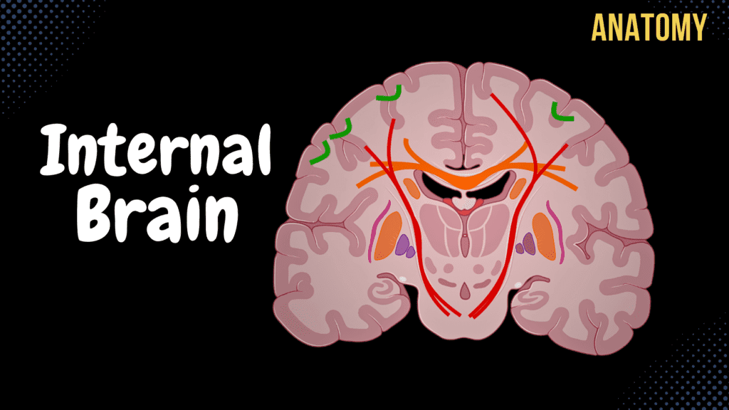
Internal Cerebrum (Association, Commissural, Projection Fibers, Basal Ganglion) Official Links Instagram Youtube Jki-discord Notes & Illustrations Quizzes Summary & Transcript Notes ☆ Members Only Go to PDF Notes Illustrations ☆ Members Only Go to Illustrations 12345678910 Internal Cerebrum – QUIZ Test your understanding with 10 random multiple-choice questions from the question bank. You're in the preview mode. Note: All elements work correctly on the front end. 1 / 10 Which tract in the posterior limb of the internal capsule transmits motor signals to the body? A) Corticospinal tract B) Acoustic radiation C) Corticonuclear tract D) Optic radiation The corticospinal tract is essential for voluntary motor control of the body. 2 / 10 What structure connects the hippocampus to the mammillary bodies? A) Cingulum B) Subthalamic nucleus C) Fornix D) Corpus callosum The fornix is a major white matter tract connecting these structures for memory functions. 3 / 10 Which structure separates the caudate nucleus from the lentiform nucleus? A) Internal capsule B) External capsule C) Cingulum D) Corpus callosum The internal capsule separates the caudate nucleus medially from the lentiform nucleus laterally. 4 / 10 What structure is the primary output nucleus of the basal ganglia? A) Putamen B) Caudate nucleus C) Substantia nigra D) Globus pallidus internus The globus pallidus internus sends inhibitory signals to the thalamus to regulate movement. 5 / 10 Which structure connects the hippocampus to the mammillary bodies? A) Subthalamic nucleus B) Fornix C) Cingulum D) Anterior commissure The fornix links the hippocampus with mammillary bodies, supporting memory consolidation. 6 / 10 What type of fibers connect cortical regions within the same hemisphere? A) Corticospinal fibers B) Commissural fibers C) Association fibers D) Projection fibers Association fibers link regions within the same hemisphere, ensuring integration of cortical functions. 7 / 10 What condition results from damage to the posterior limb of the internal capsule? A) Visual field defect B) Aphasia C) Auditory processing loss D) Hemiplegia Damage to this region can cause hemiplegia, or paralysis on one side of the body. 8 / 10 What forms the lateral wall of the lateral ventricles? A) Lentiform nucleus B) Corpus callosum C) Caudate nucleus D) Thalamus The caudate nucleus constitutes the lateral wall of the lateral ventricles. 9 / 10 Which fibers form the corona radiata? A) Commissural fibers B) Association fibers C) Corticonuclear fibers D) Projection fibers The corona radiata consists of ascending and descending projection fibers spreading to/from the cortex. 10 / 10 Which fibers are involved in communication between Wernicke’s and Broca’s areas? A) Corpus callosum B) Inferior longitudinal fasciculus C) Arcuate fasciculus D) Internal capsule The arcuate fasciculus connects these areas to facilitate language comprehension and speech production. Your score is The average score is 0% Description Telencephalon Pallium Cerebral Cortex White Matter of the Brain (Corpus Medullare Telencephali) Subpallium Basal Ganglion White Matter of the Cerebrum Association Fibers Commissural Fibers Projection Fibers Association Fibers Short Association Fibers Arcuate Fibers (Fibrae Arcuatae) Long Association Fibers Superior Longitudinal Fasciculus (Fasciculus Longitudinalis Superior) Inferior Longitudinal Fasciculus (Fasciculus Longitudinalis Inferior) Uncinate Fasciculus (Fasciculus Uncinatus) Cingulum From Frontal and Parietal Lobe to Parahippocampal Gyrus (Gyrus Parahippocampalis) Papez Circuit Commissural Fibers Corpus Callosum Splenium Truncus Genu Rostrum Minor Forceps / Frontal Forceps (Forceps Frontalis s. minor) Major Forceps / Occipital Forceps (Forceps Occipitalis s. major) Radiation of Corpus Callosum (Radiatio Corporis Callosi) Commissure of Fornix Connects two parts of Fornix together Crus of Fornix Body of Fornix Column of Fornix Anterior Commissural Fibers (Commissura Anterior) Projection Fibers Internal Capsule (Capsula Interna) Anterior Limb (Crus Anterius) Frontopontine Tract (Tractus Frontopontineus) Genu Corticonuclear Tract (Corticobulbar Tract) Posterior Limb (Crus Posterius) Corticospinal Tract (Tractus Corticospinalis) Thalamocortical Fibers (Fasciculi Thalamocorticales) Temporopontine Tract Parietopontine Tract Occipitopontine Tract Optic Radiation (Radiatio Optica) Acoustic Radiation (Radiatio Acustica) Basal Ganglia Caudate Nucleus (Nucleus Caudatus) Head (Caput) Body (Corpus) Tail (Cauda) Putamen Striatum Globus Pallidus Externus Globus Pallidus Internus Lentiform Nucleus Thalamus (Ventral Anterior and Ventral Lateral) Subthalamic Nucleus Substantia Nigra Amygdaloid Body Claustrum Sources used in this video Memorix Anatomy 2nd Edition by Hudák Radovan (Author), Kachlík David (Author), Volný Ondřej (Author) Biorender University notes and lectures Transcript Introduction0:03What’s up. Meditay here. Let’s now cover the Internal structures of the cerebrum,0:08underneath the cerebral Cortex. So the central nervous system0:11consists of two parts: the encephalon and the spinal cord. The encephalon0:16is then further divided into specific parts. We have the brainstem, which consists of0:20the medulla, pons, and the midbrain or the mesencephalon. We have the Cerebellum back here,0:25then the diencephalon and the Telencephalon. Our focus in this video is going to be the0:29telecephalon, which is this blue part here. But If we change this picture into a little0:34more realistic one, we’ll find the spinal cord, the medulla, Pons and the Cerebellum.0:39And then the Telencephalon would be the whole blue area right here.0:43Let’s now make a vertical section just like this, cut it, and then look at the brain from0:48this perspective. We’ll see this. SO this is what we call a coronal section of the brain,0:54and what we can see here is Pons and the Midbrain, which are a part of the brainstem, and the0:58diencephalon. The rest of the tissue you see in front of you now is what is referred to as the1:02Telencephalon, which is Latin for the cerebrum. When you look at the cerebrum, you’ll notice1:07straight away that it consist of two hemispheres. So the Right Hemisphere, and the Left Hemisphere.1:12And each of these two hemispheres is divided into two specific parts: the Pallium1:18and the Sub-Pallium. The Pallium is sort of the two outermost layers of the brain,1:23which is the Cerebral Cortex, on the surface here filled with nerve cell bodies,1:28and the white matter that is just underneath the cerebral Cortex formed by myelinated axons.1:34The subpallium consists of what we call the Basal Ganglia, which are nuclei located in the1:39deep white matter of the Telencephalon. So again, in this video, we’re going to1:43focus on the White matter of the brain, and the deep grey matter structures, the basal ganglia1:48So in this video, we’re first