
Neuron Action Potential
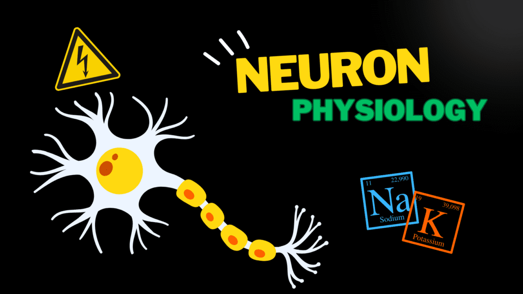
Action Potential Made Easy – Nerve Physiology Official Links Instagram Youtube Jki-discord Notes & Illustrations Quizzes Summary & Transcript 📢 Currently, there is no PDF for this video.If you’re interested in having one, feel free to send an inquiry, and I may create it in the future. BUT! There’s a quiz available in the next tab. 12345678910 Action Potentials in Neurons – QUIZ Test your understanding with 10 random multiple-choice questions from the question bank. You're in the preview mode. Note: All elements work correctly on the front end. 1 / 10 Why does saltatory conduction occur faster than continuous conduction? A) Myelin slows conduction B) Skips myelinated sections C) Nodes reduce ion flow D) Continuous depolarization Saltatory conduction skips myelinated sections, focusing activity at nodes of Ranvier. 2 / 10 During depolarization, which ion enters the neuron? A) Calcium ions B) Sodium ions C) Chloride ions D) Potassium ions Sodium ions (Na⁺) enter the neuron, making the inside more positive. 3 / 10 Which law explains that an action potential either happens fully or not at all? A) All-or-None Law B) Excitatory Law C) Depolarization Law D) Sodium-Potassium Law The All-or-None Law states that action potentials are binary events. 4 / 10 How does an action potential propagate in unmyelinated axons? A) Graded potential conduction B) Saltatory conduction C) Electrical conduction D) Continuous conduction Continuous conduction occurs in unmyelinated axons. 5 / 10 What is the typical resting potential of a neuron? A) -55 mV B) -90 mV C) -70 mV D) 0 mV The resting potential of a neuron is typically -70 mV. 6 / 10 Which ions are involved in generating the resting membrane potential? A) Only potassium B) Calcium and sodium C) Potassium and calcium D) Sodium, potassium, chloride Sodium (Na⁺), potassium (K⁺), and chloride (Cl⁻) contribute to resting potential. 7 / 10 What type of transport mechanism is used by the sodium-potassium pump? A) Passive diffusion B) Facilitated diffusion C) Primary active transport D) Secondary active transport Primary active transport is used by the sodium-potassium pump. 8 / 10 Which ion’s permeability changes first during an action potential? A) Sodium B) Potassium C) Chloride D) Calcium Sodium ion permeability increases first during depolarization. 9 / 10 Where do action potentials regenerate in myelinated axons? A) Synaptic clefts B) Nodes of Ranvier C) Axon terminals D) Internodes At the nodes of Ranvier, action potentials regenerate. 10 / 10 What prevents ion leakage in myelinated axons? A) Chloride channels B) Potassium channels C) Myelin sheath D) Sodium channels The myelin sheath prevents ion leakage. Your score is The average score is 0% Description This video covers the general physiology of a nerve, including potentials, polarizations, permeability, and excitability. Understanding these fundamental concepts is essential for grasping how nerves function and communicate through electrical and chemical signals. Basic Physiological Rules of the Cell: More negative inside the cell than on the outside. Difference in extracellular and intracellular fluid composition. Nernst Equation calculates the charged ions across a membrane. Transport Through the Cell Membrane: Passive Transport: Simple diffusion (requires small, lipophilic, and uncharged molecules). Facilitated diffusion (via ion channels and gated channels). Filtration. Electrokinetic transport. Osmosis. Active Transport: Primary Active Transport: Na/K Pump, Ca Pump. Secondary Active Transport: Na/Glucose, Na/Amino Acids, Na/H. Nerve Structure and Function: Receptive Part: Dendrites. Integrating Part: Cell body and Axon Hillock. Transmitting Part: Axon. Resting Potential: Typically -70 mV. Chemical gradient outside, Electrical Gradient inside = Electrochemical gradient outside. Sodium and potassium move through ion channels. Sodium-potassium pump: 3 sodium out, 2 potassium in. Action Potential: Voltage-Gated Channels and Excitability. Two gates on VG-Channels: Activation gate and inactivation gate. Excitability changes during an action potential. Permeability of the Cell Membrane During Action Potential: Resting state: Sodium and potassium cannot pass through gated channels. Inside the cell: Negative. Depolarization: Sodium enters the cell, making the inside positive. Repolarization: Sodium channels close, potassium channels open, leading to potassium outflux. This results in hyperpolarization (refractory period). Conduction of Action Potential Through a Nerve Axon: Continuous Conduction (Unmyelinated fibers). Saltatory Conduction (Myelinated fibers). All-Or-None Law: An action potential either occurs completely or not at all. Chemical Synapse: Ligand-gated channels or chemically gated channels. Excitatory or inhibitory responses. Transcript Introduction0:00hello and welcome to another video here I’m gonna talk about the physiology of neuron so first I’m0:06going to talk about the membrane potentials which is a differences in voltage between the inside and0:10outside of a cell and there are basically three types of potentials and neuron can be in it can0:16be in something we call resting potentials and the name doesn’t come randomly it is basically0:21when the neuron is at rest meaning it’s not doing any work and if you look at this chart a neuron0:27is at this resting potential when the voltage inside the cell is at negative 70 millivolts then0:33we also have something called graded potentials so when we receive a signal we call this stimuli0:39and that stimuli is going to stimulate which is the changes in membrane potentials and we’ll talk0:44more about this later it can be a small or a big change as you see here but as soon as we hit that0:50threshold of negative 55 millivolts and that’s gonna trigger the neuron is sending the signal0:56further away and this process we call this an action potential and that’s basically what this1:02long line is going to represent I’m also going to talk about the mechanism of impulse conduction1:08in nerve fibers we’ve got this slow continuous conduction in unmonitored nerve fibers and it’s1:16quite slow compared to the saltatory conduction in an myelinated nerve fiber so before I go detailed1:23into this topics I want to go through the basic physiological rules to kind of get a better graspBasic Physiological Rules of a Cell1:29of what’s typically happens in a cell so if you feel like you’re comfortable with the physiology1:33of a cell you can just skip this part and go right over to make membrane potentials so there1:40are a couple of things you need to keep in
Watch This to Memorize ALL 12 Cranial Nerves
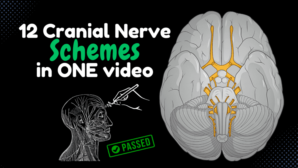
Watch This to Memorize ALL 12 Cranial Nerves Official Links Instagram Youtube Jki-discord Notes & Illustrations Quizzes Summary & Transcript Notes ☆ Members Only Go to PDF Notes Illustrations ☆ Members Only Go to Illustrations 12345678910 12 Cranial Nerve Summary – QUIZ Test your understanding with 10 random multiple-choice questions from the question bank. You're in the preview mode. Note: All elements work correctly on the front end. 1 / 10 Which cranial nerve is purely sensory? A) Facial Nerve B) Vestibulocochlear Nerve C) Trochlear Nerve D) Accessory Nerve The vestibulocochlear nerve (CN VIII) is a purely sensory nerve responsible for hearing and balance. 2 / 10 What does the internal acoustic meatus transmit? A) Glossopharyngeal Nerve B) Hypoglossal Nerve C) CN VII and CN VIII D) Trigeminal Nerve The internal acoustic meatus transmits both the facial nerve (CN VII) and the vestibulocochlear nerve (CN VIII). 3 / 10 Which cranial nerve is purely sensory for hearing and balance? A) Accessory Nerve B) Vestibulocochlear Nerve C) Glossopharyngeal Nerve D) Facial Nerve The vestibulocochlear nerve (CN VIII) is purely sensory, responsible for hearing and balance. 4 / 10 Which cranial nerve provides taste sensation to the posterior third of the tongue? A) Vagus Nerve B) Hypoglossal Nerve C) Glossopharyngeal Nerve D) Facial Nerve The glossopharyngeal nerve (CN IX) is responsible for taste in the posterior third of the tongue. 5 / 10 What is the function of the vestibular part of the vestibulocochlear nerve (CN VIII)? A) Balance and Equilibrium B) Hearing C) Speech D) Facial Movements The vestibular part of CN VIII is responsible for maintaining balance and equilibrium. 6 / 10 What is the function of the oculomotor nerve (CN III)? A) Eye Abduction B) Taste Sensation C) Sensory for Vision D) Eye Movements The oculomotor nerve innervates most extraocular muscles and is responsible for eye movements, pupil constriction, and accommodation. 7 / 10 Which cranial nerve passes through the jugular foramen along with CN IX and CN XI? A) Facial Nerve B) Trigeminal Nerve C) Vagus Nerve D) Hypoglossal Nerve The vagus nerve (CN X) exits the skull through the jugular foramen along with CN IX (Glossopharyngeal) and CN XI (Accessory). 8 / 10 Which cranial nerve innervates the pharyngeal plexus? A) Vagus Nerve B) Hypoglossal Nerve C) Trigeminal Nerve D) Facial Nerve The vagus nerve (CN X) contributes motor fibers to the pharyngeal plexus, which innervates most pharyngeal muscles. 9 / 10 What is the function of the pharyngeal plexus of the vagus nerve? A) Sensory to Pharynx B) Motor to Tongue C) Motor to Larynx D) Motor to Pharynx and Soft Palate The pharyngeal plexus of the vagus nerve provides motor innervation to the pharynx and soft palate. 10 / 10 Which cranial nerve is responsible for smell? A) Olfactory Nerve B) Facial Nerve C) Trochlear Nerve D) Optic Nerve The olfactory nerve (CN I) is the cranial nerve associated with the sense of smell. Your score is The average score is 0% Description This video compiles all the cranial nerve schemes shown in previous videos into one comprehensive guide. It is designed to help you study each cranial nerve more efficiently by summarizing their pathways, functions, and anatomical details. Whether you’re preparing for an exam or simply reinforcing your knowledge, this video serves as a quick and effective study tool. Hope it helps! 😊 Included Cranial Nerve Schemes: Olfactory Nerve (Cranial Nerve I) Optic Nerve (Cranial Nerve II) Oculomotor Nerve (Cranial Nerve III) Trochlear Nerve (Cranial Nerve IV) Trigeminal Nerve (Cranial Nerve V) Abducens Nerve (Cranial Nerve VI) Facial Nerve (Cranial Nerve VII) Vestibulocochlear Nerve (Cranial Nerve VIII) Glossopharyngeal Nerve (Cranial Nerve IX) Vagus Nerve (Cranial Nerve X) Accessory Nerve (Cranial Nerve XI) Hypoglossal Nerve (Cranial Nerve XII) This video covers: Cranial nerve functions and pathways Key anatomical structures Important clinical correlations Sources Singh, I. (2017). Human Neuroanatomy (10th ed.). Kozlowski, T. (2017). Memorix Anatomy: The Complete Study Guide. 2nd ed. Thieme Medical Publishers. University lectures and notes. Additional sources (shown in the original videos for each cranial nerve). Programs Complete Anatomy Biorender PowerPoint Camtasia 2023 Pictures and Visuals Used under licensed permission. Transcript Introduction0:00Alright so I’m making this video just to give you a quick repetition of each cranial nerve. I’m not gonna talk about all the canals and branches in detail, we already0:08talked about then in the 12 previous videos. This video is dedicated to basically give you one place where you’ll find all the schemes for each cranial nerve. So this video0:18might be a little long, but I promise if you know all these schemes, you basically have a good grasp of each cranial nerve. And what I recommend you is to redraw each of these0:27schemes several times until you can recall them easier. That’s what I did when I had to memorize them. Knowing these schemes will guarantee you enough knowledge on each cranial0:36nerve. Now before I start, I want you to understand a concept regarding the cranial nerves, a general division of the cranial nerves thatCranial Nerve Fibers0:45will help you make sense out of how the fibres are distributed. A cranial nerve can have motor fibers, sensory fibers, or parasympathetic motor fibers.0:55So cranial nerves can be sensory, motor or mixed nerves. They’re called mixed nerves1:00if they have both sensory and motor functions. Cranial nerve motor are further subdivided1:06into functional components according to their targets. So they can be General Somatic Efferent1:13(GSE), supplying skeletal muscles of somatic origin like the extraocular muscles. It can1:18be Special Visceral Efferent (SVE), supplying skeletal muscles derived from the pharyngeal1:23arch like the facial muscles, and General Visceral Efferent (GVE) fibres, giving off1:28preganglionic fibres to ganglia that contribute to the cranial parasympathetic outflow. Postganglionic1:36fibres of those peripheral ganglia further supply the smooth muscles and glands of various1:41organs and salivary glands. Sensory fibres are further subdivided according1:46to where the sensations are coming from, so it’s subdivided as Visceral or Somatic,1:52and visceral is further subdivided into general and special. So sensory fibres are classified as General visceral afferent
CN 12: Hypoglossal Nerve
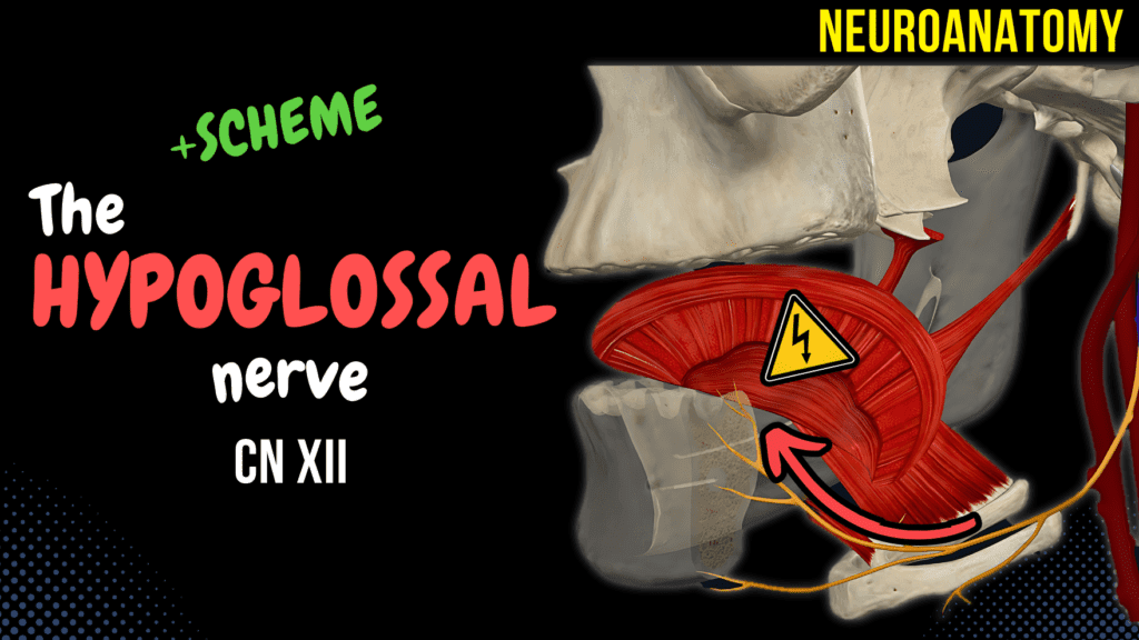
CN 12: Hypoglossal Nerve Official Links Instagram Youtube Jki-discord Notes & Illustrations Quizzes Summary & Transcript Notes ☆ Members Only Go to PDF Notes Illustrations ☆ Members Only Go to Illustrations 12345678910 Hypoglossal Nerve – QUIZ Test your understanding with 10 random multiple-choice questions from the question bank. You're in the preview mode. Note: All elements work correctly on the front end. 1 / 10 Which intrinsic tongue muscle is responsible for altering the shape of the tongue? A) Superior longitudinal muscle B) Styloglossus C) Hyoglossus D) Genioglossus The superior longitudinal muscle, innervated by the hypoglossal nerve, alters the shape of the tongue. 2 / 10 Which tongue muscle is primarily responsible for retracting and elevating the tongue? A) Genioglossus B) Styloglossus C) Hyoglossus D) Palatoglossus The styloglossus, innervated by the hypoglossal nerve, retracts and elevates the tongue. 3 / 10 Which muscle is responsible for protruding the tongue? A) Hyoglossus B) Genioglossus C) Styloglossus D) Superior longitudinal muscle The genioglossus muscle, innervated by the hypoglossal nerve, is responsible for protruding the tongue. 4 / 10 What role does the hypoglossal nerve play in swallowing? A) Controls pharyngeal constriction B) Controls epiglottis closure C) Controls tongue movement D) Controls soft palate elevation The hypoglossal nerve controls tongue movement essential for the swallowing mechanism. 5 / 10 Which nerve supplies sensory innervation to the posterior one-third of the tongue? A) Hypoglossal nerve B) Glossopharyngeal nerve C) Trigeminal nerve D) Vagus nerve The posterior one-third of the tongue is innervated by the glossopharyngeal nerve, not the hypoglossal nerve. 6 / 10 Which muscle depresses the sides of the tongue? A) Genioglossus B) Superior longitudinal muscle C) Hyoglossus D) Styloglossus The hyoglossus muscle, innervated by the hypoglossal nerve, depresses the sides of the tongue. 7 / 10 Which part of the tongue receives innervation from the hypoglossal nerve? A) Tip of the tongue B) Posterior one-third C) Entire tongue except palatoglossus D) Lateral borders The hypoglossal nerve innervates the entire tongue except the palatoglossus. 8 / 10 What is the role of the meningeal branch of the hypoglossal nerve? A) Supplies the dura mater B) Regulates parotid gland C) Innervates pharyngeal muscles D) Supplies the infrahyoid muscles The meningeal branch provides sensory fibers to the dura mater of the posterior cranial fossa. 9 / 10 Which fibers join the hypoglossal nerve to supply the geniohyoid muscle? A) C2 and C3 B) C4 C) C1 D) C2 Fibers from C1 travel with the hypoglossal nerve to innervate the geniohyoid muscle. 10 / 10 What happens to the tongue during long-term hypoglossal nerve injury? A) Fasciculations and atrophy B) Deviation without atrophy C) No changes observed D) Increases tongue tone Long-term hypoglossal nerve injury causes tongue fasciculations and atrophy. Your score is The average score is 0% Description Hypoglossal Nerve Overview The hypoglossal nerve is a purely motor nerve responsible for controlling the movement of the tongue. It plays a crucial role in tongue mobility and articulation, making it essential for speech and swallowing. Nucleus Nucleus of the hypoglossal nerve (nucleus nervi hypoglossi): A somatomotor nucleus. Course Leaves the cranial cavity via the hypoglossal canal. Passes between the internal jugular vein and the internal carotid artery. Forms an arch in the carotid triangle and continues into the submandibular triangle. Enters the root of the tongue where it gives off its terminal branches. Branches Lingual branches (rami linguales): Innervate the muscles of the tongue, except for the palatoglossus (innervated by CN X). Association with the Cervical Plexus Motor fibers of the ventral branches of the first and second cervical spinal nerves join the hypoglossal nerve. Some of these fibers leave the hypoglossal nerve via the superior root to join the inferior root, forming the ansa cervicalis (plexus cervicalis). Other fibers travel via the hypoglossal nerve to supply the geniohyoid muscle and give off a meningeal branch for the dura mater in the inferior part of the posterior cranial fossa. Sources Singh, I. (2017). Human Neuroanatomy (10th ed.). Kozlowski, T. (2017). Memorix Anatomy: The Complete Study Guide. 2nd ed. Thieme Medical Publishers. University lectures and notes. Programs Complete Anatomy Biorender PowerPoint Camtasia 2023 Pictures and Visuals Used under licensed permission. Transcript Introduction0:06What’s up, Taim Talks Med here. Let’s continue our Cranial nerve series. Cranial nerves are0:12twelve pairs of nerves that exit the brain and the brainstem, and in this segment,0:16we’ll talk detailed about the twelfth cranial nerve, the hypoglossal nerve.0:21And we’ll do that by first drawing a quick scheme of the hypoglossal nerve pathway.0:26We will talk about the distribution of nuclei and the course of this nerve. And then go through the0:31branches and its associated nerves. Awesome. Now the hypoglossal nerveHypoglossal Nerve Scheme0:37is relatively easy. It is purely a motor nerve, responsible for controlling the movement of the0:43tongue. Which means that it plays a crucial role in tongue mobility and articulation,0:50making it essential for speech and swallowing. The hypoglossal nerve originates from the nucleus0:56of the hypoglossal nerve, located within medulla oblongata. The nerve is going to give off somatic1:03efferent fibers, or motor fibers, that’re going to go out from the brainstem in the sulcus between1:09the olive and pyramid. We call it the pre-olivary groove. It then travels a short distance within1:16the cranial cavity, and then exits the skull through the hypoglossal canal. As the nerve1:22is now outside the cranium, it travels between the internal carotid artery and the internal1:28jugular vein, to then form an arch we call arch of the hypoglossal nerve. It then goes towards1:35the tongue and give off lingual branches to supply the muscles of the tongue, which1:39primarily include the extrinsic muscles, you know the genioglossus, Styloglossus and hyoglossus. And1:46the intrinsic muscles, which are the superior and inferior longitudinal lingual muscles, and1:52the vertical and transverse muscles of the tongue. The hypoglossal nerve is closely related with the1:58cervical plexus, in that the anterior ramus of the 1st and 2nd cervical spinal nerves are going to2:05send some motor fibres towards the hypoglossal nerve, that travels with the hypoglossal nerve2:11to supply the geniohyoid muscle. When you study
CN 11: Accessory Nerve
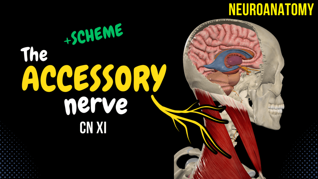
CN 11: Accessory Nerve Official Links Instagram Youtube Jki-discord Notes & Illustrations Quizzes Summary & Transcript Notes ☆ Members Only Go to PDF Notes Illustrations ☆ Members Only Go to Illustrations 12345678910 Accessory Nerve – QUIZ Test your understanding with 10 random multiple-choice questions from the question bank. You're in the preview mode. Note: All elements work correctly on the front end. 1 / 10 What is the function of the internal branch of the accessory nerve? A) Balance coordination B) Reflex arc C) Sensory innervation D) Motor fibers for pharynx and larynx The internal branch joins the vagus nerve and contributes to the pharyngeal and laryngeal muscles. 2 / 10 What symptom results from damage to the external branch of the accessory nerve? A) Ptosis B) Vocal cord paralysis C) Dysphagia D) Winged scapula Damage causes weakness in the trapezius, leading to a winged scapula. 3 / 10 What is the contribution of the accessory nerve to the pharyngeal plexus? A) Motor innervation B) Parasympathetic innervation C) Reflex coordination D) Sensory innervation It provides motor fibers to the plexus, which innervates most pharyngeal muscles. 4 / 10 What is the primary action of the sternocleidomastoid muscle innervated by the accessory nerve? A) Elevating the neck B) Lifting the scapula C) Rotating the head D) Shrugging shoulders It rotates the head to the opposite side and flexes the neck laterally. 5 / 10 How does the accessory nerve interact with the vagus nerve? A) Independent pathway B) Reflex control C) Sensory relay D) Motor fiber contribution The internal branch of the accessory nerve joins the vagus nerve to contribute to the pharyngeal plexus. 6 / 10 What is the role of the spinal accessory nucleus in accessory nerve function? A) Sensory feedback B) Reflex control C) Motor fiber generation D) Balance regulation It provides motor fibers to the spinal root of the accessory nerve. 7 / 10 What anatomical region is at risk of accessory nerve injury during surgery? A) Mandibular fossa B) Posterior triangle C) Anterior neck region D) Base of the skull The posterior triangle of the neck is at risk during surgeries. 8 / 10 Which foramen is used by the accessory nerve to exit the cranium? A) Foramen magnum B) Jugular foramen C) Hypoglossal canal D) Foramen rotundum The accessory nerve exits the cranium via the jugular foramen. 9 / 10 Which anatomical structure is at risk during neck surgeries involving the accessory nerve? A) Anterior triangle B) Foramen magnum C) External jugular vein D) Posterior triangle The posterior triangle of the neck poses a risk to the accessory nerve. 10 / 10 What is the relationship between the accessory and vagus nerves? A) They share the same nucleus B) They share sensory functions C) They collaborate in motor functions D) They are entirely separate nerves The accessory nerve joins the vagus nerve via its internal branch to contribute motor fibers to the pharyngeal plexus. Your score is The average score is 0% Description Accessory Nerve Overview The accessory nerve provides motor function to the sternocleidomastoid and the trapezius. It also joins the vagus nerve to innervate muscles in the pharynx, larynx, and soft palate. Nucleus and Course Nucleus ambiguus: Gives off the cranial root (radix cranialis). Located in the medulla oblongata. Spinal nucleus of the accessory nerve (nucleus spinalis nervi accessorii): Spinal roots (radix spinalis) arise from spinal segments C1–C6 lateral to the anterior horn, in the lateral funiculus. These roots ascend laterally to the spinal cord between the motor and sensory roots of the spinal nerve. They reach the posterior cranial fossa via the foramen magnum. When the cranial root and the spinal root meet, they form the trunk of the accessory nerve (truncus nervi accessorii), which leaves the cranium through the jugular foramen. Branches Internal branch (ramus internus): Joins the vagus nerve and participates in the pharyngeal branch and the recurrent laryngeal nerve of the vagus nerve. Innervates the pharynx, larynx, and muscles of the soft palate. External branch (ramus externus): Receives fibers from the anterior ramus of spinal nerves C2, C3, and C4 (of the cervical plexus). Innervates the sternocleidomastoid and trapezius muscles. Sources Singh, I. (2017). Human Neuroanatomy (10th ed.). Kozlowski, T. (2017). Memorix Anatomy: The Complete Study Guide. 2nd ed. Thieme Medical Publishers. Programs Complete Anatomy Biorender PowerPoint Camtasia 2023 Pictures and Visuals Used under licensed permission. Transcript Introduction0:06What’s up, Taim Talks Med here. Let’s continue our Cranial nerve series. Cranial nerves are0:11twelve pairs of nerves that exit the brain and the brainstem, and in this segment,0:16we’ll talk detailed about the eleventh cranial nerve, the accessory nerve.0:21And we’ll do that by first drawing a quick scheme of the accessory nerve pathway.0:25We will talk about the distribution of nuclei and the course of this nerve. And then go through the0:30two main branches that this nerve give off. Awesome, so the accessory nerve is purely aAccessory Nerve Scheme0:38motor nerve. It provides motor functions to the sternocleidomastoid and the trapezius,0:44and it goes together with the vagus nerve to innervate muscles in the pharynx, larynx and0:49soft palate. So it’s accessory to the vagus nerve. Now, this nerve has two main nuclei. Just like the0:58glossopharyngeal and the vagus nerve, this nerve is also has motor fibers coming from the nucleus1:03ambiguous, located in the medulla oblongata, And, we have the spinal accessory nuclei.1:10These are nuclei that lie in the spinal cord, in the 6 upper segments of the spinal cord,1:15lateral to the anterior horns of the grey matter. The nucleus ambiguous give rise to the cranial1:22root of the accessory nerve, while the spinal accessory nucleus give rise to the spinal root1:27of the accessory nerve. The cranial root is going to go out from the brainstem through1:32the retro-olivary groove, while the spinal root is going to go up, enter the cranium through foramen1:39magnus, and fuse with the cranial root of the accessory nerve, which then form the trunk of1:45the accessory nerve. The trunk of this nerve is then going to go through the jugular foramen,1:51and split
CN 10: Vagus Nerve
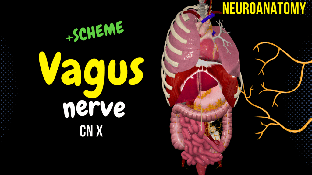
CN 10: Vagus Nerve Official Links Instagram Youtube Jki-discord Notes & Illustrations Quizzes Summary & Transcript Notes ☆ Members Only Go to PDF Notes Illustrations ☆ Members Only Go to Illustrations 12345678910 Vagus Nerve – QUIZ Test your understanding with 10 random multiple-choice questions from the question bank. You're in the preview mode. Note: All elements work correctly on the front end. 1 / 10 Which foramen does the vagus nerve pass through to exit the skull? A) Foramen magnum B) Jugular foramen C) Superior orbital fissure D) Hypoglossal canal The vagus nerve exits the skull through the jugular foramen, along with the glossopharyngeal and accessory nerves. 2 / 10 Which ganglion is associated with parasympathetic fibers of the vagus nerve? A) Superior ganglion B) Nodose ganglion C) Otic ganglion D) Celiac ganglion The parasympathetic fibers of the vagus nerve synapse at the celiac ganglion, which regulates abdominal organ functions. 3 / 10 The vagus nerve provides parasympathetic innervation to which part of the gastrointestinal tract? A) Entire gastrointestinal tract B) Distal third of transverse colon C) Proximal two-thirds of transverse colon D) Rectum The vagus nerve provides parasympathetic innervation up to the proximal two-thirds of the transverse colon. 4 / 10 The recurrent laryngeal nerve loops under which structure on the left side? A) Subclavian artery B) Pulmonary artery C) Aortic arch D) Thoracic duct The left recurrent laryngeal nerve loops under the aortic arch, ascending in the tracheoesophageal groove. 5 / 10 The vagus nerve innervates which portion of the intestines? A) Rectum B) Entire large intestine C) Distal third of the transverse colon D) Proximal two-thirds of the transverse colon The vagus nerve innervates the proximal two-thirds of the transverse colon through parasympathetic fibers. 6 / 10 Which branch of the vagus nerve supplies the laryngeal mucosa below the vocal cords? A) External branch of superior laryngeal nerve B) Pharyngeal branch C) Recurrent laryngeal nerve D) Internal branch of superior laryngeal nerve The recurrent laryngeal nerve provides sensory innervation to the laryngeal mucosa below the vocal cords. 7 / 10 What is the motor role of the vagus nerve in the larynx? A) Cricothyroid tension B) Vocal cord abduction C) Laryngeal elevation D) Motor control of intrinsic muscles The vagus nerve innervates the intrinsic muscles of the larynx (except cricothyroid) via the recurrent laryngeal nerve. 8 / 10 The vagus nerve provides sensory innervation to which area of the external ear? A) Parts of the external ear B) Tragus C) Entire external ear D) Helix The auricular branch (Arnold’s nerve) of the vagus nerve provides sensory innervation to parts of the external ear, external acoustic meatus, and external tympanic membrane. 9 / 10 What is the role of the superior laryngeal nerve? A) Both sensory and motor innervation B) Sensory innervation to the thorax C) Sensory innervation below the vocal cords D) Motor innervation to intrinsic laryngeal muscles The superior laryngeal nerve provides sensory innervation above the vocal cords and motor innervation to the cricothyroid muscle via its two branches. 10 / 10 Which structure does the vagus nerve pass through at the diaphragm? A) Caval opening B) Esophageal hiatus C) Aortic hiatus D) Foramen magnum The vagus nerve passes through the esophageal hiatus to reach the abdomen. Your score is The average score is 0% Description Cranial Nerve 10 Overview The vagus nerve has the longest course of all the cranial nerves, extending from the head to the abdomen. It has a parasympathetic component that helps regulate functions such as heart rate, digestion, and respiratory rate. Additionally, it has a sensory component that relays information from various visceral organs and a motor component responsible for speech and swallowing. Functional Components of the Vagus Nerve Special visceral efferent fibers (SVE): Somatomotor fibers of the vagus nerve. General visceral efferent fibers (GVE): Provide parasympathetic innervation. General visceral afferent fibers (GVA): Visceral sensory fibers of the vagus nerve. Special visceral afferent fibers (SVA): Responsible for taste and visceral sensations. General somatic afferent fibers (GSA): General sensory fibers. Nuclei of the Vagus Nerve Nucleus ambiguus: Motor nucleus. Posterior nucleus of the vagus nerve (nucleus posterior nervi vagi): Parasympathetic nucleus. Nuclei of the solitary tract (nuclei tractus solitarii): Viscerosensory nucleus. Gustatory nucleus (nucleus gustatorius): Special sensory nucleus. Spinal nucleus of the trigeminal nerve (nucleus spinalis nervi trigemini): Somatosensory nucleus. Course of the Vagus Nerve Intracranial Course The vagus nerve emerges as a series of rootlets in a groove between the olive and inferior cerebellar peduncle. It traverses the posterior cranial fossa and exits the skull through the jugular foramen. The superior sensory ganglion of the nerve is located in the jugular foramen. Extracranial Course The inferior ganglion of the vagus lies below the jugular foramen. Just below the inferior ganglion, the cranial root of the accessory nerve joins the vagus nerve to distribute along its pharyngeal and laryngeal branches. In the neck, the vagus lies in the carotid sheath with the internal jugular vein and common carotid arteries. The right vagus runs on the posterior surface of the esophagus, contributing to the esophageal plexus. It enters the abdomen through the esophageal opening in the diaphragm, supplying the stomach, duodenum, liver, kidneys, small and large intestines up to the proximal two-thirds of the transverse colon. It has a wide distribution in the abdomen via the celiac, superior mesenteric, and renal plexuses. The left vagus runs anterior to the esophageal plexus, then enters the abdomen, supplying the stomach, liver, duodenum, and head of the pancreas. Branches of the Vagus Nerve Branches in the Jugular Fossa Meningeal branch (ramus meningeus) Auricular branch (ramus auricularis) Branches in the Neck Pharyngeal branches (rami pharyngei): Contribute to the pharyngeal plexus (plexus pharyngeus). Superior laryngeal nerve (nervus laryngeus superior): Internal branch (ramus internus) – sensory branch. External branch (ramus externus) – mixed branch. Recurrent laryngeal nerve (nervus laryngeus recurrens): Tracheal branches (rami tracheales). Esophageal branches (rami oesophagei). Pharyngeal branches (rami pharyngei). Inferior laryngeal nerve (nervus laryngeus inferior). Superior cervical cardiac branches (rami cardiaci cervicales superiores) forming the
CN 9: Glossopharyngeal nerve
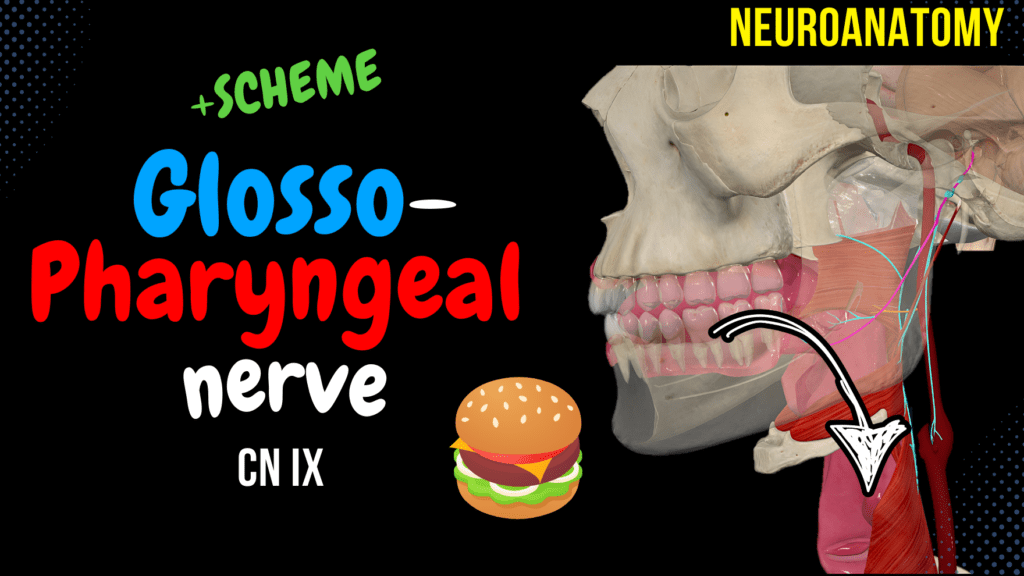
CN 9: Glossopharyngeal Nerve Official Links Instagram Youtube Jki-discord Notes & Illustrations Quizzes Summary & Transcript Notes ☆ Members Only Go to PDF Notes Illustrations ☆ Members Only Go to Illustrations 12345678910 Glossopharyngeal Nerve – QUIZ Test your understanding with 10 random multiple-choice questions from the question bank. You're in the preview mode. Note: All elements work correctly on the front end. 1 / 10 Which nucleus innervates the stylopharyngeus muscle? A) Nucleus ambiguus B) Spinal nucleus of trigeminal nerve C) Inferior salivatory nucleus D) Nucleus of the solitary tract The nucleus ambiguus is the motor nucleus that innervates the stylopharyngeus, assisting in swallowing and speech. 2 / 10 What happens when the glossopharyngeal nerve is lesioned? A) Increased salivation B) Loss of taste C) Tongue deviation D) Paralysis of facial muscles Loss of taste from the posterior third of the tongue and impaired salivation are key symptoms of glossopharyngeal nerve damage. 3 / 10 What is the main motor branch of the glossopharyngeal nerve? A) Stylopharyngeal branch B) Tympanic nerve C) Pharyngeal branches D) Lingual branches The stylopharyngeal branch innervates the stylopharyngeus muscle, aiding in swallowing. 4 / 10 What type of fibers does the tympanic nerve carry? A) Motor fibers B) Taste fibers C) Parasympathetic and sensory D) Only sensory fibers The tympanic nerve carries parasympathetic fibers to the tympanic plexus and sensory fibers to the middle ear mucosa. 5 / 10 Which branch innervates the stylopharyngeus muscle? A) Lingual branches B) Tonsillar branches C) Stylopharyngeal branch D) Pharyngeal branches The stylopharyngeal branch provides motor innervation to the stylopharyngeus muscle, aiding swallowing and speech. 6 / 10 What is the otic ganglion’s function? A) Regulates blood pressure B) Controls swallowing C) Sensory innervation of pharynx D) Parasympathetic to parotid gland The otic ganglion relays parasympathetic fibers to the parotid gland for saliva secretion. 7 / 10 What is the tympanic nerve’s function? A) Motor to pharyngeal muscles B) Sensory to carotid sinus C) Innervates middle ear D) Taste sensation The tympanic nerve innervates the mucosa of the middle ear and forms the tympanic plexus. 8 / 10 Which branch supplies sensory fibers to the tonsils? A) Tonsillar branches B) Tympanic nerve C) Lingual branches D) Pharyngeal branches The tonsillar branches provide sensory innervation to the palatine tonsils and nearby structures. 9 / 10 Which nucleus processes sensory input from the carotid sinus? A) Gustatory nucleus B) Inferior salivatory nucleus C) Nucleus of the solitary tract D) Nucleus ambiguus The nucleus of the solitary tract processes sensory input from the carotid sinus and carotid body. 10 / 10 What is the function of the general visceral efferent fibers? A) Sensory innervation of pharynx B) Salivation control C) Contraction of stylopharyngeus D) Taste conduction These fibers provide parasympathetic innervation to the parotid gland for saliva production. Your score is The average score is 0% Description Glossopharyngeal Nerve Overview The glossopharyngeal nerve plays a crucial role in transmitting sensory information from the back of the throat, taste sensations, and saliva production. It is also involved in swallowing and speaking. Functional Components of the Glossopharyngeal Nerve Special visceral efferent fibers (SVE) (branchial motor): Supply motor innervation to the stylopharyngeus muscle, which elevates the larynx and pharynx during speaking and swallowing. General visceral efferent fibers (GVE) (visceral motor): Provide parasympathetic innervation to the parotid glands. Fibers originate in the inferior salivatory nucleus, travel with the tympanic nerve through the foramen ovale, and synapse at the otic ganglion. General visceral afferent fibers (GVA) (visceral sensory): Carry sensory information from the carotid sinus and carotid body. Special visceral afferent fibers (SVA): Provide taste afferents from the posterior third of the tongue. General somatic afferent fibers (GSA) (general sensory): Provide sensory innervation to the upper pharynx, inner surface of the tympanic membrane, and the posterior third of the tongue. Nuclei of the Glossopharyngeal Nerve Nucleus ambiguus: Motor nucleus Inferior salivatory nucleus (nucleus salivatorius inferior): Parasympathetic visceromotor nucleus Nuclei of the solitary tract (nuclei tractus solitarii): Viscerosensory nucleus Gustatory nucleus (nucleus gustatorius): Special sensory nucleus Spinal nucleus of the trigeminal nerve (nucleus spinalis nervi trigemini): Somatosensory nucleus Course of the Glossopharyngeal Nerve Intracranial Course The glossopharyngeal nerve emerges from the medulla as a series of rootlets between the olive and the inferior cerebellar peduncle. It traverses the posterior cranial fossa and exits through the jugular foramen (foramen jugulare). Extracranial Course The superior and inferior ganglia of the glossopharyngeal nerve are situated at the exit, after which it gives off its side branches. Branches of the Glossopharyngeal Nerve Tympanic nerve (nervus tympanicus) Lesser petrosal nerve (nervus petrosus minor) Carotid branch (ramus sinus carotici) Lingual branches (rami linguales) Stylopharyngeal branch (ramus stylopharyngeus) Pharyngeal branches (rami pharyngei) Tonsillar branches (rami tonsillares) Sources Singh, I. (2017). Human Neuroanatomy (10th ed.). Kozlowski, T. (2017). Memorix Anatomy: The Complete Study Guide. 2nd ed. Thieme Medical Publishers. Neuroanatomy, Cranial Nerve 9 (Glossopharyngeal). Kathryn Thomas; Katrina Minutello; Joe M Das. Pictures and Visuals Complete Anatomy Biorender PowerPoint Camtasia 2021 Transcript Introduction0:06What’s up, Taim Talks Med here.0:08Let’s continue our Cranial nerve series.0:11Cranial nerves are twelve pairs of nerves that exit the brain and the brainstem, and0:15in this segment, we’ll talk detailed about the nineth cranial nerve, the glossopharyngeal0:20nerve.0:21And we’ll do that by first making a quick scheme of the glossopharyngeal nerve pathway.0:26Then we’ll break down the functional components of this nerve along with it’s respective0:31nuclei in the medulla.0:32We’ll go through the course and distribution by going through its intracranial course first,0:38then the extracranial course and its branches.0:42just a quick note before we start.0:44I’ve created a brand new Instagram account where I’ll be posting quizzes, channel updates,0:49and glimpses into my everyday life.0:51I’d truly appreciate it if you could give it a follow – your support means the so0:55much to me!0:56Alright let’s continue to the video.Glossopharyngeal Nerve Scheme0:58Alright, let’s start making a little scheme and then we’ll talk about it in detail.1:03Now, the glossopharyngeal nerve plays a crucial role in transmitting sensory information from1:09the back of the throat, taste sensations and saliva production, and is involved in
CN 8: Vestibulocochlear Nerve
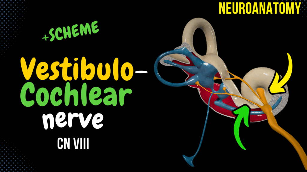
CN 8: Vestibulocochlear Nerve Official Links Instagram Youtube Jki-discord Notes & Illustrations Quizzes Summary & Transcript Notes ☆ Members Only Go to PDF Notes Illustrations ☆ Members Only Go to Illustrations 12345678910 Vestibulocochlear Nerve – QUIZ Test your understanding with 10 random multiple-choice questions from the question bank. You're in the preview mode. Note: All elements work correctly on the front end. 1 / 10 What is the role of the medial vestibular nucleus in balance? A) Maintains head posture B) Localizes sound C) Adjusts eye movements D) Processes auditory signals The medial vestibular nucleus sends fibers to control posture and head movements. 2 / 10 Which nerve division transmits signals related to balance? A) Optic division B) Temporal branch C) Vestibular division D) Cochlear division The vestibular division transmits balance signals from the inner ear to the brain. 3 / 10 What is the function of the lateral lemniscus in the auditory pathway? A) Controls balance B) Transmits auditory signals C) Stabilizes gaze D) Amplifies sound The lateral lemniscus transmits auditory signals from the cochlear nuclei to the inferior colliculus. 4 / 10 Which tract conveys auditory reflex signals? A) Tectospinal tract B) Corticospinal tract C) Vestibulospinal tract D) Spinothalamic tract The tectospinal tract conveys reflex signals for head and neck movement in response to sound. 5 / 10 Which semicircular canal detects rotational motion in the horizontal plane? A) Vertical semicircular canal B) Posterior semicircular canal C) Superior semicircular canal D) Lateral semicircular canal The lateral semicircular canal detects horizontal rotational motion. 6 / 10 Where are otolith organs located? A) Semicircular canals B) Cochlea C) Utricle and saccule D) Ampullae Otolith organs are located in the utricle and saccule. 7 / 10 Which fibers mediate the vestibulo-ocular reflex? A) Tectospinal tract B) Lateral lemniscus C) Vestibulospinal tract D) Medial longitudinal fasciculus The medial longitudinal fasciculus mediates the vestibulo-ocular reflex. 8 / 10 Where do the auditory radiations terminate? A) Superior olivary complex B) Medial geniculate body C) Primary auditory cortex D) Inferior colliculus Auditory radiations terminate in the primary auditory cortex. 9 / 10 What type of neurons form the spiral ganglion? A) Bipolar neurons B) Unipolar neurons C) Multipolar neurons D) Sensory neurons The spiral ganglion contains bipolar neurons for hearing. 10 / 10 Which structure connects the inner ear to the middle ear? A) Oval window B) Tympanic membrane C) Eustachian tube D) Vestibule The oval window connects the middle ear to the cochlea. Your score is The average score is 0% Description Correction! The tectospinal tract originates from the superior colliculus, not the inferior colliculus. However, the inferior colliculus sends auditory inputs to the superior colliculus, which then gives rise to the tract (Singh, I., 2017, p. 76). Apologize for this minor mistake. Cranial Nerve 8 Purely sensory nerve, consists of a vestibular part (maintaining equilibrium/balance), and a cochlear part (facilitates hearing). Inner Ear Anatomy Osseous labyrinth and Membranous labyrinth Vestibule Utricle Saccule Ampullae Semicircular canals Semicircular ducts Cochlea Cochlear duct Perilymph vs. Endolymph Perilymph: Between Osseous and Membranous labyrinth. Similar to extracellular fluid. High in sodium and low in potassium. Endolymph: Inside Membranous labyrinth. Similar to cytosol. Low in sodium and high in potassium. Vestibular System Ampulla contains hair cells with stereocilia that detect linear movement. In utricle and saccule, otolith organs with otoliths and hair cells detect movements in horizontal and vertical planes. Cochlear System Cochlea consists of Scala Tympani, Scala Vestibuli, and Scala Media. When the oval window is depressed, it creates waves that travel through the cochlea, vibrating the basilar membrane and generating neural activity through the spiral organ of Corti. Vestibulocochlear Nerve Vestibular ganglion: Consists of superior and inferior divisions. Spiral (cochlear) ganglion: Located inside the cochlea, several ganglia send out fibers that unite to form the cochlear division. Vestibular and cochlear nerves unite to form the vestibulocochlear nerve and travel through the internal acoustic meatus. Vestibular Nerve Pathways Vestibular Nuclear Complex Consists of superior, lateral, medial, and inferior nuclei. Vestibulo-Ocular Reflex (VOR) Happens through the medial longitudinal fasciculus. Fibers from the medial vestibular nuclei connect to the contralateral 6th cranial nerve, which sends fibers to the contralateral 3rd and 4th cranial nerves. Vestibulo-Spinal Pathway Vestibulospinal reflex maintains posture and resists gravity. Lateral vestibular nuclei send out the lateral vestibulospinal tract. Medial vestibular nuclei send out the medial vestibulospinal tract, which travels within the medial longitudinal fasciculus (MLF). Vestibulo-Cerebellar Pathway Inferior vestibular nuclei send fibers through the inferior cerebellar peduncle to the flocculonodular lobe and vermis. Fibers also come directly from the vestibulocochlear nerve. Cerebellum sends fibers toward the vestibular nuclear complex to influence the vestibulospinal tract. Vestibulo-Cerebral Pathway Efferent vestibular projections to bilateral ventral posterior thalamic nuclei. Cortical regions involved in vestibular processing: Frontal eye fields: Control eye movements and receive vestibular motion information. Primary somatosensory cortex (Areas 2v and 3a): Maps body location and movement signals. PIVC (Parieto-Insular Vestibular Cortex): Responds to body and head motion information. Posterior parietal cortex: Motion perception, responds to both visual and vestibular motion cues. Hippocampus and parahippocampal regions: Spatial orientation and navigation functions. Cochlear Nerve (Hearing Pathway) Synapses with ventral and dorsal cochlear nuclei. Lateral lemniscus (lemniscus lateralis) – Crossing fibers form the trapezoid body. Superior olivary complex helps localize sound direction. Some fibers synapse in the trapezoid nucleus. Some fibers synapse in the nuclei of the lateral lemniscus. Fibers project to the inferior colliculus, then travel via the inferior brachium to the medial geniculate body. Most fibers reach the medial geniculate body without relaying in the inferior colliculus. The inferior colliculus controls a descending tract called the Tectospinal tract, which helps with auditory reflexes. From the medial geniculate body, auditory radiations project to the primary auditory cortex. Impulses also reach auditory association areas, Wernicke’s area, and Broca’s area. Sources Singh, I. (2017). Human Neuroanatomy (10th ed.). Kozlowski, T. (2017). Memorix Anatomy. 2nd ed. Thieme Medical Publishers. Neuroanatomy, Cranial Nerve 8. Bruno Bordoni; Nicholas L. Mankowski; Daniel T. Daly. Neuroanatomy, Auditory Pathway. Diana C. Peterson; Vamsi Reddy; Renee N. Hamel.
CN 7: Facial Nerve
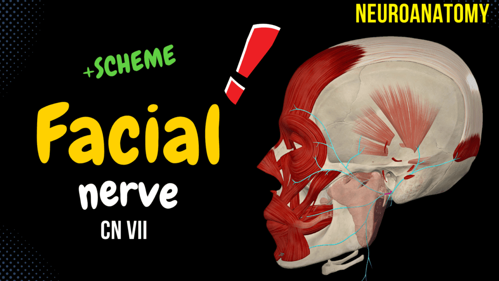
CN 7: Facial Nerve Official Links Instagram Youtube Jki-discord Notes & Illustrations Quizzes Summary & Transcript Notes ☆ Members Only Go to PDF Notes Illustrations ☆ Members Only Go to Illustrations 12345678910 Facial Nerve – QUIZ Test your understanding with 10 random multiple-choice questions from the question bank. You're in the preview mode. Note: All elements work correctly on the front end. 1 / 10 Which gland does the facial nerve NOT innervate? A) Lacrimal gland B) Submandibular gland C) Sublingual gland D) Parotid gland The parotid gland is innervated by the glossopharyngeal nerve, not the facial nerve. 2 / 10 What forms the parotid plexus? A) Facial nerve B) Trigeminal nerve C) Vagus nerve D) Hypoglossal nerve The parotid plexus is formed by the facial nerve after it enters the parotid gland and divides into terminal branches. 3 / 10 What is a characteristic of an upper motor neuron lesion affecting the facial nerve? A) Contralateral lower face paralysis B) Ipsilateral paralysis of all facial muscles C) Loss of taste in posterior tongue D) Hyperacusis UMN lesions spare the forehead because it receives bilateral cortical input. 4 / 10 Where does the facial nerve split into its terminal branches? A) Geniculate ganglion B) Parotid gland C) Internal acoustic meatus D) Stylomastoid foramen The facial nerve splits into its terminal branches within the parotid gland, forming the parotid plexus. 5 / 10 What type of fibers innervate the lacrimal gland? A) Special sensory B) Special visceral efferent C) General visceral efferent D) General somatic afferent General visceral efferent (GVE) fibers from the superior salivatory nucleus innervate the lacrimal gland. 6 / 10 Which branch of the facial nerve supplies taste to the anterior 2/3 of the tongue? A) Posterior auricular nerve B) Nerve to stapedius C) Greater petrosal nerve D) Chorda tympani The chorda tympani is responsible for transmitting taste from the anterior 2/3 of the tongue. 7 / 10 Which branch of the facial nerve is responsible for tearing? A) Greater petrosal nerve B) Chorda tympani C) Temporal branch D) Cervical branch The greater petrosal nerve is responsible for innervating the lacrimal gland to stimulate tearing. 8 / 10 Which muscle is innervated by the temporal branch of the facial nerve? A) Frontalis B) Platysma C) Buccinator D) Zygomaticus major The temporal branch of the facial nerve innervates the frontalis muscle, responsible for raising the eyebrows. 9 / 10 Which branch of the facial nerve innervates the platysma? A) Cervical branch B) Temporal branch C) Marginal mandibular branch D) Buccal branch The cervical branch of the facial nerve innervates the platysma. 10 / 10 Which nerve branch carries parasympathetic fibers to the submandibular gland? A) Greater petrosal nerve B) Buccal branch C) Lingual nerve D) Chorda tympani The chorda tympani carries parasympathetic fibers to the submandibular gland via the submandibular ganglion. Your score is The average score is 0% Description Facial Nerve Overview The facial nerve is responsible for providing motor innervation to facial muscles, as well as taste (anterior 2/3 of the tongue) and producing saliva and tears. Functional Components of the Facial Nerve Special Visceral Efferent (SVE) fibers Nucleus: Motor nucleus Supplies muscles of facial expression General Visceral Efferent (GVE) fibers Nucleus: Superior salivatory nucleus To submandibular ganglion for salivary glands and pterygopalatine ganglion for the lacrimal gland Special Visceral Afferent (SVA) fibers From the anterior 2/3 of the tongue to the geniculate ganglion to the nucleus of the solitary tract General Somatic Afferent (GSA) fibers External ear to the geniculate ganglion to the spinal nucleus of the trigeminal nerve Nuclei of the Facial Nerve Motor nucleus of facial nerve (nucleus nervi facialis) – SVE fibers Somatomotor nucleus for innervation of all facial muscles, platysma, stylohyoid, and posterior belly of the digastric Superior salivatory nucleus (nucleus salivatorius superior) – GVE fibers Visceromotor nucleus for parasympathetic innervation of the lacrimal, nasal, palatine, nasopharyngeal, submandibular, sublingual, and lingual glands Gustatory nucleus (nucleus gustatorius) – SVA fibers Special sensory nucleus for the nuclei of the solitary tract – receives impulses from the chorda tympani Spinal nucleus of the trigeminal nerve (nucleus spinalis nervi trigemini) – GSA fibers Somatosensory fibers from the facial nerve synapse in this nucleus Parts of the Facial Nerve Motor root/Facial nerve (somatomotor fibers) Sensory root/Intermediate nerve (nervus intermedius) Consists of visceromotor fibers (for glands) and special sensory fibers (taste) Course of the Facial Nerve Within the brainstem, it turns around the nucleus of the abducens nerve, elevating the facial colliculus on the floor of the fourth ventricle. Leaves the pons at the pontocerebellar angle and runs in the posterior cerebral fossa. Enters the internal acoustic canal and passes through the facial canal / Fallopian canal (canalis nervi facialis). Runs ventrolaterally in the labyrinthic part (pars labyrinthica), takes a 90° turn at the geniculum of the facial canal (geniculum canalis nervi facialis). Continues within the facial canal and exits the skull via the stylomastoid foramen (foramen stylomastoideum). Enters the parotid gland, where it splits into inferior and superior branches, forming the parotid plexus (plexus intraparotideus), which gives rise to terminal branches for facial muscles. Intracranial Branches Greater petrosal nerve (nervus petrosus major), comes from the geniculate ganglion (ganglion geniculi) Reaches the pterygopalatine fossa, terminating in the pterygopalatine ganglion – brings parasympathetic fibers from the superior salivatory nucleus and some taste fibers. Nerve to stapedius (nervus stapedius) Chorda tympani Joins the lingual nerve to reach the submandibular ganglion. Provides parasympathetic innervation for the submandibular and sublingual glands and the lingual glands in the anterior 2/3 of the tongue. Extracranial Branches Stylohyoid branch (ramus stylohyoideus) Digastric branch (ramus digastricus) Posterior auricular nerve (nervus auricularis posterior) Occipital branch (ramus occipitalis) Auricular branch (ramus auricularis) Branches forming the parotid plexus Temporal branches (rami temporales) Zygomatic branches (rami zygomatici) Buccal branches (rami buccales) Marginal mandibular branch (ramus marginalis mandibulae) Cervical branch (ramus colli) Clinical Relevance Dorsal facial nucleus: Supplies upper face muscles. Ventral facial nucleus: Supplies lower face muscles. Upper motor neuron lesion: Causes paralysis of the middle and lower halves
CN 6: Abducens Nerve
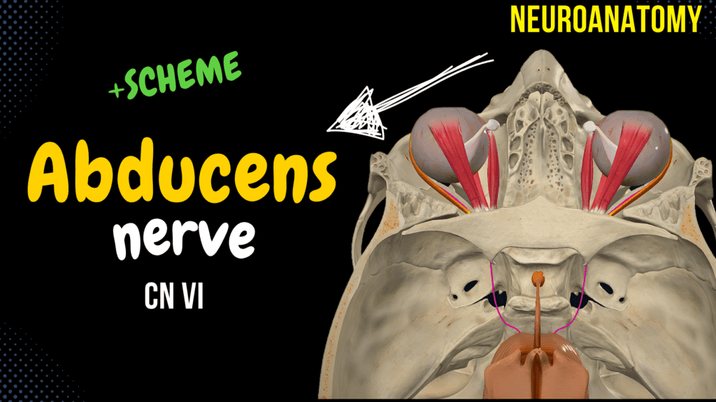
CN 6: Abducens Nerve Official Links Instagram Youtube Jki-discord Notes & Illustrations Quizzes Summary & Transcript Notes ☆ Members Only Go to PDF Notes Illustrations ☆ Members Only Go to Illustrations 12345678910 Abducens Nerve – QUIZ Test your understanding with 10 random multiple-choice questions from the question bank. You're in the preview mode. Note: All elements work correctly on the front end. 1 / 10 What does damage to the lateral rectus muscle result in? A) Medial deviation B) Upward deviation C) Lateral deviation D) Downward deviation Paralysis of the lateral rectus muscle causes the affected eye to deviate medially (esotropia). 2 / 10 What forms the facial colliculus in the pons? A) Spinal trigeminal nucleus B) Trochlear nucleus C) Oculomotor nucleus D) Abducens nucleus The abducens nerve nucleus is encircled by the fibers of the facial nerve, forming the facial colliculus. 3 / 10 What is the most common cause of abducens nerve palsy? A) Meningitis B) Increased intracranial pressure C) Trochlear nerve lesion D) Optic neuritis Increased intracranial pressure is a common cause of abducens nerve palsy, leading to nerve compression. 4 / 10 Which muscle is responsible for lateral gaze? A) Medial rectus B) Superior rectus C) Lateral rectus D) Inferior oblique The lateral rectus muscle, innervated by the abducens nerve, controls lateral gaze. 5 / 10 Which muscle does the abducens nerve control? A) Medial rectus B) Lateral rectus C) Inferior rectus D) Superior oblique The abducens nerve innervates the lateral rectus muscle, which abducts the eye. 6 / 10 What is the result of increased intracranial pressure on the abducens nerve? A) Medial strabismus B) Ptosis C) Horizontal diplopia D) Exotropia Increased intracranial pressure can compress the abducens nerve, leading to an inability to abduct the eye. 7 / 10 What happens if the abducens nerve is damaged? A) Nystagmus B) Lateral strabismus C) Medial strabismus D) Ptosis Damage to the abducens nerve causes medial strabismus, where the eye turns inward due to paralysis of the lateral rectus muscle. 8 / 10 How does the abducens nerve pass into the orbit? A) Inferior orbital fissure B) Foramen rotundum C) Optic canal D) Superior orbital fissure It passes through the superior orbital fissure within the common tendinous ring. 9 / 10 Which cranial nerve works in conjunction with the abducens nerve for gaze? A) Facial nerve B) Optic nerve C) Trochlear nerve D) Oculomotor nerve The oculomotor nerve (CN III) works with the abducens nerve to coordinate horizontal gaze. 10 / 10 Which pathway connects the abducens and oculomotor nuclei? A) Medial longitudinal fasciculus B) Reticular formation C) Optic tract D) Superior colliculus The medial longitudinal fasciculus connects the abducens and oculomotor nuclei for coordinated eye movements. Your score is The average score is 0% Description Abducens Nerve Overview The abducens nerve is purely a motor nerve supplying the lateral rectus muscle, which is involved in the abduction of the eye. Abducens/Abducent Nerve Nucleus Nucleus of abducens nerve (nucleus nervi abducentis) The facial nerve loops around this nucleus, forming the facial colliculus (colliculus facialis). A somatomotor nucleus located in the pons that innervates the lateral rectus muscle. Course Emerges from the medullopontine sulcus between the pons and medulla oblongata. Leaves the brainstem on the ventral side. Runs on the base of the skull, penetrates the dura mater, and then enters the cavernous sinus. Enters the orbit via the superior orbital fissure and common tendinous ring. Innervates the lateral rectus muscle. Function of the Lateral Rectus Muscle Abduction of the eye. Hering’s Law of Equal Innervation Coordinates both eyes together so that the medial and lateral rectus muscles contract equally. Deactivates the contralateral muscle through the medial longitudinal fasciculus. Clinical Relevance The abducens nerve can get compressed by a lesion or a rise in intracranial pressure. Compression causes paralysis of the lateral rectus muscle, resulting in medial deviation of the affected eye. This leads to a fully adducted eye at rest and an inability to abduct the eye. Sources Singh, I. (2017). Human Neuroanatomy (10th ed.). Helwany M, Bordoni B. Neuroanatomy, Cranial Nerve 1 (Olfactory) [Updated 2022 Aug 8]. In: StatPearls [Internet]. Treasure Island (FL): StatPearls Publishing; 2022 Jan-. Kozlowski, T. (2017). Memorix Anatomy: The Complete Study Guide. 2nd ed. Thieme Medical Publishers. Pictures and Visuals Complete Anatomy Biorender PowerPoint Camtasia 2021 Transcript Introduction0:01What’s up, Taim Talks Med here. Let’s continue our Cranial nerve series.0:10Cranial nerves are twelve pairs of nerves that exit the brain and the brainstem,0:15and in this segment, we’ll talk detailed about the sixth cranial nerve, which is the abducent nerve.0:20And we’ll do that by first making a quick scheme of the abducent nerve pathway.0:25Then we’ll cover the nerve in a little more detail, by nucleus and the course0:30of this nerve. Then we’ll talk a little bit about how the eyes are coordinated,0:35through something called Hering’s law, and then end with a little bit of a clinical relevance.Abducens Nerve Scheme0:42Alright, so the abducent nerve is a purely motor nerve, supplying the lateral rectus0:47muscle involved in abduction of the eye. Alright so here’s the scheme. We got the0:53nucleus of the abducent nerve in Pons. And we got the nerve. The nerve will travel through0:59the medullopontine sulcus, or the junction between the medulla and pons. It’ll pierce the dura mater1:05and travel through the cavernous sinus, then it’ll go through the superior orbital fissure,1:12and then through the common tendinous ring, where it’ll innervate the lateral rectus muscle.1:18So the way this works is imagine looking to the right. Signals from the right abducent nucleus1:24travel through the abducent nerve, activating the lateral rectus muscle of the right eye. This1:30causes the right eye to move outward—a movement known as abduction. Naturally you don’t want1:36the left abducent nerve to work at the same time, otherwise you’ll end up looking like this guy. So1:40here’s the marvel: to maintain balanced eye movement and prevent double vision,1:45we have Hering’s Law of Equal Innervation. We’ll talk a little bit more about this later. But1:51that’s the general outline of this nerve. Let’s dive a little
CN 5: Trigeminal Nerve
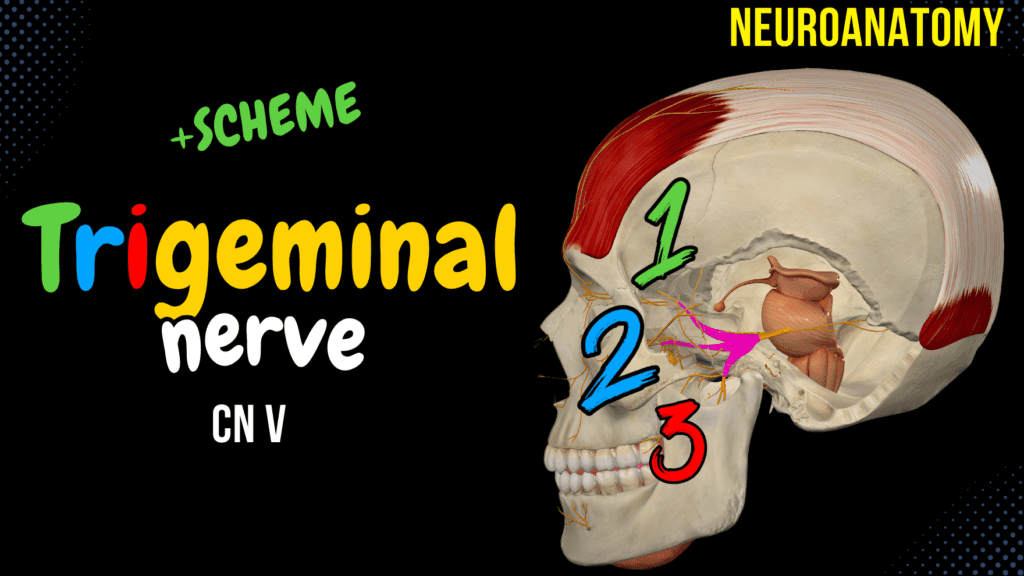
CN 5: Trigeminal Nerve Official Links Instagram Youtube Jki-discord Notes & Illustrations Quizzes Summary & Transcript Notes ☆ Members Only Go to PDF Notes Illustrations ☆ Members Only Go to Illustrations 12345678910 Trigeminal Nerve – QUIZ Test your understanding with 10 random multiple-choice questions from the question bank. You're in the preview mode. Note: All elements work correctly on the front end. 1 / 10 What is the sensory role of the infraorbital nerve? A) Upper teeth B) Parotid gland C) Tongue D) Lower eyelid and upper lip The infraorbital nerve supplies the lower eyelid, upper lip, and nasal skin. 2 / 10 Which nerve supplies the skin of the forehead? A) Buccal nerve B) Zygomatic nerve C) Supraorbital nerve D) Infraorbital nerve The supraorbital nerve, a branch of the frontal nerve (V1), supplies the skin of the forehead. 3 / 10 Which branch of the mandibular nerve provides sensory innervation to the anterior tongue? A) Inferior alveolar nerve B) Lingual nerve C) Buccal nerve D) Mental nerve The lingual nerve provides general sensory innervation to the anterior two-thirds of the tongue. 4 / 10 What is the primary function of the mandibular nerve (V3)? A) Parasympathetic motor innervation B) Special sensory (taste) C) Sensory to the upper face D) Mixed sensory and motor innervation The mandibular nerve is responsible for both sensory (e.g., lower face) and motor (e.g., mastication) functions. 5 / 10 What is the clinical significance of the trigeminal ganglion? A) Origin of motor fibers B) Sensory ganglion C) Relays motor signals D) Parasympathetic ganglion The trigeminal ganglion houses the cell bodies of sensory neurons for the trigeminal nerve. 6 / 10 Which branch of the trigeminal nerve carries parasympathetic fibers to the parotid gland? A) Lingual nerve B) Auriculotemporal nerve C) Mental nerve D) Inferior alveolar nerve The auriculotemporal nerve carries parasympathetic fibers from the otic ganglion to the parotid gland. 7 / 10 Which branch of the trigeminal nerve provides the afferent limb of the corneal reflex? A) Maxillary nerve B) Infraorbital nerve C) Mandibular nerve D) Ophthalmic nerve The ophthalmic nerve (V1) mediates the afferent limb of the corneal reflex. 8 / 10 What is the role of the chorda tympani nerve in relation to the trigeminal nerve? A) Taste to posterior tongue B) Taste to anterior tongue C) Motor to mastication D) Pain from face The chorda tympani carries taste fibers from the facial nerve and joins the lingual nerve of the mandibular branch. 9 / 10 What is the motor role of the mandibular nerve? A) Facial expression B) Muscles of mastication C) Chewing and swallowing D) Eye movement The mandibular nerve provides motor innervation to the muscles of mastication, including the masseter, temporalis, and pterygoids. 10 / 10 Which nerve supplies the hard palate? A) Lesser palatine nerve B) Nasopalatine nerve C) Greater palatine nerve D) Infraorbital nerve The greater palatine nerve, a branch of the maxillary nerve, supplies the hard palate. Your score is The average score is 0% Description Course of the Trigeminal Nerve Distribution of Nuclei in the Brainstem Mesencephalic nucleus of trigeminal nerve (nucleus mesencephalicus nervi trigemini): Receives proprioceptive information from the masticatory, mimetic, and extra-ocular muscles. Principal nucleus of trigeminal nerve (nucleus principalis nervi trigemini): Receives information about fine touch, vibration, and some proprioception. Spinal nucleus of trigeminal nerve (nucleus spinalis nervi trigemini): Receives information about crude touch, pain, and temperature. Motor nucleus of trigeminal nerve (nucleus motorius nervi trigemini): Innervates the muscles of the first pharyngeal arch, including: Masticatory muscles (masseter, temporalis, lateral and medial pterygoid) Tensor tympani Tensor veli palatini Anterior belly of the digastric Mylohyoid Trigeminal Nerve Course Leaves the anterior aspect of the pons. Sensory root from the trigeminal ganglion, motor root goes with the mandibular branch. Ophthalmic Nerve (nervus ophthalmicus) (V1) Tentorial Branch (ramus tentorius) Lacrimal nerve (nervus lacrimalis) Frontal nerve (nervus frontalis) Supratrochlear nerve (nervus supratrochlearis) Supraorbital nerve (nervus supraorbitalis) Medial branch (ramus medialis) Lateral branch (ramus lateralis) Nasociliary nerve (nervus nasociliaris) Communicating branch with ciliary ganglion (ramus communicans cum ganglio ciliari) Long ciliary nerves (nervi ciliares longi) Posterior ethmoidal nerve (nervus ethmoidalis posterior) Anterior ethmoidal nerve (nervus ethmoidalis anterior) Infratrochlear nerve (nervus infratrochlearis) Maxillary Nerve (nervus maxillaris) (V2) Meningeal Branch (ramus meningeus) Zygomatic nerve (nervus zygomaticus) Zygomaticofacial branch (ramus zygomaticofacialis) Zygomaticotemporal branch (nervus zygomaticotemporalis) Infraorbital nerve (nervus infraorbitalis) Inferior palpebral branches (rami palpebrales inferiores) Superior labial branches (rami labiales superiores) External nasal branches (rami nasales externi) Internal nasal branches (rami nasales interni) Posterior superior alveolar branches (rami alveolares superiores posteriores) Superior dental branches (rami dentales superiores) Superior gingival branches (rami gingivales superiores) Anterior superior alveolar branches and middle superior alveolar branch (rami alveolares superiores anteriores et ramus alveolaris superior medius) Superior dental branches (rami dentales superiores) Superior gingival branches (rami gingivales superiores) Posterior nasal branches (rami nasales posteriores) Greater palatine nerve (nervus palatinus major) and lesser palatine nerves (nervi palatini minores) Pharyngeal nerve (nervus pharyngeus) Mandibular Nerve (nervus mandibularis) (V3) Meningeal branch (ramus meningeus) Buccal nerve (nervus buccalis) Auriculotemporal nerve (nervus auriculotemporalis) Inferior alveolar nerve (nervus alveolaris inferior) Mental nerve (nervus mentalis) Lingual nerve (nervus lingualis) Sources Singh, I. (2017). Human Neuroanatomy (10th ed.). Helwany M, Bordoni B. Neuroanatomy, Cranial Nerve 1 (Olfactory) [Updated 2022 Aug 8]. In: StatPearls [Internet]. Treasure Island (FL): StatPearls Publishing; 2022 Jan-. Kozlowski, T. (2017). Memorix Anatomy: The Complete Study Guide. 2nd ed. Thieme Medical Publishers. Pictures and Visuals Complete Anatomy Biorender PowerPoint Camtasia 2021 Transcript Introduction0:06What’s up, Taim Talks Med here. Let’s continue our Cranial nerve series. Cranial0:11nerves are twelve pairs of nerves that exit the brain and the brainstem, and in this segment,0:16we’ll talk detailed about the fifth cranial nerve, which is the Trigeminal nerve.0:20And we’ll do that by first making a quick scheme of the trochlear pathway to get an0:25overview of it. Then we’ll cover the nerve in a little more0:28detail, by first talking about the distribution of the trigeminal nerve nuclei within the0:32brainstem. Then we’ll talk detailed about each branch of the trigeminal nerve, which0:37is the ophthalmic nerve, maxillary