
Sepsis & Septic Shock – Symptoms, Pathophysiology, Diagnosis, Treatment
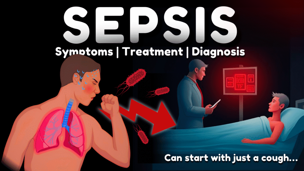
Sepsis & Septic Shock – Symptoms, Pathophysiology, Diagnosis, Treatment Official Links Instagram Youtube Jki-discord Notes & Illustrations Quizzes Summary & Transcript 📢 Currently, there is no PDF for this video.If you’re interested in having one, feel free to send an inquiry, and I may create it in the future. BUT! There’s a quiz available in the next tab. 12345678910 SEPSIS – QUIZ Test your understanding with 10 random multiple-choice questions from the question bank. You're in the preview mode. Note: All elements work correctly on the front end. 1 / 10 Which lab test supports a diagnosis of tissue hypoxia in sepsis? A) Hematocrit B) Lactate C) BUN D) CRP Elevated lactate is a marker of tissue hypoperfusion and anaerobic metabolism. 2 / 10 What happens during disseminated intravascular coagulation (DIC) in sepsis? A) Only bleeding increases B) Fibrinogen levels rise C) Only platelets decrease D) Clotting and bleeding simultaneously Widespread clotting and bleeding occurs due to impaired coagulation. 3 / 10 What role do cytokines play in sepsis? A) Boost clotting only B) Stimulate oxygen delivery C) Increase red blood cells D) Drive systemic inflammation They mediate inflammation, contributing to vasodilation, fever, and tissue damage. 4 / 10 What does low urine output in a septic patient suggest? A) Liver failure B) Dehydration from exercise C) Impaired kidney function D) Electrolyte loss It may indicate kidney hypoperfusion or acute kidney injury. 5 / 10 What is the first step before starting antibiotics in sepsis treatment? A) CT scan B) Lumbar puncture C) Nasal swab D) Blood cultures Blood cultures should be drawn before antibiotics to guide therapy. 6 / 10 Which of the following is a parasitic cause of sepsis? A) Candida albicans B) Staphylococcus aureus C) Plasmodium falciparum D) Influenza A Malaria (Plasmodium spp.) is a parasitic infection that can lead to sepsis. 7 / 10 Which score uses respiratory rate, blood pressure, and mental status to identify sepsis risk? A) CURB-65 B) qSOFA C) SOFA D) GCS The qSOFA score is a quick bedside tool used outside the ICU. 8 / 10 Which of the following is an early sign of organ dysfunction in sepsis? A) Confusion B) Hiccups C) Jaundice D) Rash Altered mental status often appears early and is part of qSOFA. 9 / 10 What does an elevated lactate level indicate in sepsis? A) High blood sugar B) Kidney failure C) Tissue hypoxia D) Normal metabolism Lactate >2 mmol/L indicates tissue hypoperfusion and cellular hypoxia. 10 / 10 What is the initial fluid resuscitation dose for sepsis? A) 10 mL/kg B) 1 L over 6 hours C) 100 mL/hr D) 30 mL/kg The recommended dose is 30 mL/kg IV fluids within the first hour. Your score is The average score is 0% Description Sepsis Overview:Sepsis is a life-threatening condition where the body’s immune system reacts excessively to an infection, causing tissue and organ damageIn 2017, there were 48.9 million cases globally and 11 million deaths.Sepsis affects anyone but is more common in the elderly, young, pregnant women, or those with chronic health conditions. Definition:Sepsis occurs when an infection triggers a chain reaction in the body Pathophysiology of Sepsis:1.Infection triggers immune response: The immune system overreacts to infections (bacterial, viral, fungal, or parasitic)2-Release of cytokines: Cells release IL-1, IL-6, TNF-α, and other mediators, causing inflammation.3.Vasodilation and permeability: Blood vessels dilate and leak, leading to hypotension4-Decreased cardiac output: Blood volume drops, reducing oxygen delivery to organs5.Tissue hypoxia and acidosis: Lack of oxygen leads to lactic acid build-up.6.Coagulation: Impaired blood clotting results in DIC.7-Multi-organ dysfunction: Key organs (heart, lungs, kidneys) are affected.8-Septic shock: Persistent low blood pressure without treatment can lead to septic shock. Causes of Sepsis:-Bacterial infections: Staphylococcus aureus, Escherichia coli, and Pseudomonas aeruginosa are common.-Viral infections: Influenza and COVID-19.-Fungal infections: Candida and Aspergillus.-Parasitic infections: Malaria (Plasmodium).-Infection sources: Pneumonia, urinary tract infections, abdominal infections, and skin infections.-Healthcare-associated infections: Infections from catheters, ventilators, or surgery.-Weakened immunity: People with compromised immune systems (chemotherapy, organ transplants) are more vulnerable. Clinical Features (TIME mnemonic):T – Temperature: High fever or low temperatureI – Infection signs: Symptoms vary (e.g., cough, shortness of breath, abdominal pain)M – Mental status: Confusion or difficulty wakingE – Extremely ill: Severe discomfort, pain, and shortness of breath Clinical Criteria for Diagnosing Sepsis:1,Infection suspected (bacterial, viral, fungal, or parasitic)2.qSOFA score:Systolic BP ≤ 100 mmHgRespiratory rate ≥ 22 breaths/minAltered mental statusqSOFA score ≥ 2 suggests higher risk3.NEWS score: NEWS ≥ 5 predicts sepsis risk based on RR, BP, and oxygen levels. Treatment for Sepsis:Blood cultures: Obtain before starting antibiotics.Lactate levels: Elevated lactate (≥ 2 mmol/L) indicates tissue hypoxia.Urine output: Monitor for over 0.5 mL/kg/h to assess kidney function.IV fluids: Start 30 mL/kg within the first hour to treat hypotension.Oxygen therapy: Maintain oxygen saturation ≥ 94% (unless chronic respiratory issues are present).Antibiotics: Administer broad-spectrum antibiotics within the first hour.Vasopressors: If fluids fail to restore BP, use vasopressors (e.g., norepinephrine) to maintain a MAP ≥ 65 mmHg.Monitoring: Track blood pressure, lactate, and urine output to ensure effective treatment. Clinical Scenario Example:Assessment: Patient with low BP (88 mmHg), rapid breathing, and confusion.Treatment: Start IV fluids, monitor BP, administer vasopressors if needed, give antibiotics immediately. Transcript Introduction0:00Sepsis is a serious condition that happens when the body’s immune system has an extreme response0:05to an infection, and this extreme immune response causes damage to its own tissue and organs.0:11A study done in 2017 showed that there were 48.9 million cases worldwide, and 11 million0:19sepsis-related deaths in the same year. It’s a very deadly condition and it’s crucial to be able0:25to recognize it early, and know what to do. Sepsis can affect anyone, but people who0:30are older, very young, pregnant or have other health problems are at higher risk.0:34So, in this video, we’re going to answer the question what is sepsis?Content0:38And we’re going to do that by going through the definition. Then go through the pathophysiology0:43for Sepsis, basically what happens within the body. Then we’re going to0:47talk a little bit about the causes. After that we’ll briefly go through0:51what the symptoms of Sepsis are using
What Are Joints? Classification, Types & Clinical Anatomy Explained
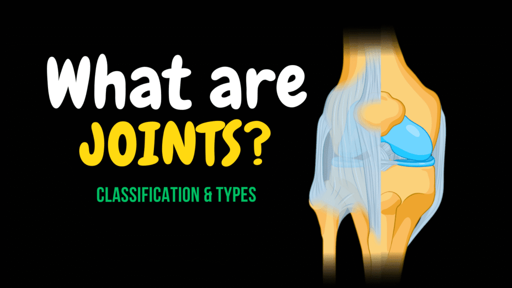
What Are Joints? Classification, Types & Clinical Anatomy Explained Official Links Instagram Youtube Jki-discord Notes & Illustrations Quizzes Summary & Transcript Notes ☆ Members Only Go to PDF Notes Illustrations ☆ Members Only Go to Illustrations 12345678910 Joints Overview – QUIZ Test your understanding with 10 random multiple-choice questions from the question bank. You're in the preview mode. Note: All elements work correctly on the front end. 1 / 10 Which ligament connects the distal tibia and fibula? A) Posterior tibiofibular ligament B) Cruciate ligament C) Anterior talofibular ligament D) Collateral ligament The posterior tibiofibular ligament is part of the syndesmosis joint. 2 / 10 What joint allows childbirth-related pelvic expansion? A) Hip joint B) Sacroiliac joint C) Pubic symphysis D) Intervertebral joint Hormonal changes increase flexibility at the pubic symphysis. 3 / 10 Which synovial joint type allows flexion, extension, abduction, adduction, and rotation? A) Pivot B) Hinge C) Saddle D) Ball-and-socket Ball-and-socket joints allow movement in all three planes. 4 / 10 The glenoid labrum is found in which joint? A) Knee joint B) Elbow joint C) Hip joint D) Shoulder joint The glenoid labrum deepens the socket of the shoulder joint. 5 / 10 The term “arthrology” refers to: A) Joint dislocations B) The study of joints C) Inflammation of joints D) Surgical repair of joints Arthrology is the study of joints. 6 / 10 What is the function of the menisci in the knee joint? A) Lubricate the joint B) Deepen the socket C) Allow rotation D) Absorb shock Menisci are fibrocartilaginous pads that absorb shock in weight-bearing joints. 7 / 10 Which joint type has flat articular surfaces and allows gliding? A) Ball-and-socket B) Plane C) Pivot D) Saddle Plane joints allow gliding movements. 8 / 10 Which structure connects bone to bone in a joint? A) Tendons B) Labrum C) Menisci D) Ligaments Ligaments stabilize joints by connecting bones. 9 / 10 The carpometacarpal joint of the thumb is an example of: A) Ellipsoid joint B) Hinge joint C) Saddle joint D) Plane joint This joint is a saddle joint enabling thumb opposition. 10 / 10 Which joint type consists of bones connected by hyaline cartilage? A) Symphysis B) Synchondrosis C) Gomphosis D) Syndesmosis Synchondroses are primary cartilaginous joints made of hyaline cartilage. Your score is The average score is 0% Description This video is about joint classification, structure, function, and clinical relevance. Topics covered in this video: • What are joints?• Joint classification based on structure and function• Types of joints in the human body• Examples of fibrous, cartilaginous, and synovial joints• Subtypes of each joint category with real anatomical examples• Clinical relevance of joints (e.g., high ankle sprains, TMJ, arthritis)• Supporting structures: ligaments, bursae, menisci, labrum, fat pads• Functional mobility: synarthrosis, amphiarthrosis, diarthrosis• Synovial joint types: – Ball-and-socket joint (shoulder, hip) – Ellipsoid joint (wrist) – Saddle joint (thumb) – Hinge joint (elbow, knee, fingers) – Pivot joint (atlantoaxial, radioulnar) – Plane joint (acromioclavicular, vertebral facet joints) Joint classification explained:• Fibrous joints – sutures (suturae), syndesmoses, gomphoses• Cartilaginous joints – synchondroses (hyaline cartilage), symphyses (fibrocartilage)• Synovial joints – contain a joint cavity filled with synovial fluid – include articular cartilage, synovial membrane, joint capsule – supported by ligaments, tendons, labrum, bursae, menisci Clinical anatomy references include:• Atlantoaxial joint (articulatio atlantoaxialis mediana)• Glenohumeral joint (articulatio humeri)• Temporomandibular joint (articulatio temporomandibularis)• Proximal radioulnar joint (articulatio radioulnaris proximalis)• Pubic symphysis (symphysis pubica)• Costochondral joints (junctiones costochondrales)• Intervertebral discs (disci intervertebrales)• Distal tibiofibular syndesmosis (syndesmosis tibiofibularis distalis) Whether you’re a medical student or revising anatomy for clinical practice, this video breaks down complex arthrology in a visual, memorable way. Transcript 0:00Joints. They come in many forms, but at their core, joints exist to link bones0:05together and allow movement. Some let you rotate your arm in every direction,0:10some move just a little bit, and some, like the joints in your skull, don’t move at all.0:16You don’t really think about them, until something goes wrong. When joints wear down,0:21become inflamed, or stop working properly, even the simplest movements can become difficult.0:27So, why are some joints flexible while others are completely rigid? What makes0:32one joint allow movement while another barely moves at all? And how do we actually classify0:38all the different joints in the body? In this video, we’ll start by answering0:43the fundamental question – what are joints? Then, we’ll go through all the joints in the body and0:49classify them based on their structure and function. As we go through them,0:54we’ll also highlight their clinical relevance, understanding how joint problems develop and0:58what makes them vulnerable to damage. Hey everyone, my name is Taim. I’m a1:02medical doctor, and I make animated medical lectures to make different topics in medicine1:06visually easier to understand. If you’d like a PDF version or a quiz of this presentation, you can1:11find it on my website, along with organized video lectures to help with your studies.1:15Alright, let’s get started! So what are joints?What are Joints?1:19Just, in simple terms, The point at which two bones lay adjacent to each1:24other (with or without the ability to move) is called a joint. Let’s visualize this.1:30Here we see a bone. Here is another bone. The point at which they lay adjacent to1:35each other is a joint. Here are two bones, between them, a joint. Here are two bones,1:41between them a joint. And even, I’ll surprise you now. Even between your skull bones, is a joint.1:48So joints come in different shape, and they are structurally and functionally different.1:54For example. The shoulder joint is1:56called the glenohumeral joint. Structurally, we call this a Synovial joint, Functionally,2:02it’s a Diarthrosis, since it’s a freely movable joint. And we subclassify it as a ball-and-socket2:09joint, which provides free rotational movement. Between the articular surfaces of vertebrae,2:15we got the facet joint, or zygapophyseal joints. They are synovial joints,2:20functionally movable so diarthrosis as well, but subclassified as plane joint,2:26allowing only gliding movements. Okay, let’s take another example, in the skull we got2:32sutures. Or lambdoid suture is what we’re pointing at specifically now. Structurally,2:37it’s a strong fibrous joint. And functionally, a Synarthrosis, meaning a joint that does
Skull Bones: Viscerocranium (Facial Skeleton + Hyoid Bone)
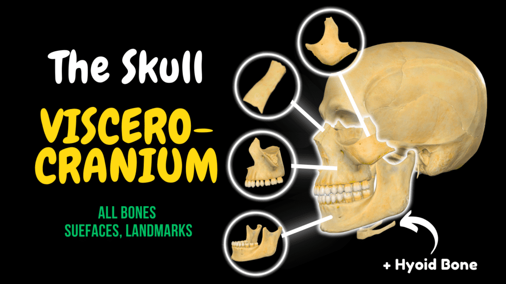
Skull Bones: Viscerocranium (Facial Skeleton + Hyoid Bone) Official Links Instagram Youtube Jki-discord Notes & Illustrations Quizzes Summary & Transcript Notes ☆ Members Only Go to PDF Notes Illustrations ☆ Members Only Go to Illustrations 12345678910 Viscerocranium – QUIZ Test your understanding with 10 random multiple-choice questions from the question bank. You're in the preview mode. Note: All elements work correctly on the front end. 1 / 10 Which facial bones articulate medially at the internasal suture? A) Nasal bones B) Lacrimal bones C) Maxillae D) Zygomatic bones The two nasal bones meet medially at this suture. 2 / 10 What muscle attaches to the nasal bone? A) Levator labii superioris B) Procerus and nasalis C) Orbicularis oculi D) Buccinator The procerus and nasalis attach to the nasal bones and control facial expressions. 3 / 10 What structure articulates with the vomer to form the nasal septum? A) Ethmoidal crest B) Inferior nasal concha C) Conchal crest D) Nasal crest The nasal crest of the maxilla articulates with the vomer. 4 / 10 The ethmoidal crest of the palatine bone is the attachment site for: A) Vomer B) Middle nasal concha C) Maxilla D) Inferior nasal concha The ethmoidal crest articulates with the middle nasal concha. 5 / 10 What passes through the nasal foramen of the nasal bone? A) Emissary veins B) Zygomatic nerve C) Ethmoidal artery D) Greater palatine vessels Small emissary veins pass through the nasal foramen. 6 / 10 What structure forms the prominence of the cheek? A) Zygomatic bone B) Frontal process C) Maxilla D) Lacrimal bone The zygomatic bone forms the lateral cheek contour. 7 / 10 What foramen transmits nerves to the cheek and temple? A) Infraorbital foramen B) Supraorbital foramen C) Mandibular foramen D) Zygomaticoorbital foramina The zygomaticoorbital foramina carry zygomatic nerve branches. 8 / 10 What bone forms the posterior-inferior part of the nasal septum? A) Vomer B) Maxilla C) Ethmoid bone D) Inferior nasal concha The vomer contributes to the posterior-inferior portion of the septum. 9 / 10 The sphenopalatine foramen transmits which of the following? A) Sphenopalatine artery and nasopalatine nerve B) Greater palatine vessels C) Zygomatic nerve D) Infraorbital nerve The sphenopalatine artery and nasopalatine nerve pass through it into the nasal cavity. 10 / 10 Which bone forms the lower jaw? A) Mandible B) Palatine C) Maxilla D) Zygomatic The mandible is the only movable bone of the facial skeleton. Your score is The average score is 0% Description This video is about the viscerocranium.Bones of the skull:• Neurocranium – surrounds the brain• Viscerocranium – forms the facial skeleton Bones included in the viscerocranium:• Maxilla (maxilla) – paired• Zygomatic bone (os zygomaticum) – paired• Palatine bone (os palatinum) – paired• Nasal bone (os nasale) – paired• Lacrimal bone (os lacrimale) – paired• Inferior nasal concha (concha nasalis inferior) – paired• Vomer (vomer) – unpaired• Mandible (mandibula) – unpaired• Hyoid bone (os hyoideum) – unpaired Maxilla:• Body (corpus maxillae)• Maxillary sinus (sinus maxillaris)• Frontal process• Zygomatic process (processus zygomaticus maxillae)• Alveolar process (processus alveolaris)• Palatine process (processus palatinus)• Infraorbital margin• Infraorbital groove (sulcus infraorbitalis)• Infraorbital canal (canalis infraorbitalis)• Infraorbital foramen (foramen infraorbitale)• Canine fossa (fossa canina)• Canine eminence (eminentia canina)• Nasal notch (incisura nasalis)• Maxillary tuberosity (tuber maxillae)• Alveolar foramina (foramina alveolaria)• Nasal crest (crista nasalis)• Conchal crest (crista conchalis)• Ethmoidal crest (crista ethmoidalis)• Lacrimal notch (incisura lacrimalis)• Lacrimal groove (sulcus lacrimalis)• Greater palatine groove (sulcus palatinus major)• Dental alveoli (alveoli dentales maxillae)• Interalveolar septa (septa interalveolaria)• Interradicular septa (septa interradicularia)• Palatine grooves (sulci palatini)• Incisive canal (canalis incisivus) Zygomatic bone:• Frontal process (processus frontalis ossis zygomatici)• Temporal process (processus temporalis ossis zygomatici)• Orbital surface (facies orbitalis)• Zygomaticofacial foramen (foramen zygomaticofaciale)• Zygomaticotemporal foramen (foramen zygomaticotemporale)• Zygomaticoorbital foramina (foramina zygomaticoorbitalia)• Zygomatic arch (arcus zygomaticus) Palatine bone:• Horizontal plate (lamina horizontalis)• Perpendicular plate (lamina perpendicularis)• Pyramidal process (processus pyramidalis)• Sphenoidal process (processus sphenoidalis)• Orbital process (processus orbitalis)• Nasal surface (facies nasalis)• Palatine surface (facies palatina)• Maxillary surface (facies maxillaris)• Nasal crest (crista nasalis)• Conchal crest (crista conchalis)• Ethmoidal crest (crista ethmoidalis)• Sphenopalatine notch (incisura sphenopalatina)• Greater palatine groove (sulcus palatinus major)• Greater palatine canal (canalis palatinus major)• Lesser palatine foramina (foramina palatina minora)• Greater palatine foramen (foramen palatinum majus) Lacrimal bone:• Lacrimal groove (sulcus lacrimalis)• Lacrimal fossa (fossa sacci lacrimalis) Nasal bone:• Internasal suture (sutura internasalis)• Frontonasal suture (sutura frontonasalis)• Nasal foramen (foramen nasale)• Ethmoidal groove (sulcus ethmoidalis) Inferior nasal concha (concha nasalis inferior):• Separate bone, distinct from ethmoid conchae• Increases nasal surface area• Aids in humidification and filtration of air Vomer:• Posterior part of nasal septum• Articulates with:– Perpendicular plate of ethmoid (lamina perpendicularis ossis ethmoidalis)– Maxilla– Palatine bone– Sphenoid bone Mandible:• Body (corpus mandibulae)• Ramus (ramus mandibulae)• Angle (angulus mandibulae)• Coronoid process (processus coronoideus)• Condylar process (processus condylaris)• Head of mandible (caput mandibulae)• Neck of mandible (collum mandibulae)• Mandibular notch (incisura mandibulae)• Fovea for lateral pterygoid (fovea pterygoidea)• Mental foramen (foramen mentale)• Alveolar part (pars alveolaris mandibulae)• Mental protuberance (protuberantia mentalis)• Mental tubercles (tubercula mentalia)• Oblique line• Mandibular symphysis (symphysis mandibulae)• Superior mental spines (spinae mentales superiores)• Inferior mental spines (spinae mentales inferiores)• Mylohyoid line• Sublingual fossa (fovea sublingualis)• Submandibular fossa (fovea submandibularis)• Mylohyoid groove (sulcus mylohyoideus)• Mandibular foramen (foramen mandibulae)• Mandibular canal (canalis mandibulae)• Digastric fossa (fossa digastrica) Hyoid bone (os hyoideum):• Body (corpus ossis hyoidei)• Greater horns (cornua majora)• Lesser horns (cornua minora) Sources:Standring S. (2020). Gray’s Anatomy, 42nd ed.Tubbs RS, Shoja MM, Loukas M. (2016). Bergman’s Encyclopedia of Human Anatomic Variation.White TD, Folkens PA. (2005). The Human Bone Manual. Programs: Complete Anatomy, Biorender, Powerpoint Transcript Introduction0:00In the previous video, we covered the bones of the neurocranium, the part of the skull0:04that protects the brain. But what about the bones that shape our face, hold our teeth,0:08and form the structure of our nasal cavity and orbits? That’s where the viscerocranium comes in.0:14While the neurocranium safeguards the brain, the viscerocranium is responsible for everything we0:20recognize in a person’s face – from the curve of the cheekbones to the structure of the jaw.0:25In this video, we’ll go
Skull Bones: Neurocranium (Frontal, Parietal, Temporal, Occipital, Sphenoid, Ethmoid)
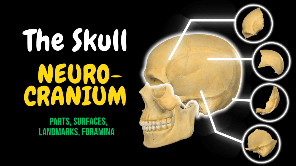
Skull Bones: Neurocranium (Frontal, Parietal, Temporal, Occipital, Sphenoid, Ethmoid) Official Links Instagram Youtube Jki-discord Notes & Illustrations Quizzes Summary & Transcript Notes ☆ Members Only Go to PDF Notes Illustrations ☆ Members Only Go to Illustrations 12345678910 Neurocranium – QUIZ Test your understanding with 10 random multiple-choice questions from the question bank. You're in the preview mode. Note: All elements work correctly on the front end. 1 / 10 What are the main parts of the occipital bone? A) Squamous, basilar, and two lateral parts B) Temporal, sphenoidal, and nuchal C) Body, lesser wing, greater wing, and pterygoid D) Squamous, mastoid, petrous, and occipital condyles The occipital bone has four parts: one squamous, one basilar, and two lateral parts. 2 / 10 Which of the following foramina transmit the mandibular nerve? A) Foramen ovale B) Foramen spinosum C) Jugular foramen D) Foramen rotundum The mandibular nerve passes through the foramen ovale. 3 / 10 Which two structures are associated with the superior and inferior temporal lines? A) Parotid gland and stylomastoid artery B) Epicranial aponeurosis and mastoid notch C) Trapezius and sternocleidomastoid D) Temporal fascia and temporalis muscle The superior line marks the fascia attachment, while the inferior is origin for the temporalis muscle. 4 / 10 What passes through the parietal foramen? A) Parietal emissary vein B) Middle meningeal artery C) Superior temporal vein D) Supraorbital nerve The parietal emissary vein passes through the parietal foramen, connecting extracranial veins to intracranial sinuses. 5 / 10 What are the contents of the foramen magnum? A) Optic chiasm and oculomotor nerve B) Facial nerve and labyrinthine artery C) Medulla, arteries, nerves, and spinal vein D) Jugular vein and glossopharyngeal nerve It contains the medulla, spinal arteries, vertebral arteries, accessory nerve root and spinal vein. 6 / 10 What nerves pass through the supraorbital notch and frontal notch? A) Infraorbital and zygomatic nerves B) Oculomotor and lacrimal nerves C) Lateral and medial supraorbital nerve branches D) Optic and trochlear nerves These notches transmit the lateral and medial branches of the supra-orbital nerve. 7 / 10 What structure is found on the internal surface of the parietal bone and houses venous blood from the brain? A) Parietal eminence B) Sagittal suture C) Groove for superior sagittal sinus D) Groove for middle meningeal artery The groove for the superior sagittal sinus contains the superior sagittal sinus, a major dural venous sinus. 8 / 10 Which part of the temporal bone contains the mastoid air cells? A) Tympanic part B) Squamous part C) Petrous part D) Mastoid process The mastoid process of the temporal bone houses mastoid air cells. 9 / 10 The anterior clinoid processes serve as attachment for which structure? A) Middle meningeal artery B) Tentorium cerebelli C) Superior oblique muscle D) Trigeminal nerve The tentorium cerebelli attaches to both anterior and posterior clinoid processes. 10 / 10 Which angle of the parietal bone lies toward the sphenoid bone? A) Sphenoid angle B) Mastoid angle C) Occipital angle D) Frontal angle The parietal bone has four angles; the sphenoid angle lies near the sphenoid bone. Your score is The average score is 0% Description This video is about the neurocranium AnatomyBones of the skull:Neurocranium: forms the protective case around the brainViscerocranium: forms the facial skeleton Neurocranium:Skullcap (calvaria):Frontal bone – os frontaleParietal bones (paired) – ossa parietaliaOccipital bone – os occipitale Cranial base (basis cranii):Temporal bones (paired) – ossa temporaliaSphenoid bone – os sphenoidaleEthmoid bone – os ethmoidale Frontal boneCoronal suture – sutura coronalisSupraorbital margin – margo supraorbitalisSupraorbital notch – incisura supraorbitalisFrontal notch – incisura frontalisZygomatic process – processus zygomaticusFrontal eminence – tuber frontaleSuperciliary arches – arcus superciliaresGlabellaFrontal crest – crista frontalisGroove for superior sagittal sinus – sulcus sinus sagittalis superiorisTemporal lines – lineae temporales (also on parietal)Lacrimal fossa – fossa glandulae lacrimalisTrochlear fovea/spine – fovea trochlearis / spina trochlearisNasal spineEthmoidal grooves (anterior/posterior) – sulci ethmoidales anterior et posteriorFrontal sinus – sinus frontalis Parietal bonesSagittal, coronal, lambdoid, squamous suturesAngles: frontal, occipital, sphenoidal, mastoid – anguli frontalis, occipitalis, sphenoidalis, mastoideusParietal eminence – tuber parietaleParietal foramen – foramen parietaleGrooves: superior sagittal sinus, sigmoid sinus, middle meningeal artery – sulci sinus sagittalis superioris, sigmoidei, arteriae meningeae mediaeTemporal lines (also on frontal) Occipital boneForamen magnumOccipital condylesHypoglossal canal – canalis nervi hypoglossiExternal/internal occipital protuberance – protuberantia occipitalis externa/internaExternal occipital crest – crista occipitalis externaNuchal lines – lineae nuchales suprema, superior et inferiorCruciform eminence – eminentia cruciformisCerebral/cerebellar fossae – fossae cerebrales et cerebellaresClivusPharyngeal tubercle – tuberculum pharyngeumSinus grooves: transverse, occipital, sigmoid, superior sagittal – sulci sinuumInferior petrosal sinus groove – sulcus sinus petrosi inferiorisJugular notch – incisura jugularis, intrajugular process – processus intrajugularisSutures: lambdoid, occipitomastoid; spheno-occipital synchondrosis Temporal boneSquamous part:Zygomatic process – processus zygomaticusMandibular fossa – fossa mandibularis, articular tubercle – tuberculum articulareSupramastoid crest – crista supramastoidea Tympanic part:External acoustic meatus – meatus acusticus externusTympanic ring – anulus tympanicusPetrotympanic fissure Petrous part:Apex – apex partis petrosaeArcuate eminence – eminentia arcuata, tegmen tympaniTrigeminal impression – impressio trigeminalisGrooves: greater/lesser petrosal nerves, superior/inferior petrosal sinusesInternal acoustic meatus – meatus acusticus internusSubarcuate fossa – fossa subarcuata, jugular notch – incisura jugularis Mastoid part:Mastoid process – processus mastoideus, mastoid notch – incisura mastoideaGroove for occipital artery – sulcus arteriae occipitalisStylomastoid foramen – foramen stylomastoideumStyloid process – processus styloideusFacial canal – canalis facialis Sphenoid boneBody – corpus ossis sphenoidalis, sella turcica with hypophyseal fossa, dorsum sellae, clinoid processes, tuberculum sellaePrechiasmatic sulcus – sulcus chiasmatis, carotid sulcus – sulcus caroticus Wings:Greater wings – alae majores: foramina rotundum, ovale, spinosumLesser wings – alae minores: optic canal – canalis opticusSuperior/inferior orbital fissures – fissurae orbitales Pterygoid processes – processus pterygoideiMedial/lateral plates – laminae medialis et lateralisPterygoid hamulus – hamulus pterygoideus, pterygoid/scaphoid fossaeSphenoidal crest – crista sphenoidalis, conchae – conchae sphenoidales, sinuses – sinus sphenoidales, rostrum Ethmoid boneCribriform plate & foramina – lamina / foramina cribrosa, crista galliPerpendicular plateEthmoidal labyrinths – labyrinthus ethmoidalis (ethmoidal cells: anterior, middle, posterior)Superior/middle nasal conchae – conchae nasalesUncinate processOrbital plate – lamina orbitalisEthmoidal foramina: anterior/posterior – foramina ethmoidalia Transcript Introduction0:00The human skull consists of 22 bones, which, in adults, are mostly connected together through0:06strong
Thoracolumbar Fascia
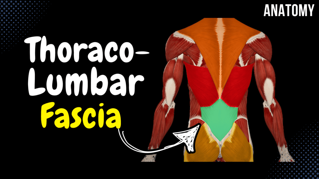
Thoracolumbar Fascia Official Links Instagram Youtube Jki-discord Notes & Illustrations Quizzes Summary & Transcript Notes ☆ Member Only Go to PDF Notes Illustrations ☆ Member Only Go to Illustrations 12345678910 Thoracolumbar Fascia – QUIZ Test your understanding with 10 random multiple-choice questions from the question bank. You're in the preview mode. Note: All elements work correctly on the front end. 1 / 10 Which muscle group does the posterior layer of the thoracolumbar fascia provide attachment for? A) Erector spinae B) Abdominal muscles C) Psoas major D) Transversospinal muscles The erector spinae group is attached to the posterior layer. 2 / 10 How many layers does the thoracolumbar fascia have? A) One B) Two C) Four D) Three The thoracolumbar fascia is divided into three layers: anterior, middle, and posterior. 3 / 10 Which fascia layer is directly involved in stabilizing the lumbar region during heavy lifting? A) Anterior layer B) Middle layer C) Deep layer D) Posterior layer The posterior layer plays a key role in stabilization during heavy lifting. 4 / 10 What is the anatomical relationship of the quadratus lumborum to the thoracolumbar fascia? A) Enclosed by the middle layer B) Unrelated to the thoracolumbar fascia C) Attached to the anterior layer D) Covered by the posterior layer The quadratus lumborum is enclosed by the middle layer. 5 / 10 Which muscles are supported by the posterior layer of the thoracolumbar fascia? A) Rectus abdominis B) Psoas major C) Erector spinae muscles D) Abdominal wall muscles The erector spinae muscles are supported by the posterior layer. 6 / 10 Which nerves pass close to the thoracolumbar fascia? A) Thoracic and cervical nerves B) Sciatic and pudendal nerves C) Iliohypogastric and ilioinguinal nerves D) Femoral and obturator nerves The iliohypogastric and ilioinguinal nerves pass close to the fascia. 7 / 10 What anatomical structure is supported by the thoracolumbar fascia during rotation? A) Lumbar vertebrae B) Sacrum C) Thoracic spine D) Pelvis The lumbar vertebrae are supported during rotation. 8 / 10 Which layer of the thoracolumbar fascia assists in lateral flexion of the spine? A) Anterior layer B) Superficial layer C) Posterior layer D) Middle layer The middle layer assists in lateral flexion. 9 / 10 Which nerve runs close to the thoracolumbar fascia in the lumbar region? A) Iliohypogastric nerve B) Subcostal nerve C) Genitofemoral nerve D) Femoral nerve The iliohypogastric nerve runs close to the thoracolumbar fascia. 10 / 10 Which muscle contributes to the formation of the posterior layer of the thoracolumbar fascia? A) Quadratus lumborum B) Transverse abdominis C) Psoas major D) Latissimus dorsi The latissimus dorsi contributes to the posterior layer. Your score is The average score is 0% Description This video is about the Thoracolumbar Fascia (Fascia Thoracolumbalis), its structure, and the muscles associated with its different layers. Thoracolumbar Fascia (Fascia Thoracolumbalis) Posterior Layer (Lamina Posterior) Associated with Deep Back Muscles Middle Layer (Lamina Media) Quadratus Lumborum Transverse Abdominal Internal Oblique External Oblique Anterior Layer (Lamina Anterior) Psoas Major Transcript Introduction0:03What’s up. Meditay here and in this video, we’ll be going through the thoracolumbar fascia.0:08The thoracolumbar fascia is a large roughly diamond shapes area of connective tissue inThoracolumbar Fascia0:13the lumbar region, as you see here. The good thing with this fascia is that it organizes0:18muscles in the lumbar region into groups and it also functions as a main attachment0:23point to large muscles like the trapezius, Latissimus Dorsi and the Gluteus Maximus.0:28And to go through this fascia, we need to look at a cross section of the back. So here’s aParts of the Thoracolumbar Fascia0:33superior view of one of the Lumbar vertebra. The thoracolumbar fascia is organized into0:39three layers. It has a Posterior Layer, which is the most superficial. It has a middle layer,0:44as you see here. And it has an anterior layer. Aight. So, the Posterior Layer, cover all0:50the deep back muscles, You know all the muscles of the transversospinal system,0:54of the spinotransverse system and the spinospinal system, this one is going to cover them.1:00Between the middle layer and the anterior layer, there’s the quadratus lumborum,1:04which is the posterior muscle of the abdomen. Then, from the anterior layer and the middle1:09layer, you’ll find the lateral abdominal muscles attached to them,1:12like the transverse abdominal muscle, and the Internal oblique muscle. The external oblique1:18is indirectly attached to the thoracolumbar fascia, not directly, as you see here.1:23And then the anterior layer is going to cover the psoas major from the posterior surface.1:28So that was everything I had for the Thoracolumabr fascia, and I hope that was helpful. Notes & Illustrations Quizzes Summary & Transcript Notes ☆ Member Only Go to PDF Notes Illustrations ☆ Member Only Go to Illustrations Thoracolumbar Fascia – QUIZ Test your understanding with 10 random multiple-choice questions from the question bank. Start Become a Member You have to become a member before you can access the Notes and the Quizzes. Membership Plans Description This video is about the Thoracolumbar Fascia (Fascia Thoracolumbalis), its structure, and the muscles associated with its different layers. Thoracolumbar Fascia (Fascia Thoracolumbalis) Posterior Layer (Lamina Posterior) Associated with Deep Back Muscles Middle Layer (Lamina Media) Quadratus Lumborum Transverse Abdominal Internal Oblique External Oblique Anterior Layer (Lamina Anterior) Psoas Major Transcript Introduction0:03What’s up. Meditay here and in this video, we’ll be going through the thoracolumbar fascia.0:08The thoracolumbar fascia is a large roughly diamond shapes area of connective tissue inThoracolumbar Fascia0:13the lumbar region, as you see here. The good thing with this fascia is that it organizes0:18muscles in the lumbar region into groups and it also functions as a main attachment0:23point to large muscles like the trapezius, Latissimus Dorsi and the Gluteus Maximus.0:28And to go through this fascia, we need to look at a cross section of the back. So here’s aParts of the Thoracolumbar Fascia0:33superior view of one of the Lumbar vertebra. The thoracolumbar fascia is organized into0:39three layers. It has a Posterior Layer, which is the most superficial. It has a middle layer,0:44as you see here. And it has an anterior layer. Aight. So, the
Deep Back Muscles
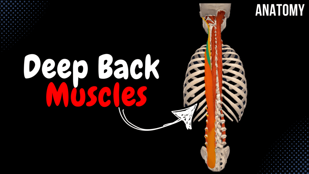
Deep Back Muscles (Division, Origin, Insertion, Function) Official Links Instagram Youtube Jki-discord Notes & Illustrations Quizzes Summary & Transcript Notes ☆ Member Only Go to PDF Notes Illustrations ☆ Member Only Go to Illustrations 12345678910 Deep Back Muscles – QUIZ Test your understanding with 10 random multiple-choice questions from the question bank. You're in the preview mode. Note: All elements work correctly on the front end. 1 / 10 Which muscle is part of the transversospinal system and inserts on the spinous process of the second vertebra above its origin? A) Intertransversarii B) Multifidi breves C) Rotatores breves D) Rotatores longi The rotatores longi muscles fit this description. 2 / 10 Which deep back muscle is the largest and most developed in the lumbar region? A) Multifidi B) Spinalis C) Interspinales D) Rotatores The multifidi muscles are largest in the lumbar region. 3 / 10 What is the function of the obliquus capitis inferior? A) Rotation of the atlas on the axis B) Extension of the thoracic spine C) Lateral flexion of the cervical spine D) Flexion of the cervical spine It rotates the atlas on the axis. 4 / 10 What is the function of the spinalis thoracis muscle? A) Extension of the thoracic spine B) Flexion of the lumbar spine C) Lateral flexion of the spine D) Rotation of the cervical spine It extends the thoracic spine. 5 / 10 What is the primary function of the semispinalis capitis? A) Rotation of the cervical spine B) Stabilization of the cervical spine C) Lateral flexion of the head D) Extension and contralateral rotation of the head and neck It extends the head and neck and rotates them contralaterally. 6 / 10 What is the function of the iliocostalis cervicis muscle? A) Stabilization of the thoracic spine B) Lateral flexion and extension of the cervical spine C) Flexion of the cervical spine D) Rotation of the cervical spine It assists with lateral flexion and extension of the cervical spine. 7 / 10 Which muscle extends and stabilizes the lumbar spine? A) Interspinales B) Longissimus C) Multifidi D) Rotatores The multifidi muscles extend and stabilize the lumbar spine. 8 / 10 Which muscle connects adjacent transverse processes in the cervical region? A) Interspinales B) Intertransversarii C) Rotatores D) Multifidi The intertransversarii muscles connect adjacent transverse processes. 9 / 10 Which muscle originates from the transverse process of C1 and inserts onto the occipital bone? A) Rectus capitis posterior minor B) Rectus capitis posterior major C) Obliquus capitis superior D) Obliquus capitis inferior This describes the obliquus capitis superior muscle. 10 / 10 What is the origin of the semispinalis thoracis? A) Transverse processes of C1-T5 B) Spinous processes of T10-L3 C) Spinous processes of T1-T8 D) Transverse processes of T6-T11 It originates from the transverse processes of T6-T11. Your score is The average score is 0% Description This video covers the deep muscles of the back, organized into different layers and muscle systems. Deep Muscles of the Back 3rd Layer Suboccipital Muscles System of Short Muscles 2nd Layer Transversospinal System 1st Layer (Superficial) Spinospinal System Spinotransverse System 1. Suboccipital Muscles System of deep muscles of the neck. Rectus Capitis Posterior Minor (Musculus Rectus Capitis Posterior Minor) Origin: Posterior Tubercle of Atlas (C1) Insertion: Occipital Bone below Inferior Nuchal Line Rectus Capitis Posterior Major (Musculus Rectus Capitis Posterior Major) Origin: Spinous Process of Axis (C2) Insertion: Occipital Bone – Inferior Nuchal Line Obliquus Capitis Superior (Musculus Obliquus Capitis Superior) Origin: Transverse Process of Atlas (C1) Insertion: Occipital Bone – Inferior Nuchal Line Obliquus Capitis Inferior (Musculus Obliquus Capitis Inferior) Origin: Spinous Process of Axis (C2) Insertion: Transverse Process of Atlas (C1) 2. System of Short Muscles Short muscles connecting adjacent vertebrae. Interspinales (Musculi Interspinales) Developed mainly in the cervical and lumbar regions. Origin: Spinous Process of vertebra below Insertion: Spinous Process of vertebra above Intertransversarii (Musculi Intertransversales) Mostly developed in the cervical region. Located between transverse processes. Origin: Transverse Process of vertebra below Insertion: Transverse Process of vertebra above 3. Transversospinal System Runs from the transverse process of a lower vertebra to the spinous process of an upper vertebra. Rotatores (Musculi Rotatores) Located in the thoracic vertebrae. Long Rotatores (Musculi Rotatores Longi) Origin: Transverse Process of vertebra below Insertion: Spinous Process of 2 vertebrae above Short Rotatores (Musculi Rotatores Breves) Origin: Transverse Process of vertebra below Insertion: Spinous Process of 1 vertebra above Multifidi (Musculi Multifidi) Fills space lateral to spinous processes. Most distinctive in the lumbar region. Short Multifidi (Musculi Multifidi Breves) Origin: Transverse Process of vertebra below Insertion: Spinous Process of 2 vertebrae above Long Multifidi (Musculi Multifidi Longi) Origin: Transverse Process of vertebra below Insertion: Spinous Process of 3 vertebrae above Semispinalis (Musculi Semispinales) Divided into thoracis, cervicis, and capitis. Semispinalis Thoracis (Musculi Semispinalis Thoracis) Origin: Transverse Process of T6-T11 Insertion: Spinous Process of C6-T4 Semispinalis Cervicis (Musculi Semispinalis Cervicis) Origin: Transverse Process of T1-T6 Insertion: Spinous Process of C2-C5 Semispinalis Capitis (Musculi Semispinalis Capitis) Origin: Transverse Process of C4-T6 Insertion: Occipital Bone – Between Inferior and Superior Nuchal Line 4. Spinospinal System Spinalis (Musculus Spinalis) Spinalis Cervicis (Musculus Spinalis Cervicis) Origin: Spinous Process of C6-T2 Insertion: Spinous Process of C2-C4 Spinalis Thoracis (Musculus Spinalis Thoracis) Origin: Spinous Process of T10-L3 Insertion: Spinous Process of T1-T8 5. Spinotransverse System Longissimus (Musculus Longissimus) Splenius (Musculus Splenius) Iliocostalis (Musculus Iliocostalis) Transcript Introduction0:03Hey What’s up. Meditay here and in this video, we’ll be covering the Deep muscles0:07of the back. Alright. Generally, the muscles of the back consist of superficial musclesDivision of the Back Muscles0:12and deep muscles. The superficial muscles consist of the The trapezius and Latissimus,0:17which are the 1st layer. The 2nd layer are the Rhomboid Major Minor and Levator0:22Scapula, and the 3rd layer of muscles consists of the Serratus Posterior superior and inferior.0:29And when you remove these three layers, you’ll finally get to the Deep muscles of the back.Division of the Deep Back Muscles0:34Now the deep muscles of the back are categorized based on their shape and structure and location.0:40Generally,
Superficial Back Muscles
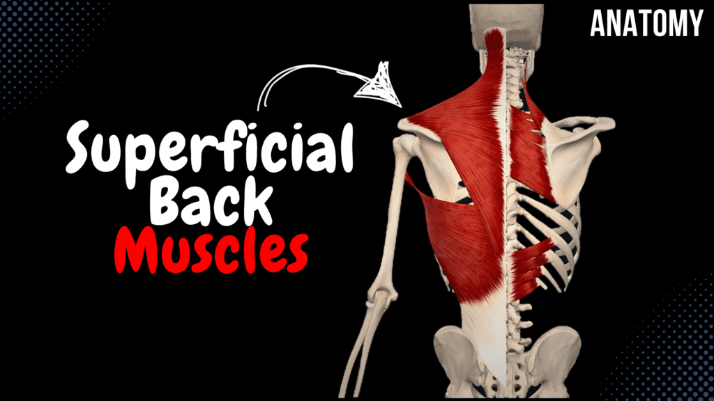
Superficial Back Muscles (Division, Origin, Insertion, Function) Official Links Instagram Youtube Jki-discord Notes & Illustrations Quizzes Summary & Transcript Notes ☆ Member Only Go to PDF Notes Illustrations ☆ Member Only Go to Illustrations 12345678910 Superficial Back Muscles – QUIZ Test your understanding with 10 random multiple-choice questions from the question bank. You're in the preview mode. Note: All elements work correctly on the front end. 1 / 10 Which muscle assists in respiration by elevating the ribs? A) Rhomboid major B) Latissimus dorsi C) Trapezius D) Serratus posterior superior The serratus posterior superior elevates the ribs, aiding in inspiration. 2 / 10 Which muscle depresses the ribs during expiration? A) Serratus posterior superior B) Rhomboid major C) Serratus posterior inferior D) Latissimus dorsi The serratus posterior inferior assists in expiration by pulling the ribs downward. 3 / 10 Which muscle forms the lower border of the posterior axillary fold? A) Serratus posterior inferior B) Rhomboid minor C) Latissimus dorsi D) Trapezius The latissimus dorsi forms the lower border of the posterior axillary fold. 4 / 10 Which superficial back muscle is innervated by the dorsal scapular nerve? A) Latissimus dorsi B) Rhomboid minor C) Serratus posterior superior D) Trapezius The rhomboids and levator scapulae are innervated by the dorsal scapular nerve. 5 / 10 Which muscle attaches to the acromion of the scapula? A) Latissimus dorsi B) Trapezius C) Rhomboid minor D) Serratus posterior inferior The trapezius inserts onto the acromion of the scapula. 6 / 10 What is the insertion point of the rhomboid minor? A) Medial border of scapula B) Spine of scapula C) Inferior angle of scapula D) Acromion process It inserts on the medial border of the scapula, above the rhomboid major. 7 / 10 Which muscle is part of the first layer of the superficial back muscles? A) Rhomboid minor B) Levator scapulae C) Serratus posterior inferior D) Trapezius The first layer includes large muscles like the trapezius. 8 / 10 Which superficial back muscle originates from the external occipital protuberance and inserts onto the clavicle? A) Trapezius B) Serratus posterior superior C) Rhomboid major D) Latissimus dorsi The trapezius originates from the external occipital protuberance and attaches to the clavicle. 9 / 10 Where does the rhomboid major originate? A) Spinous processes of T6-T12 B) Spinous processes of T1-T4 C) Lateral border of the scapula D) Superior nuchal line It originates from the spinous processes of T1-T4. 10 / 10 Which part of the trapezius is responsible for scapular elevation? A) Inferior B) Superior C) Middle D) All parts The superior part elevates the scapula. Your score is The average score is 0% Description This video is about the superficial muscles of the back, their anatomical layers, origins, insertions, and functions. Superficial Muscles of the Back Muscles of the 1st Layer Trapezius Latissimus Dorsi Muscles of the 2nd Layer Rhomboid Major Rhomboid Minor Levator Scapulae Muscles of the 3rd Layer Serratus Posterior Superior Serratus Posterior Inferior Trapezius (Musculus Trapezius) Superior Part Origin: Superior Nuchal Line External Occipital Protuberance Nuchal Ligament Insertion: Acromial End of Clavicle Acromion of Scapula Middle Part Origin: Spinous Process of C7-T3/T4 Insertion: Spine and Acromion of Scapula Inferior Part Origin: Spinous Process of T4-T12 Insertion: Spine of Scapula Latissimus Dorsi (Musculus Latissimus Dorsi) Origin: Spinous Process of T7-T12 Thoracolumbar Fascia Iliac Crest Inferior Surface of Ribs 9-12 Inferior Angle of Scapula Insertion: Crest of Lesser Tubercle (Humerus) (Crista Tuberculi Minoris Humeri) Rhomboid Major (Musculus Rhomboideus Major) Origin: Spinous Process of T1-T4 Insertion: Lower 2/3 of Medial Border of Scapula (Margo Medialis Scapulae) Function: Elevates and retracts the scapula Internal rotation of the scapula Rhomboid Minor (Musculus Rhomboideus Minor) Origin: Spinous Process of C6-C7 Insertion: Upper 1/3 of Medial Border of Scapula (Margo Medialis Scapulae) Function: Elevates and retracts the scapula Internal rotation of the scapula Levator Scapulae (Musculus Levator Scapulae) Origin: Spinous Process of C1-C4 Insertion: Superior Angle of Scapula (Angulus Superior Scapulae) Function: Elevates the scapula Serratus Posterior Superior (Musculus Serratus Posterior Superior) Origin: Spinous Process of C6-T2 Insertion: External Surface of 2nd-5th Ribs Function: Elevates the ribs (Inspiration) Serratus Posterior Inferior (Musculus Serratus Posterior Inferior) Origin: Spinous Process of T11-L2 Insertion: External Surface of 9th-12th Ribs Function: Pulls the ribs downward (Expiration) Transcript Introduction0:03Hey What’s up. Meditay here and in this video, we’ll be covering the Superficial muscles of0:08the back. Alright. Generally, the muscles of the back consist of superficial musclesDivision of the Superficial Muscles0:13and deep muscles. So let’s take a closer look at the superficial muscles.0:17These muscles are divided into layers. So, the 1st layer is the most superficial one.0:22It consist of the Trapezius, as you see here, and the Latissimus Dorsi.0:27The 2nd layer are the Rhomboid Major, Rhomboid Minor and Levator Scapula, and the 3rd layer of0:33muscles consists of the Serratus Posterior superior and Serratus Posterior inferior.0:39So these are the muscles we’re going to go through throughout this video.0:42We’ll start with the 1st Layer, the Trapezius. Now the trapezius Is this large muscleTrapezius0:48that take up the majority of your shoulders. They can also be classified as a cardiothoracic muscle,0:53in addition to being one of the superficial muscles of the back.0:57Now the Trapezius consists of 3 parts. There’s a Superior Part, a Middle part and an inferior part.1:04The superior part will originate from the Superior nuchal line, the external occipital protuberance,1:10and the nuchal ligament. It’s then going to insert1:13at the Acromial end of the clavicle, as you see here, as well as the acromion of the scapula.1:19Then we have the middle part, which originates from the spinous process of the C7-T3 or T4,1:26and it’s going to insert at the Spine of the Scapula as you see here, and the acromion.1:32Then we have the Inferior Part, which originates from the spinous process of T4-T121:38and insert at the spine of scapula as well. Ok. So what is the function of the Trapezius?1:44The Inferior part will pull the shoulder downwards, Middle and superior fibers1:49will pull the scapula towards the midline, as well as elevating the scapula. They may
Fascia of the Abdomen
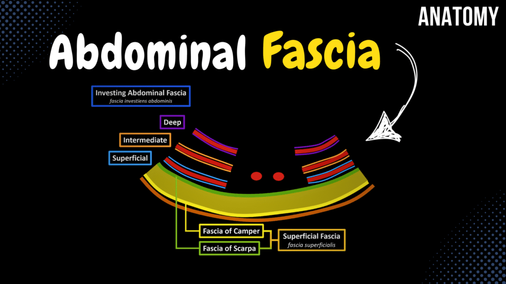
Fascia of the Abdomen (Superficial, Investing Abdominal, Endoabdominal) Official Links Instagram Youtube Jki-discord Notes & Illustrations Quizzes Summary & Transcript Notes ☆ Members Only Go to PDF Notes Illustrations ☆ Members Only Go to Illustrations 12345678910 Fascia of the Abdomen – QUIZ Test your understanding with 10 random multiple-choice questions from the question bank. You're in the preview mode. Note: All elements work correctly on the front end. 1 / 10 Which fascia surrounds the psoas major muscle? A) Psoas fascia B) Transversalis fascia C) Thoracolumbar fascia D) Iliac fascia The psoas fascia encloses the psoas major muscle. 2 / 10 Which layer of fascia forms the floor of the inguinal canal? A) Transversalis fascia B) Scarpa's fascia C) Iliac fascia D) Camper's fascia The transversalis fascia forms the floor of the inguinal canal. 3 / 10 What is the role of the vincula tendinum in tendon sheaths? A) Reduces friction B) Lubricates tendons C) Supplies nutrients to tendons D) Provides structural support Vincula tendinum supply nutrients to tendons within tendon sheaths. 4 / 10 What forms the outermost layer of the abdominal wall fascia? A) Investing abdominal fascia B) Thoracolumbar fascia C) Superficial fascia D) Endoabdominal fascia The superficial fascia forms the outermost layer of the abdominal wall fascia, comprising Camper’s and Scarpa’s layers. 5 / 10 What is the role of the transversalis fascia in hernia formation? A) Contributes to hernia formation B) Protects the lumbar spine C) Facilitates abdominal wall movement D) Stabilizes the iliac crest Weakness in the transversalis fascia can contribute to the development of inguinal hernias. 6 / 10 Which layer of fascia provides mechanical support and organization? A) Iliac fascia B) Camper's fascia C) Psoas fascia D) Scarpa's fascia Scarpa’s fascia lies beneath Camper’s fascia and provides mechanical support and structural organization. 7 / 10 What anatomical structure passes through the saphenous opening? A) Great saphenous vein B) Inferior vena cava C) Femoral artery D) Superficial epigastric vein The great saphenous vein passes through the saphenous opening in the fascia lata. 8 / 10 What is the primary anatomical location of the iliac fascia? A) Rectus sheath B) Thoracolumbar fascia C) Over the iliacus muscle D) Between internal oblique and external oblique The iliac fascia lies over the iliacus muscle, blending with the psoas fascia and extending to the pelvic brim. 9 / 10 What is the primary role of Camper’s fascia? A) Insulation and energy storage B) Stabilization of lumbar spine C) Protection from infections D) Structural support Camper’s fascia provides insulation, energy storage, and cushioning as the fatty layer of superficial abdominal fascia. 10 / 10 The transversalis fascia lies between which two structures? A) Transverse abdominal muscle and peritoneum B) Quadratus lumborum and iliac fascia C) Rectus sheath and internal oblique D) External oblique and Camper's fascia The transversalis fascia lies between the transverse abdominal muscle and the peritoneum. Your score is The average score is 0% Description This video is about the fascia of the abdomen, including its different layers and anatomical significance. Muscles of the Abdomen Lateral Group External Oblique Internal Oblique Transverse Abdominal Anterior Group Rectus Abdominis Pyramidalis Posterior Group Quadratus Lumborum Superficial Fascia (Fascia Superficialis) Fascia of Camper Fascia of Scarpa Investing Abdominal Fascia (Fascia Investiens Abdominis) Superficial Layer Intermediate Layer Deep Layer Endoabdominal Fascia (Fascia Endoabdominalis) Transversalis Fascia (Fascia Transversalis) Iliac Fascia (Fascia Iliaca) Transcript Introduction0:03What’s up. Meditay here and in this video, we’ll be going through0:06the Fascia you’ll find in the Abdomen. So the fascia of the abdomen vover theMuscles of the Abdomen0:11muscles of the abdomen from both the external and internal side. So here I’ve cut through all the0:16muscle layers of the abdomen. And remember, they consist of the external oblique, Internal Oblique0:22and Transverse Abdominal Muscle. These three at considered as the lateral abdominal muscles.0:27And we have the Anterior abdominal Muscles, like the Rectus Abdominis.0:30Our goal in this video is to go through the fascia that you’ll find wrapping around these muscles,0:36and separating them from the organs within the abdominal cavityContent0:39So In this video, we’re going to go through the Superficial Fascia,0:43we’ll go thrgouh the Investing Abdominal Fascia. And we’ll go thrgouh the Endoabdominal Fascia.0:48And to do that, we’ll have to make a transverse cut of the abdomen. Remove the upper half,0:53and look at it from this perspective. And now we’re gonna try to draw all0:57the structures and go through them as we do that. First we have the Skin.1:01And right underneath the skin layers, we’ll find the adipose tissue, or fat cells. Underneath the1:07fat cells, we can find the External Oblique. Internal Oblique, and Transverse Abdominal1:12Muscle. And in the middle here we can find the Rectus Abdominis muscle. So this is a very1:16schematic outline of the abdominal muscles. The first fascia we’re gonna go thrgouh isSuperficial Fascia1:21associated with the fat Layer. The fascia that cover the fat layer from the superficial side1:26is called Fascia of Camper. And the fascia that cover it from the inner side, is called1:31Fascia of Scarpa. The two layers of fascia together, form the so called Superficial Fascia.1:38So that’s the first one in our list. Next we have a Fascia called Investing Abdominal Fascia.Investing Abdominal Fascia1:45And as the name implies, this fascia is going to surround the abdominal muscles.1:49So the investing abdominal fascia si divided into three layers.1:54First is the Superficial investing abdominal fascia, aurrounding the external oblique.1:59Then there’s the Intermediate Investing abdominal fascia, surrounding the internal Oblique.2:04Then there’s the Deep investing abdominal fascia, surrounding the transverse abdominal muscle. Now.2:10Underneath the Deep investing abdominal fascia, that’s where you’ll find the third fascia we’reEndoabdominal Fascia2:15gonna talk about, called the endoabdominal fascia. And just underneath the endoabdominal fascia,2:21that’s where you’ll find the Parietal peritoneum, which over all of your internal organs.2:28Now, the endoabdominal fascia is actually divided into certain parts depending on the location of2:34it, So let’s look at that a little bit. So now we’re gonna draw the lateral view of he abdomen.2:38First we have the Skin, then the Fat cells, then The external oblique, Internal Oblique
Muscles of the Abdomen

Muscles of the Abdomen (Groups, Origin, Insertion, Function) Official Links Instagram Youtube Jki-discord Notes & Illustrations Quizzes Summary & Transcript Notes ☆ Members Only Go to PDF Notes Illustrations ☆ Members Only Go to Illustrations 12345678910 Abdominal Muscles – QUIZ Test your understanding with 10 random multiple-choice questions from the question bank. You're in the preview mode. Note: All elements work correctly on the front end. 1 / 10 Where does the rectus abdominis originate? A) Pubic symphysis and crest B) Iliac crest C) Xiphoid process D) Costal cartilages of ribs 5–7 The rectus abdominis originates from the pubic symphysis and pubic crest. 2 / 10 What is the function of the cremaster muscle in males? A) Compresses abdominal contents B) Elevates the testis C) Flexes the hip D) Stabilizes the pelvis The cremaster muscle elevates the testis and scrotum to regulate temperature. 3 / 10 The pyramidalis muscle is absent in approximately what percentage of people? A) 20% B) 25% C) 10% D) 5% The pyramidalis is absent in around 20% of the population. 4 / 10 What is the function of the external oblique muscle during bilateral contraction? A) Laterally flexes the trunk B) Compresses abdominal contents C) Flexes the trunk D) Rotates the trunk The external oblique flexes the trunk during bilateral contraction. 5 / 10 Which abdominal muscle is considered part of the posterior group? A) External oblique B) Quadratus lumborum C) Transverse abdominal D) Internal oblique The quadratus lumborum belongs to the posterior abdominal group. 6 / 10 The cremaster muscle is formed by fibers of which two muscles? A) Quadratus lumborum and external oblique B) Rectus abdominis and external oblique C) Internal oblique and transverse abdominal D) Pyramidalis and rectus abdominis The cremaster muscle is formed by the internal oblique and transverse abdominal muscles. 7 / 10 Which abdominal muscle inserts into the linea alba and the pubic crest? A) Transverse abdominal B) Internal oblique C) Pyramidalis D) External oblique The transverse abdominal muscle inserts into these structures. 8 / 10 Which layer of fascia lies directly under Camper’s fascia? A) Transversalis fascia B) Investing fascia C) Thoracolumbar fascia D) Scarpa's fascia Scarpa’s fascia is the membranous layer beneath Camper’s fascia. 9 / 10 What is the insertion point of the external oblique muscle? A) Linea alba and iliac crest B) Xiphoid process C) Pubic symphysis D) Inguinal ligament The external oblique inserts into the linea alba and the iliac crest. 10 / 10 Which abdominal muscle tenses the linea alba? A) Rectus abdominis B) Internal oblique C) Transverse abdominal D) Pyramidalis The pyramidalis muscle tenses the linea alba. Your score is The average score is 0% Description Small correction: The rectus abdominis originates from the pubic crest and symphysis and inserts into the xiphoid process and costal cartilages of ribs 5–7. I mistakenly swapped the origin and insertion in the video. Apologies for the error! Muscles of the Abdomen Lateral Group External Oblique Internal Oblique Transverse Abdominal Anterior Group Rectus Abdominis Pyramidalis Posterior Group Quadratus Lumborum Posterior Group Quadratus Lumborum (Musculus Quadratus Lumborum) Origin: Iliac Crest Iliolumbar Ligament Insertion: 12th Rib Costal Process of L1-L4 Function: Extension of Trunk (Bilateral Contraction) Lateroflexion of Trunk (Unilateral Contraction) Anterior Group Pyramidalis (Musculus Pyramidalis) Origin: Superior Pubic Ramus Insertion: Linea Alba Function: Tenses Linea Alba and Strengthens the Rectus Sheath Rectus Abdominis (Musculus Rectus Abdominis) Origin: Pubic Crest Pubic Symphysis Insertion: Xiphoid Process Costal Cartilage of 5th – 7th Rib Function: Ventral Flexion of Trunk Expiration Muscle Lateral Group Transverse Abdominal (Musculus Transversus Abdominis) Origin: Inguinal Ligament Iliac Crest Thoracolumbar Fascia 7th – 12th Ribs Insertion: Linea Alba Function: Rotation of the Trunk Expiration Muscle Internal Oblique (Musculus Obliquus Internus Abdominis) Origin: Inguinal Ligament Iliac Crest Thoracolumbar Fascia Insertion: Linea Alba 10th – 12th Ribs Function: Ventral Flexion of Trunk (Bilateral Contraction) Tilts the Trunk to the Side (Unilateral Contraction) Expiration Muscle Cremaster (Musculus Cremaster) Formed by: Internal Oblique and Transverse Abdominal Muscles External Oblique (Musculus Obliquus Externus Abdominis) Origin: 5th – 12th Ribs Insertion: Linea Alba Iliac Crest Function: Ventral Flexion of Trunk (Bilateral Contraction) Tilts the Trunk to the Side (Unilateral Contraction) Expiration Muscle Transcript Introduction0:03What’s up. Meditay here and in this video, we’ll be going through the0:07muscles you’ll find in the abdominal region. So here you see the anterior view of the Abdomen.Division of the Abdominal Muscles0:12And here I’ve cut through all the muscles of the abdomen in order to se them all. The most0:17external muscle of the abdomen is the External Oblique Muscle.0:21Then there’s the Internal Oblique muscle, and then the Transverse Oblique Muscle. These three muscles0:27are a part of the Lateral Abdominal Muscles, or lateral groups. So the muscles of the abdomen0:31are organized into three groups. And these are the lateral group. Then we have the Rectus Abdominis,0:37and a tiny muscle called Pyramidalis. These two are considered the Anterior Group of muscles.0:44And then the Posterior group has only one muscle, which is this one,0:48called the quadratus lumborum. So, these are the muscles we’re going to focus on in this video.0:53All of the muscles of the abdomen corporate in their function.0:57They corporate in flexing your abdomen, rotating it from one side to another and so on. All of them1:03also corporate with the diaphragm during breathing, and they do that by regulating1:08the intra abdominal pressure by either squeeze your abdomen, to push your air out, or releasing1:13the pressure off from your abdomen to allow the diaphragm to expand volume of the thoracic cavity1:19to push air into the lungs. Awesome. Now let’s start with the posterior group.1:23Then we’ll do the anterior group and then end with the lateral group. So the Quadratus Lumborum,Quadratus Lumborum1:28again, is situated on the posterior abdominal wall, as you see here. It originates from the1:34Iliac crest and the Iliolumbar ligament. And it inserts at the 12th rib, as well as1:39the costal processes of L1 to L4. And its function is either extension of the trunk during bilateral1:46contraction, or Lateral flexion of the trunk, if only one side
Topography of the Thigh and Leg
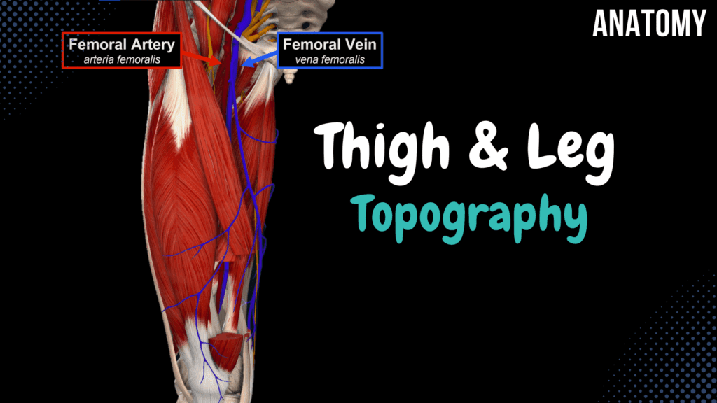
Topography of the Thigh and Leg (Femoral Triangle, Adductor Canal, Popliteal Fossa) Official Links Instagram Youtube Jki-discord Notes & Illustrations Quizzes Summary & Transcript Notes ☆ Member Only Go to PDF Notes Illustrations ☆ Member Only Go to Illustrations 12345678910 Topography of the Thigh and Leg – QUIZ Test your understanding with 10 random multiple-choice questions from the question bank. You're in the preview mode. Note: All elements work correctly on the front end. 1 / 10 Which nerve is located in the muscular space of the inguinal region? A) Femoral nerve B) Genitofemoral nerve C) Obturator nerve D) Lateral cutaneous nerve of the thigh The lateral cutaneous nerve of the thigh is located in the muscular space. 2 / 10 What are the boundaries of the femoral triangle? A) Iliopsoas, Pectineus, Sartorius B) Inguinal ligament, Gracilis, Sartorius C) Adductor Magnus, Gracilis, Iliopsoas D) Inguinal ligament, Sartorius, Adductor Longus The femoral triangle is bounded by the inguinal ligament, sartorius muscle, and adductor longus. 3 / 10 Which artery passes through the adductor hiatus? A) Tibial artery B) Femoral artery C) Popliteal artery D) Deep femoral artery The femoral artery passes through the adductor hiatus. 4 / 10 Which artery supplies the anterior compartment of the leg? A) Fibular artery B) Popliteal artery C) Posterior tibial artery D) Anterior tibial artery The anterior tibial artery supplies the anterior compartment of the leg. 5 / 10 What structure forms the superior boundary of the adductor canal? A) Adductor longus B) Rectus femoris C) Sartorius D) Vastus medialis The sartorius muscle forms the superior boundary of the adductor canal. 6 / 10 What forms the roof of the popliteal fossa? A) Semimembranosus B) Popliteal fascia and skin C) Gastrocnemius muscle D) Crural fascia The roof of the popliteal fossa is formed by the popliteal fascia and skin. 7 / 10 What structure passes through the adductor hiatus? A) Fibular nerve B) Femoral artery and vein C) Tibial artery D) Popliteal nerve The femoral artery and vein pass through the adductor hiatus. 8 / 10 Which structure is located within the femoral triangle? A) Popliteal artery B) Deep fibular nerve C) Femoral artery D) Obturator artery The femoral triangle contains the femoral artery, vein, and nerve. 9 / 10 Which muscle forms the anterior boundary of the adductor canal? A) Adductor magnus B) Sartorius C) Vastus medialis D) Gracilis The sartorius muscle forms the anterior boundary of the adductor canal. 10 / 10 Which structure connects the femoral triangle to the adductor canal? A) Apex of the femoral triangle B) Adductor hiatus C) Cribriform fascia D) Sartorius The apex of the femoral triangle connects it to the adductor canal. Your score is The average score is 0% Description This video covers the topography of the thigh and leg, including key anatomical structures, compartments, and their contents. Topography of the Thigh Femoral Triangle (Trigonum Femorale) Boundaries: Sartorius Muscle Adductor Longus Inguinal Ligament (Ligamentum Inguinale) Floor: Iliopsoas Pectineus Mnemonic: NAVEL (Nerve, Artery, Vein, Empty Space, Lymphatics) Adductor Canal (Canalis Adductorius) Contents: Femoral Artery (Arteria Femoralis) Femoral Vein (Vena Femoralis) Boundaries: Sartorius Muscle Vastus Medialis Adductor Magnus Starts: Femoral Triangle Ends: Popliteal Fossa Popliteal Fossa (Fossa Poplitea) Boundaries: Semimembranosus and Semitendinosus (Medial Upper Border) Biceps Femoris (Lateral Upper Border) Gastrocnemius Medial Head (Medial Lower Border) Gastrocnemius Lateral Head (Lateral Lower Border) Contents: Popliteal Fascia (Fascia Poplitea) Topography of the Leg Crural Fascia (Fascia Cruris) Contents: Anterior Tibial Artery and Vein Deep Fibular Nerve Fibular Artery and Vein Posterior Tibial Artery and Vein Tibial Nerve Transcript Introduction0:03In the last video, we covered the main topographical openings of the Hip. Now0:08let’s do the topography of the Thigh and the Topography of the Leg.Thigh Topography Overview0:11So the topography of the thigh consists of the Femoral Triangle,0:14Adductor Canal and the Popliteal Fossa. So let’s start with the femoral triangle.Femoral Triangle0:19The femoral triangle is a region in the anterior thigh of a triangular zone0:23that will help ou identify many structures within this part of our body. And ot help you remember0:29the sequence of the structures within the femoral triangle, I like to use the mnemonic Navel.0:34Which will help remember the order from the lateral moving medially, that the femoral nerve0:39is the most lateral structure within the space. Followed by the femoral artery. The femoral vein0:44and then the lymphatics. So let’s now take a closer look at the boundaries of this area.0:49The first thing that we’re gonna see is that the lateral border is gonna be0:52formed by the sartorius muscle. The superior border is going to0:56be the inguinal ligament, and the medial border is going to be one of the adductor1:00muscles. The adductor Longus muscle. In other words, The base of the triangle1:05is actually going to be the inguinal ligament. And the apex is directed inferiomedially.1:10Deep to the contents. The floor of this area is going to be made by the Iliopsoas laterally,1:16and the pectineus medially, as you see here. So that was the Femoral Triangle.Adductor Canal1:21Now let’s cover a canal called the adductor canal. The adductor canal is a special region within the1:27thigh, that is going to allow a passage for the femoral artery and the femoral vein to run down1:33through the thigh. And once they reach the end of the thigh, they’re going to go posteriorly1:38into a region called the popliteal fossa as these two vessels become,1:42he popliteal artery and the popliteal vein. To better visualize the canal, I’ve cut a window1:48through two of the muscles on the anterior compartment of the thigh.1:51The first of which is the sartorius muscle. As you see here. This muscle is going to form the roof of1:56the adductor canal. Meaning that the structures that is going to within this space, is going to2:01lie deep to the sartorius muscle. Therefore, I needed to cut a window through this muscle2:06to allow us to see the path of those vessels. I’ve also cut through the vastus medialis, which is one2:11of the large quadriceps muscles, which is going to be on the medial aspect of the knee.