Ureter, Urinary Bladder & Urethra

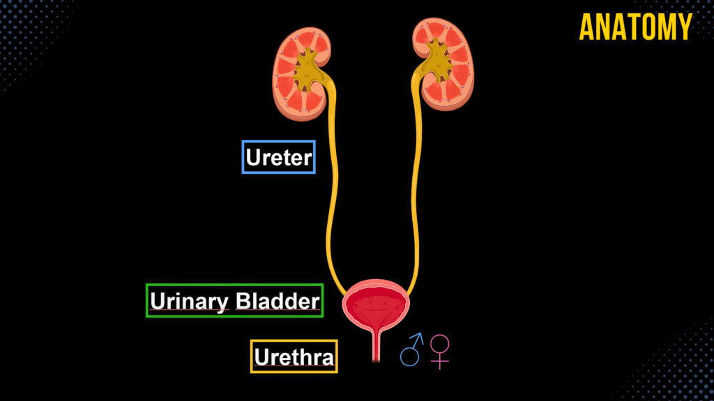
Ureter, Urinary Bladder & Urethra (Structures & Walls) Official Links Instagram Youtube Jki-discord Notes & Illustrations Quizzes Summary & Transcript Notes ☆ Member Only Go to PDF Notes Illustrations ☆ Member Only Go to Illustrations 12345678910 Ureter Urinary Bladder and Urethra – QUIZ Test your understanding with 10 random multiple-choice questions from the question bank. You're in the preview mode. Note: All elements work correctly on the front end. 1 / 10 Which muscle layer forms the detrusor muscle in the urinary bladder? A) Tela submucosa B) Tunica adventitia C) Tunica mucosa D) Tunica muscularis The tunica muscularis, consisting of three muscle layers, forms the detrusor muscle. 2 / 10 What is the function of umbrella cells in the urinary bladder? A) Regulate urine pH B) Transport urine C) Allow stretching of bladder D) Produce mucous Umbrella cells are specialized for stretching and protecting the bladder lining. 3 / 10 What prevents backflow of urine from the bladder into the ureters? A) Detrusor muscle contraction B) Oblique entry of ureters C) Transitional epithelium D) Urethral sphincters The oblique entry of the ureters into the bladder and the ureteric orifices prevent backflow. 4 / 10 Which structure forms the base of the urinary bladder? A) Apex of bladder B) Trigone of bladder C) Vesical crest D) Fundus of bladder The trigone of the bladder forms its base, bordered by the ureteric orifices and internal urethral orifice. 5 / 10 Which part of the urinary bladder contains no folds in the mucosa? A) Apex of bladder B) Body of bladder C) Fundus of bladder D) Trigone of bladder The trigone of the bladder has no folds in the mucosa. 6 / 10 Which layer of the ureter wall provides structural support? A) Tunica mucosa B) Tunica adventitia C) Tela submucosa D) Tunica muscularis The tunica adventitia provides structural support to the ureter wall. 7 / 10 Which part of the male urethra passes through the corpus spongiosum? A) Urethral crest B) Spongy urethra C) Membranous urethra D) Prostatic urethra The spongy urethra runs through the corpus spongiosum of the penis. 8 / 10 Which structure in the male urethra contains openings for the ejaculatory ducts? A) Spongy urethra B) Membranous urethra C) Prostatic urethra D) Urethral crest The prostatic urethra contains the openings of the ejaculatory ducts. 9 / 10 What forms the internal urethral sphincter? A) Lamina propria B) Submucosa of urethra C) Middle circular muscle fibers D) Outer longitudinal muscle fibers The internal urethral sphincter is formed by the middle circular layer of the tunica muscularis. 10 / 10 Where is the ureter’s upper narrowing located? A) Intramural part B) Junction with renal pelvis C) At the line terminalis D) At the ureteric orifice The ureter’s upper narrowing is at the junction with the renal pelvis. Your score is The average score is 0% Description Ureter, Urinary Bladder, and Male/Female Urethra This video covers the anatomy and function of the ureter, urinary bladder, and male and female urethra, including their structures, layers, and clinical significance. PS! At 6:23, I incorrectly stated that the Median Umbilical Ligament is a remnant of the umbilical arteries. It is, in fact, a remnant of the fetal urachus. 1. Ureter Function: Transports urine from the renal pelvis to the bladder via peristaltic movement. Course of the Ureter: Enters the lesser pelvis and crosses structures: In Males: Crosses the ductus deferens. In Females: Passes between the broad ligament (Ligamentum Latum Uteri) and the cardinal ligament (Ligamentum Cardinale Uretri). Opens into the bladder as the ureteric orifices (Ostium Ureteris). Parts of the Ureter: Abdominal Part (Pars Abdominalis). Pelvic Part (Pars Pelvica). Intramural Part (Pars Intramuralis). Diameter: 7-8 mm. Narrowings of the Ureter: Upper: Junction with the renal pelvis. Middle: Line terminalis. Lower: Intramural part. Layers of the Ureter Wall: Tunica Mucosa: Inner lining. Muscularis: Lower 1/3: Three muscle layers (inner longitudinal, middle circular, outer longitudinal). Upper 2/3: Two muscle layers (inner longitudinal, outer circular). Tunica Adventitia: Outer connective tissue. 2. Urinary Bladder (Vesica Urinaria) Function: Stores 250-500 ml of urine. Location: Empty bladder lies behind the pubic symphysis; a full bladder extends above it. Parts of the Bladder: Apex of Bladder (Apex Vesicae). Body of Bladder (Corpus Vesicae). Fundus of Bladder (Fundus Vesicae). Trigone of Bladder (Trigonum Vesicae). Right and Left Ureteric Orifices (Ostium Ureteris Dextrum et Sinistrum). Internal Urethral Orifice (Ostium Urethrae Internum). Interureteric Crest (Plicae Interureterica). Layers of the Bladder Wall: Tunica Mucosa: Lined with transitional epithelium. Tela Submucosa: Loose connective tissue; no folds in the trigone. Tunica Muscularis (Detrusor Muscle): Inner longitudinal layer. Middle circular layer (forms the internal urethral sphincter). Outer longitudinal layer. Tunica Adventitia or Serosa: Adventitia if facing the pelvis. Serosa if facing the peritoneum. 3. Male Urethra (Urethra Masculina) Length: ~18-20 cm. Parts of the Male Urethra: Prostatic Urethra (Pars Prostatica): Contains the urethral crest, seminal colliculus, and prostatic utricle. Receives openings of ejaculatory and prostatic ducts. Membranous Urethra (Pars Membranacea): Between the prostate and the bulb of the penis. Surrounded by the external urethral sphincter. Spongy Urethra (Pars Spongiosa): Located inside the corpus spongiosum. Receives the duct of the bulbourethral gland. Narrowings of the Male Urethra: Internal Urethral Orifice. Membranous Part. External Urethral Orifice. Enlargements of the Male Urethra: Prostatic Urethra. Spongy Urethra. Navicular Fossa. 4. Female Urethra (Urethra Femina) Length: ~4 cm. Location: Opens posterior to the clitoral glans. Walls of the Female Urethra: Tunica Mucosa: Contains urethral lacunae. Tunica Muscularis: Internal Urethral Sphincter. External Urethral Sphincter. 5. Clinical Relevance Kidney Stones (Urolithiasis): Commonly lodged at ureter narrowings. Urinary Tract Infections (UTIs): More common in females due to shorter urethra. Benign Prostatic Hyperplasia (BPH): Can obstruct the prostatic urethra, causing urinary retention. Sources: Biorender. University notes and lectures. Transcript Introduction0:01[Music]0:04what’s up0:04mediterra here let’s talk about the0:06anatomy of the urinary system0:07in this segment we will be talking about0:09the anatomy of the ureter the urinary0:11bladder and the urethra in both male and0:13female0:14so the urinary system consists of all0:16the organs involved in handling the0:18urine0:19and these are the kidneys the ureter
Kidneys

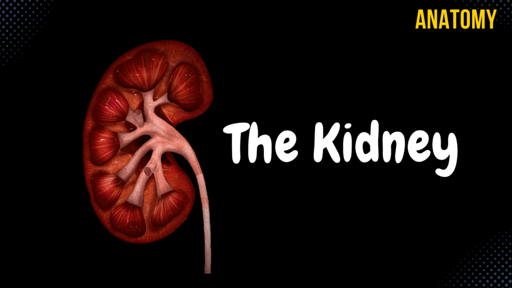
Kidneys (Functions, Structures, Coverings, Nephron) Official Links Instagram Youtube Jki-discord Notes & Illustrations Quizzes Summary & Transcript Notes ☆ Member Only Go to PDF Notes Illustrations ☆ Member Only Go to Illustrations 12345678910 Kidneys – QUIZ Test your understanding with 10 random multiple-choice questions from the question bank. You're in the preview mode. Note: All elements work correctly on the front end. 1 / 10 Which structure is found between the renal pyramids? A) Medullary rays B) Renal papillae C) Renal columns D) Renal lobes Renal columns are extensions of the cortex that separate the renal pyramids. 2 / 10 What is the apex of a renal pyramid called? A) Medulla B) Renal column C) Cortex D) Renal papilla The apex of a renal pyramid is called the renal papilla, where urine flows into the minor calyces. 3 / 10 What is the approximate thickness of the renal cortex? A) 15-20 mm B) 11-15 mm C) 4-11 mm D) 2-5 mm The renal cortex is approximately 4-11 mm thick. 4 / 10 Which anatomical structure collects urine from multiple collecting ducts? A) Renal pelvis B) Renal pyramid C) Renal papilla D) Minor calyx The minor calyx collects urine from multiple collecting ducts and funnels it into the major calyces. 5 / 10 Which structure anchors the kidneys to surrounding tissues? A) Perinephric fat B) Renal fascia C) Paranephric fat D) Fibrous capsule The renal fascia anchors the kidney and adrenal gland to surrounding structures. 6 / 10 What is the function of the renal pelvis? A) Filters plasma B) Anchors the kidney C) Produces hormones D) Funnels urine into the ureter The renal pelvis collects urine from the major calyces and funnels it into the ureter. 7 / 10 What is the terminal part of the nephron that delivers urine into the renal pelvis? A) Collecting duct B) Loop of Henle C) Renal corpuscle D) Proximal convoluted tubule The collecting duct collects urine from the nephron and empties into the renal pelvis. 8 / 10 Which structure of the kidney contains renal corpuscles? A) Renal medulla B) Renal cortex C) Renal sinus D) Renal pyramid The renal cortex contains renal corpuscles, proximal and distal convoluted tubules. 9 / 10 What forms the apex of the renal pyramid? A) Renal column B) Renal papilla C) Renal hilum D) Renal lobe The renal papilla forms the apex of the renal pyramid and drains urine into the minor calyces. 10 / 10 What is the primary site of plasma filtration in the kidney? A) Loop of Henle B) Collecting duct C) Bowman’s capsule D) Glomerulus Plasma filtration occurs in the glomerulus, a network of capillaries in the renal corpuscle. Your score is The average score is 0% Description Kidney Anatomy: Topography, Structures, and Function This video covers a comprehensive overview of the kidneys, including their topography, functions, external and internal structures, and nephron anatomy. 1. Topography of the Kidney The right kidney is slightly lower than the left. Both kidneys start at T12. Left kidney: Ends at L2. Right kidney: Ends at L3. 2. Functions of the Kidney Plasma Filtration: Through nephrons. Excretion: Removal of metabolic waste. Acid-Base Homeostasis: Regulates pH balance. Hormone Production: Erythropoietin, Renin. Vitamin D Metabolism: Activation of Vitamin D. 3. External Structures Weight: 120-200 grams. Size: 10-13 cm long, 5-6 cm wide, 4 cm thick. Inferior Pole (Extremitas Inferior). Superior Pole (Extremitas Superior): Suprarenal gland located on top. Lateral Border (Margo Lateralis). Medial Border (Margo Medialis): Contains the hilum (Hilum Renalis). 4. Coverings of the Kidney Fibrous Capsule (Capsula Fibrosa). Perinephric Fat (Capsula Adiposa). Renal Fascia (Fascia Renalis): Anterior Layer: Prerenal Layer (Lamina Prerenalis) / Fascia of Toldt. Posterior Layer: Retrorenal Layer (Lamina Retrorenalis) / Fascia of Zuckerkandl. Both layers fuse laterally. Peritoneum. 5. Internal Structures Renal Sinus Sinus Renalis: Fat-filled space inside the kidney. Renal Pelvis (Pelvis Renalis): Central collecting region. Renal Calyces (Calyces Renales): Divided into: Minor Renal Calyces (Calyx Renales Minor). Major Renal Calyces (Calyx Renales Major). Renal Medulla Renal Pyramids (Pyramides Renales). Renal Lobes (Lobi Renales). Renal Papillae (Papillae Renales): Tip of pyramids. Renal Medullary Rays (Radii Medullares): Contain nephrons’ loops. Loop of Henle and Collecting Ducts. Openings of Papillary Ducts (Foramina Papillaria). Renal Cortex Thickness: 4-11 mm. Renal Columns (Columna Renalis): Extension of the cortex into the medulla. Contains Renal Corpuscles, Proximal & Distal Convoluted Tubules. 6. Renal Lobe & Nephron Renal Corpuscle (Corpusculum Renis) Glomerulus: Network of capillaries. Afferent Arteriole (Vas Afferens): Brings blood in. Efferent Arteriole (Vas Efferens): Drains blood out. Glomerular Capsule (Capsula Glomeruli): Encases glomerulus. Nephron Tubules Proximal Convoluted Tubule (Tubulus Proximalis): Reabsorbs nutrients. Has a convoluted part and a straight part. Loop of Henle (Ansa Nephroni): Descending Limb. Ascending Limb. Distal Convoluted Tubule (Tubulus Distalis): Fine-tunes ion balance. Collecting Duct (Ductus Colligentes): Final urine concentration. Papillary Duct (Ductus Papillares): Empties into minor calyx. 7. Clinical Relevance Polycystic Kidney Disease (PKD): Genetic disorder causing multiple cysts in the kidney. Kidney Stones (Nephrolithiasis): Crystallized minerals causing obstruction. Acute Kidney Injury (AKI): Sudden loss of kidney function. Chronic Kidney Disease (CKD): Progressive decline in kidney function. Sources Used: Memorix Anatomy (2nd Edition) – Hudák Radovan, Kachlík David, Volný Ondřej. Complete Anatomy by 3D4Medical. Biorender. University notes and lectures. Transcript Introduction0:03What’s up.0:04Meditay here.0:05Let’s talk about the anatomy of the urinary system.0:07In this segment, we will be talking about the anatomy of the Kidneys.0:10Alright, so the Urinary system consists of all the organs involved in handling the urine.0:15These are the Kidneys, the Ureter, the Urinary bladder, and the urethra.0:20Our goal is to cover the anatomy of all these structures you see here, step by step, and0:25we’ll start with the Kidneys In this video, we’re first going to talk about0:28the functions of the kidneys.0:30Then we’ll talk about the external structures and the coverings of the kidneys.0:34After that, we’ll open up the kidney and cover the internal structures.0:39When we’re done with the kidneys, we’ll talk about the general anatomy of the nephron,0:42which is the functional unit of the kidney.Topography of Kidney0:45Alright, so here you see
