
Male Genital System
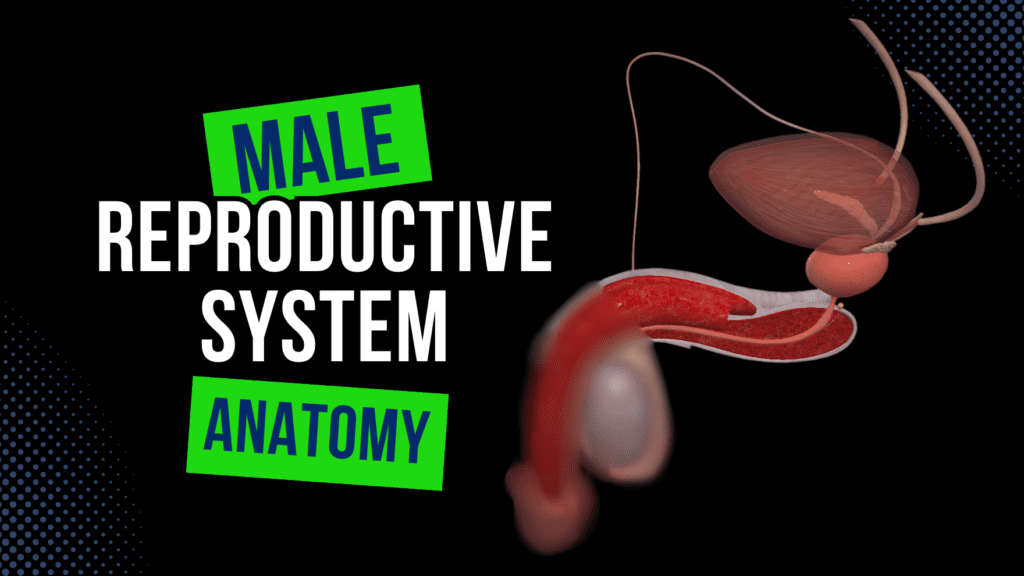
Male Genital System (Internal & External) Official Links Instagram Youtube Jki-discord Notes & Illustrations Quizzes Summary & Transcript Notes ☆ Member Only Go to PDF Notes Illustrations ☆ Member Only Go to Illustrations 12345678910 Male Genital System – QUIZ Test your understanding with 10 random multiple-choice questions from the question bank. You're in the preview mode. Note: All elements work correctly on the front end. 1 / 10 What structure carries sperm from the epididymis to the ejaculatory duct? A) Pampiniform plexus B) Ductus deferens C) Tunica vaginalis D) Prostate gland The ductus deferens transports sperm from the epididymis to the ejaculatory duct. 2 / 10 What structure anchors the penis to the pubic symphysis? A) Tunica albuginea B) Fundiform ligament C) Dartos fascia D) Suspensory ligament The suspensory ligament anchors the penis to the pubic symphysis. 3 / 10 What is the main role of Sertoli cells? A) Regulate blood supply B) Secrete testosterone C) Produce ejaculate fluid D) Regulate spermatogenesis Sertoli cells provide structural support and regulate spermatogenesis in the seminiferous tubules. 4 / 10 What is the name of the structure where the ductus deferens and seminal gland ducts join? A) Spongy urethra B) Prostatic urethra C) Membranous urethra D) Ejaculatory duct The ductus deferens and seminal gland ducts join to form the ejaculatory duct. 5 / 10 Which artery supplies blood to the ductus deferens? A) Artery to ductus deferens B) External iliac artery C) Pampiniform plexus D) Testicular artery The artery to the ductus deferens arises from the internal iliac artery and supplies the ductus deferens. 6 / 10 What is the function of the urethral crest in the prostate? A) Facilitates ejaculation B) Produces sperm C) Provides structural support D) Stores sperm The urethral crest provides structural support to the prostatic urethra. 7 / 10 What layer of the testis is responsible for the formation of septa and lobules? A) Cremaster muscle B) Tunica albuginea C) Vascular layer D) Internal spermatic fascia The tunica albuginea is a dense connective tissue layer that extends inward to form septa, dividing the testis into lobules. 8 / 10 Which artery primarily supplies the testis? A) Testicular artery B) Pampiniform plexus C) External iliac artery D) Internal iliac artery The testicular artery arises from the abdominal aorta and supplies the testis. 9 / 10 What is the primary function of the prostate gland? A) Facilitates sperm maturation B) Produces nourishing fluid for sperm C) Produces hormones D) Regulates testicular function The prostate gland produces a fluid that nourishes and protects sperm. 10 / 10 What does the seminal gland secrete? A) Mucus B) Spermatogenic fluid C) Testosterone D) Fructose-rich fluid The seminal gland secretes a fluid rich in fructose and prostaglandins to nourish sperm. Your score is The average score is 0% Description Complete Overview of Male Genital Organs (Internal & External) This video provides a comprehensive breakdown of male internal and external genital anatomy, covering structures, histology, functions, and clinical relevance. 1. Internal Genital Organs Testis Key Features: Male reproductive gland responsible for sperm production. Leydig Cells: Produce testosterone. Sertoli Cells: Structural support and aid spermatogenesis. Prenatal Development: Originates in the lumbar region. Descends and takes abdominal layers, forming scrotal layers. External Structures: Upper Pole (Extremitas Superior). Lower Pole (Extremitas Inferior). Anterior Border (Margo Anterior). Posterior Border (Margo Posterior). Lateral Surface (Facies Lateralis). Medial Surface (Facies Medialis). Internal Structures: Tunica Albuginea. Vascular Layer (Tunica Vasculosa). Septa of Testis (Septula Testis). Lobules of Testis (Lobuli Testis). Seminiferous Tubules (Tubuli Seminiferi Contorti & Recti). Rete Testis. Efferent Ductules (Ductuli Efferentes Testis). Epididymis Composed of tightly packed tubules; stores sperm. Head (Caput Epididymidis), Body (Corpus Epididymidis), Tail (Cauda Epididymidis). Coverings & Fixation of Testes & Epididymis Tunica Vaginalis: Visceral (Epiorchium) & Parietal Layer (Periorchium). Fascial Layers: Internal Spermatic Fascia, Cremaster, External Spermatic Fascia. Dartos Fascia (Tunica Dartos). Spermatic Cord (Funiculus Spermaticus) Contains Ductus Deferens, Testicular Artery, Pampiniform Plexus, Lymph Vessels. Ductus Deferens Pathway for sperm transport. Divided into Scrotal, Funicular, Inguinal, & Pelvic Parts. Seminal Gland (Glandula Vesiculosa) Produces ejaculate fluid. Joins ductus deferens to form Ejaculatory Duct. Prostate (Prostata) Structure: Base & Apex. Posterior & Anterior Surfaces. Prostatic Urethra. Histological Zones: Peripheral, Central, Transitional, & Anterior Fibromuscular Zones. Posterior Features: Urethral Crest, Seminal Colliculus, Prostatic Utricle (Fetal Remnant). Openings of Ejaculatory Ducts. Bulbo-Urethral Glands (Glandulae Bulbourethrales) Empties into the spongy urethra. Male Urethra (Urethra Masculina) Prostatic, Membranous, and Spongy Urethra. 2. External Genital Organs Penis External Structures: Root (Bulb & Crura). Body (Corpora Cavernosa & Corpus Spongiosum). Glans Penis (Corona, Collum, Prepuce, Frenulum). Internal Structures: Corpora Cavernosa & Corpus Spongiosum. Tunica Albuginea. Buck’s Fascia (Deep Fascia of Penis). Scrotum Contains layers derived from abdominal structures: Tunica Vaginalis, Internal & External Spermatic Fasciae, Cremaster Muscle, Dartos Fascia. 3. Clinical Relevance Testicular Torsion: Twisting of the spermatic cord leading to ischemia. Benign Prostatic Hyperplasia (BPH): Enlargement of the prostate affecting urination. Cryptorchidism: Failure of testis to descend. Sources Used: Memorix Anatomy (2nd Edition) – Hudák Radovan, Kachlík David, Volný Ondřej. Complete Anatomy by 3D4Medical. Biorender. Snell, Richard S. (2012). Clinical Anatomy by Regions (9th Edition). University notes and lectures. Transcript Introduction0:03What’s up.0:04Let’s go ahead and cover the male genital system.0:07We will cover the most important aspect of the male genitals, and hopefully, in the end,0:12you’ll have a pretty good understanding about this topic0:15Alright.0:16The male genital organs are divided into the internal genital organs and the external genital0:22organs.0:23We’ll cover both of these in detail, And we’ll start with the internal genitalInternal Genital Organs0:27organs So the internal genital organs consist of0:31the testes, epididymis, ductus deferens, seminal gland, Ejaculatory Duct, Prostate, Male urethra,0:40and the bulbourethral glands.0:43Alright, let’s follow this arrangement, starting with the testis.Testis0:47Now, of the whole male reproductive system, the primary functional cells the male has0:54are Leydig and Sertoli cells.0:56And both of them are found in the testes.1:00So the testicle or testis is the male reproductive gland or gonad in all animals, not just humans.1:07Leydig cells produce testosterone, which is a steroid hormone that binds to intracellular1:14receptors and regulates protein synthesis.1:18And what
Female Genital System
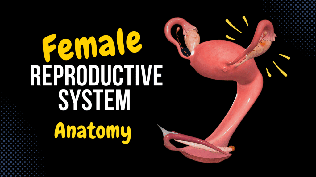
Female Genital System (Internal & External) Official Links Instagram Youtube Jki-discord Notes & Illustrations Quizzes Summary & Transcript Notes ☆ Member Only Go to PDF Notes Illustrations ☆ Member Only Go to Illustrations 12345678910 Female Genital System – QUIZ Test your understanding with 10 random multiple-choice questions from the question bank. You're in the preview mode. Note: All elements work correctly on the front end. 1 / 10 What is the purpose of the broad ligament in the female genital system? A) Facilitate uterine contractions B) Supply blood to ovaries C) Separate abdominal organs D) Support internal organs The broad ligament is a double fold of peritoneum that supports the uterus, uterine tubes, and ovaries, helping maintain their position. 2 / 10 Which lymph nodes drain the external genitalia in females? A) Para-aortic B) Lumbar C) Internal iliac D) Superficial inguinal The superficial inguinal lymph nodes primarily drain the external genitalia, including the labia majora and minora. 3 / 10 What is the function of the zona pellucida in oocyte development? A) Provides nutrients B) Supports the corpus luteum C) Facilitates sperm binding D) Produces follicular fluid The zona pellucida surrounds the oocyte and facilitates sperm binding, protecting the developing oocyte. 4 / 10 Which veins primarily drain the blood from the ovaries? A) Vaginal venous plexus B) Ovarian veins C) Uterine venous plexus D) Pudendal veins The right ovarian vein drains into the inferior vena cava, while the left ovarian vein drains into the left renal vein. 5 / 10 What is the histological structure of the vaginal epithelium? A) Stratified squamous B) Simple columnar C) Simple cuboidal D) Transitional epithelium The vaginal epithelium is composed of stratified squamous non-keratinized epithelium, which stores glycogen. 6 / 10 What is the histological lining of the uterine cavity? A) Transitional epithelium B) Simple columnar C) Simple cuboidal D) Stratified squamous The uterine cavity is lined by simple columnar epithelium that forms the endometrium. 7 / 10 What type of follicle is ready for ovulation? A) Primary follicle B) Graafian follicle C) Secondary follicle D) Primordial follicle The Graafian follicle, or tertiary follicle, contains the secondary oocyte and is ready for ovulation. 8 / 10 What are the main functions of the ovaries? A) Produce oocytes and hormones B) Transport gametes C) Regulate blood supply D) Provide structural support The ovaries are responsible for gametogenesis and the production of hormones such as estrogen and progesterone. 9 / 10 What are the structural layers of the uterine wall? A) Endometrium, myometrium, perimetrium B) Myocardium, epimysium C) Epimetrium, perimysium D) Serosa, stroma The uterine wall is composed of the endometrium (mucosa), myometrium (muscular layer), and perimetrium (serosa). 10 / 10 What are the functional layers of the endometrium during the menstrual cycle? A) Endometrial, stromal layers B) Mucosal, serosal layers C) Functional, basal layers D) Perimetrium, myometrium The functional layer of the endometrium undergoes cyclic changes and is shed during menstruation, while the basal layer regenerates it. Your score is The average score is 0% Description Complete Overview of Female Internal and External Genital Organs This video provides a detailed anatomical breakdown of the internal and external genital structures, including their histology, functions, and clinical relevance. Small Correction: At 7:06, the ovarian artery and vein run within the suspensory ligament of the ovary, not the ligament of the ovary as originally stated. 1. Internal Genital Organs Ovary Inner Structures: Tunica Albuginea (Tunica albuginea ovarii) Ovarian Cortex (Cortex ovarii) Ovarian Medulla (Medulla ovarii) Follicles External Structures: Mesovarian Border (Margo mesovaricus) Free Border (Margo liber) Tubal Extremity (Extremitas tubaria) Uterine Extremity (Extremitas uterina) Medial Surface (Facies medialis) Lateral Surface (Facies lateralis) Fixation of the Ovary: Ligament of Ovary (Ligamentum ovarii proprium) Suspensory Ligament of Ovary (Ligamentum suspensorium ovarii) Mesovarium Growth and Maturation of Follicles Primordial Follicle: Contains primary oocyte, single layer of follicular cells. Primary Follicles: After puberty (FSH stimulation), granulosa cells form. Secondary Follicle: Zona pellucida, follicular fluid, theca folliculi, estrogen production. Graafian Follicle: Mature follicle containing a secondary oocyte, released during ovulation. Corpus Luteum: Hormone-secreting structure post-ovulation. Corpus Albicans: Remnant of corpus luteum. Uterine Tube (Tuba Uterina) Parts of the Uterine Tube: Infundibulum (Infundibulum tubae uterinae) Abdominal Ostium (Ostium abdominale) Fimbriae (Fimbriae tubae uterina) Ovarian Fimbria (Fimbria ovarica) Ampulla (Ampulla tubae uterinae) Isthmus (Isthmus tubae uterinae) Uterine Part (Pars uterina) – Uterine Ostium (Ostium uterinum) Walls of Uterine Tube: Tunica Mucosa Tunica Muscularis Tunica Serosa Mesosalpinx Uterus Parts: Positioned in anteflexion. Fundus of Uterus (Fundus uteri). Body of Uterus (Corpus uteri). Intestinal (Posterior) Surface (Facies intestinalis). Vesical (Anterior) Surface. Cervix (Portio supravaginalis cervicis and vaginal part). Internal Structures (Histology): Endometrium: Functional and basal layers, simple columnar epithelium, uterine glands. Myometrium: Smooth muscle layers, vascularized middle layer. Perimetrium: Outer serous layer. Fixation of the Uterus Peritoneal Folds: Broad Ligament (Ligamentum latum uteri) Mesovarium Mesosalpinx Mesometrium Parametrial Ligaments: Cardinal Ligament (Ligamentum cardinale uteri). Vesico-uterine Ligament. Pubocervical Ligament. Uterosacral Ligaments. Recto-uterine Ligament. Round Ligament of Uterus (Ligamentum teres uteri). 2. Vagina Vaginal Orifice (Ostium Vaginae). Vestibule (Vestibulum Vaginae). Vaginal Canal (Canalis Vaginalis). Vaginal Fornices (Anterior, Posterior, and Lateral). Vaginal Wall (Histology): Mucosa – Forms transverse folds. Muscularis Layer. Clinical Relevance: Imperforate Hymen: May require medical intervention. Recto-uterine Pouch (Pouch of Douglas): Important for fluid accumulation. 3. External Genital Organs Vestibule: Lesser and Greater Vestibular Glands (Bartholin’s Glands). Labia Minora (Labia Minora Pudendi). Clitoris: Glans, Body, Crura, Prepuce. Bulb of Vestibule (Bulbus Vestibuli). Labia Majora (Labia Majora Pudendi). Mons Pubis. Sources Used: Memorix Anatomy (2nd Edition) – Hudák Radovan, Kachlík David, Volný Ondřej. Complete Anatomy by 3D4Medical. Biorender. Transcript Introduction0:03What’s up. Meditay Here. Let’s finally talk about the female genital system. Now, this is going to0:08be a longer video than usual. It’s not because the female genital system is necessarily difficult,0:14but it’s because I want to cover everything in detail so that you understand the fundamentals of the female genital system. I’ve put some timestamps on the bottom of the video,0:23so you can use that if it’s any specific topic you came here for. Alright. So the