
Fascia of the Head and Neck
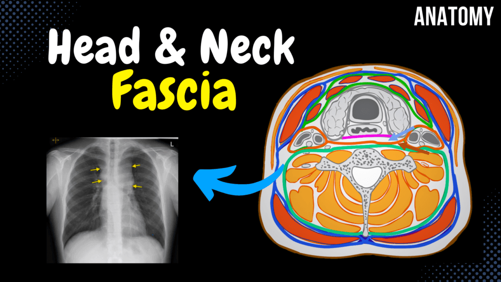
Fascia of the Head and Neck (Groups, Attachment Points, Arrangement) Official Links Instagram Youtube Jki-discord Notes & Illustrations Quizzes Summary & Transcript Notes ☆ Member Only Go to PDF Notes Illustrations ☆ Member Only Go to Illustrations 12345678910 Fascia of Head and Neck – QUIZ Test your understanding with 10 random multiple-choice questions from the question bank. You're in the preview mode. Note: All elements work correctly on the front end. 1 / 10 Which fascia covers the temporal muscle and attaches to the superior temporal line? A) Masseteric fascia B) Buccopharyngeal fascia C) Pretracheal fascia D) Temporal fascia The temporal fascia covers the temporal muscle and attaches to the superior temporal line. 2 / 10 What is the structural role of the superficial cervical fascia? A) Covers neurovascular structures B) Connects with thoracic fascia C) Separates skin and platysma D) Supports deep muscles The superficial cervical fascia separates skin and platysma from underlying structures. 3 / 10 Which cervical fascia layer encloses the infrahyoid muscles? A) Prevertebral fascia B) Temporal fascia C) Buccopharyngeal fascia D) Pretracheal fascia The pretracheal layer of cervical fascia encloses the infrahyoid muscles. 4 / 10 Where does the temporal fascia begin? A) Coronoid process B) Inferior temporal line C) Superior temporal line D) Zygomatic arch The temporal fascia begins at the superior temporal line. 5 / 10 Which muscle lies superficial to the superficial cervical fascia? A) Platysma B) Sternocleidomastoid C) Trapezius D) Omohyoid The platysma lies superficial to the superficial cervical fascia. 6 / 10 What is the function of the cervical fascia? A) Muscle elevation B) Stabilization and separation C) Oxygen exchange D) Synovial fluid transfer The cervical fascia stabilizes and separates neck muscles and organs, forming compartments. 7 / 10 Which fascia serves as a pathway for lymphatic drainage in the neck? A) Buccopharyngeal fascia B) Superficial cervical fascia C) Deep cervical fascia D) Temporal fascia The deep cervical fascia, including the pretracheal and prevertebral layers, aids in lymphatic drainage. 8 / 10 Which fascia covers the deep cervical muscles? A) Prevertebral layer B) Temporal fascia C) Masseteric fascia D) Superficial cervical fascia The prevertebral layer of cervical fascia covers the deep cervical muscles. 9 / 10 Which fascia of the neck contributes to the compartmentalization of neck structures? A) Buccopharyngeal fascia B) Pretracheal fascia C) Temporal fascia D) Masseteric fascia The pretracheal fascia contributes to the compartmentalization of the neck structures. 10 / 10 Which fascia of the neck covers the deep cervical muscles? A) Temporal fascia B) Superficial cervical fascia C) Prevertebral fascia D) Buccopharyngeal fascia The prevertebral layer of cervical fascia covers the deep cervical muscles. Your score is The average score is 0% Description This video covers the fascia of the head and neck, including its functions, types, and anatomical distribution. Fascia Functions Stabilizes and separates muscles from other internal organs Forms compartments Passage for nerves, blood vessels, and lymph Storage medium for fat and water Three Types of Fascia Superficial Fascia Deep Fascia Visceral Fascia Fascia of the Head Temporal Fascia Starts at the Superior Temporal Line Splits into two layers: Deep Layer: Lamina Profunda Superficial Layer: Lamina Superficialis Parotid Fascia Covers the Parotid Gland Masseteric Fascia Covers the Masseter Muscle Buccopharyngeal Fascia Covers the Buccinator Muscle Extends behind the pharynx Fuses with the Pterygomandibular Raphe Fascia of the Neck Cervical Fascia Superficial Layer Covers the surface of the neck Envelops the Sternocleidomastoid and Trapezius Platysma is superficial to this fascia Pretracheal Layer Envelops the Infrahyoid Muscles Forms the Carotid Sheath (surrounds the neurovascular bundle) Prevertebral Layer Covers the Deep Cervical Muscles Transcript Introduction0:03Hey what’s up. Meditay here and in this video, we’re gonna look at the main fascia covering the0:08head and neck. But first, If I would ask you, what s a fascia? And how do we categorize them, wouldWhat is a Fascia?0:14you be able to answer? Because these ae important things to understand before you actually learn0:19about the different fascia we have in the body. So a fascia is just a connective tissue0:24surrounding structures within the body. So here is a muscle, just a raw muscle within our body.0:30And here is a fascia. It surrounds the muscle. Now why do we need them?0:34Well one thing is that fascia stabilizes and separates muscles from other internal organs.0:40Fascia form compartments. Specially in clinics if you get patients with edema0:48within the compartment that the fascia forms, we’ll get the so called compartment syndrome,0:53which could be very dangerous as blood supply may get cut off due to the pressure.0:57Fascia also forms a passage for nerves, blood vessels and lymph. And this is also important1:03to keep in mind. Specially in people with chronic neck pain who are on constant pain medications. It1:09doesn’t necessarily have to be your muscle that’s still, it could also be the fascia. So massage and1:13stretching exercises are important factors which can stretch the fascia and help loosening it up.1:19Fascia also function as a storage medium for fat and water. And lastly. There are1:25three types of fasciae that you need to know. These are Superficial fascia,1:30Deep fascia, and Visceral Fascia. Ok. So here is the skin without removing1:35any layers. If you remove just the layers of the skin, you’ll see a superficial fascia,1:41located right underneath the skin. And then when you remove the superficial fascia, you’ll see1:46the deep fascia. The deep fascia is actually the that can surround individual muscles and groups of1:52muscles to separate into compartments. And when we talk about fascia within the body, it’s most1:58often the deep fascia we’re talking about. SO when you remove the deep fascia,2:03and enough muscle and bone to see an organ, we’ll see the visceral fascia,2:08that surrounds the organs within our body. Here we see the fascia covering the lungs, called pleura.2:14So that is the three types of fasciae we have And if we go back here. This fascia I showed you2:20earlier, was a deep fascia. Alright. So finally- In this video, we’re first going to look at theContent2:26fascia of the head, which consist of the Temporal
Muscles of the Neck
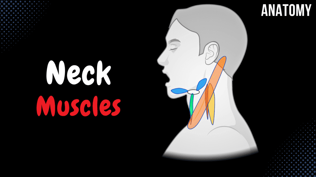
Muscles of the Neck (Groups, Origin, Insertion, Function) Official Links Instagram Youtube Jki-discord Notes & Illustrations Quizzes Summary & Transcript Notes ☆ Member Only Go to PDF Notes Illustrations ☆ Member Only Go to Illustrations 12345678910 Muscles of the Neck – QUIZ Test your understanding with 10 random multiple-choice questions from the question bank. You're in the preview mode. Note: All elements work correctly on the front end. 1 / 10 Where does the scalenus posterior muscle insert? A) Transverse processes of C6 B) First rib C) Lateral clavicle D) Second rib The scalenus posterior muscle inserts on the second rib. 2 / 10 Which muscle inserts at the basilar part of the occipital bone and originates from the atlas? A) Scalenus medius B) Rectus capitis anterior C) Longus capitis D) Rectus capitis lateralis The rectus capitis anterior muscle inserts at the basilar part of the occipital bone and originates from the lateral mass of the atlas (C1). 3 / 10 What is the function of the sternothyroid muscle? A) Elevates the thyroid cartilage B) Depresses the mandible C) Depresses the thyroid cartilage D) Rotates the thyroid cartilage The sternothyroid depresses the thyroid cartilage. 4 / 10 Which muscle has both superior and inferior bellies and originates from the scapula? A) Platysma B) Sternohyoid C) Thyrohyoid D) Omohyoid The omohyoid muscle has superior and inferior bellies, with the inferior belly originating from the scapula. 5 / 10 Where does the scalenus medius muscle originate? A) Superior surface of first rib B) First rib C) Transverse processes of C1/C2-C7 D) Lateral border of clavicle The scalenus medius originates from the transverse processes of C1/C2 to C7. 6 / 10 What is the insertion of the sternocleidomastoid muscle? A) Clavicle B) Mastoid process and superior nuchal line C) External occipital protuberance D) Manubrium of sternum The sternocleidomastoid muscle inserts on the mastoid process and superior nuchal line. 7 / 10 What is the function of the scalenus anterior muscle? A) Elevates the second rib B) Depresses the clavicle C) Elevates the first rib D) Rotates the head laterally The scalenus anterior aids in elevating the first rib during inspiration and laterally flexes the neck. 8 / 10 Where does the platysma muscle originate? A) Pectoral and deltoid fascia B) Clavicle C) Sternum D) Base of mandible The platysma muscle originates from the pectoral and deltoid fascia. 9 / 10 What is the origin of the longus capitis muscle? A) Lateral mass of atlas B) Transverse processes of C5-C7 C) Transverse processes of C1-C4 D) Transverse processes of C3-C6 The longus capitis originates from the transverse processes of C3-C6. 10 / 10 What is the insertion of the rectus capitis anterior muscle? A) Lateral part of occipital bone B) Inferior border of mandible C) Basilar part of occipital bone D) Atlas (C1) transverse process The rectus capitis anterior inserts on the basilar part of the occipital bone. Your score is The average score is 0% Description This video covers the Division of the Neck Muscles, including their origins, insertions, and classifications. Division of the Neck Muscles Deep Muscles of the Neck Lateral Muscles of the Neck Suprahyoid Muscles Infrahyoid Muscles Craniothoracal Muscles Deep Muscles of the Neck Longus Capitis (Musculus Longus Capitis) Origin: Transverse Processes of C3-C6 (Proc. transversus vertebrae cervicalis III – VI) Insertion: Basilar part of Occipital Bone (Pars basilaris ossis occipitalis) Longus Colli (Musculus Longus Colli) Superior Oblique Part Origin: Transverse Processes of C3-C5 (Proc. transversus vertebrae cervicalis III – V) Insertion: Anterior Tubercle of Atlas C1 (Tuberculum anterius atlantis) Vertical Part Origin: C5-T3 Body Insertion: C2-C4 Body Inferior Oblique Part Origin: T1-T3 Body Insertion: C5-C6 Transverse Process Rectus Capitis Anterior (Musculus Rectus Capitis Anterior) Origin: Lateral Mass of Atlas C1 (Massa lateralis atlantis) Insertion: Basilar Part of Occipital Bone (Pars basilaris ossis occipitalis) Rectus Capitis Lateralis (Musculus Rectus Capitis Lateralis) Origin: Transverse Process of Atlas C1 (Processus transversus atlantis) Insertion: Lateral Part of Occipital Bone (Pars lateralis ossis occipitalis) Lateral Muscles of the Neck Scalenus Anterior (Musculus Scalenus Anterior) Origin: Transverse Process of C3-C6 (Proc. transversus vertebrae cervicalis III – VI) Insertion: Scalenus Tubercle on 1st Rib (Tuberculum m. scaleni costa primae) Scalenus Medius (Musculus Scalenus Medius) Origin: Transverse Process of C1/C2-C7 (Proc. transversus vertebrae cervicalis I/II – VII) Insertion: Surface of 1st Rib (behind scalenus anterior) (Facies superior costae prima) Scalenus Posterior (Musculus Scalenus Posterior) Origin: Transverse Process of C5-C7 (Proc. transversus vertebrae cervicalis V – VII) Insertion: Surface of 2nd Rib (Costae II) Suprahyoid Muscles Digastricus (Musculus Digastricus) Posterior Belly: Origin: Mastoid Notch of Temporal Bone (Incisura mastoidea) Insertion: Tendon inserting at hyoid bone Anterior Belly: Origin: Tendon inserting at hyoid bone Insertion: Digastric Fossa of Mandible (Fossa digastrica) Infrahyoid Muscles Sternohyoid (Musculus Sternohyoideus) Origin: Sternum and Clavicle Insertion: Hyoid Bone Craniothoracal Muscles Sternocleidomastoid (Musculus Sternocleidomastoideus) Origin: Sternum + Clavicle Insertion: Mastoid Process + Superior Nuchal Line Trapezius (Musculus Trapezius) Superior Part Origin: Superior Nuchal Line, External Occipital Protuberance, Nuchal Ligament Insertion: Acromial End of Clavicle, Acromion of Scapula Middle Part Origin: Spinous Process of C7-T3/T4 Insertion: Spine and Acromion of Scapula Inferior Part Origin: Spinous Process of T4-T12 Insertion: Spine of Scapula Platysma Origin: Pectoral and Deltoid Fascia Insertion: Base of Mandible, Lower Lip, Skin around Mouth Transcript Introduction0:00Hey what’s up.0:04Meditay here and in this video, we’re gonna cover all the muscles of the neck.0:08Alright so The muscles of the neck can be divided into0:105 groups based on their anatomical location.0:13And keep in mind the classification of the neck muscles may vary depending on the source0:18you’re studying from, but all the muscles are the same.0:21So first are the deep muscles of the neck.0:24Those are the deepest close to the vertebra.0:26Then we have the Lateral Muscles of the neck.0:29The other two groups are related with the hyoid bone.0:32The suprahyoid muscles are located above the hyoid bone, and the infrahyoid muscles are0:36located below the hyoid bone.0:38And notice that both groups attach to the hyoid bone.0:42Then we have the cardiothoracic muscles, and they go from the cranium to
Muscles of Facial Expression
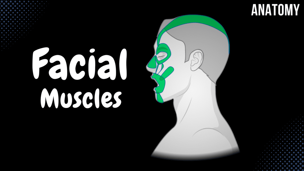
Muscles of Facial Expression (Parts, Origin, Insertion, Function) Official Links Instagram Youtube Jki-discord Notes & Illustrations Quizzes Summary & Transcript Notes ☆ Member Only Go to PDF Notes Illustrations ☆ Member Only Go to Illustrations 12345678910 Muscles of Facial Expression – QUIZ Test your understanding with 10 random multiple-choice questions from the question bank. You're in the preview mode. Note: All elements work correctly on the front end. 1 / 10 Which muscle originates from the temporal fascia and inserts into the auricle? A) Occipitofrontalis B) Auricularis posterior C) Buccinator D) Auricularis anterior The auricularis anterior muscle originates from the temporal fascia and inserts into the auricle. 2 / 10 Which muscle inserts into the auricle and originates from the temporal bone? A) Auricularis posterior B) Auricularis anterior C) Orbicularis oris D) Corrugator supercilii The auricularis posterior muscle originates from the temporal bone and inserts into the auricle. 3 / 10 What is the origin of the occipital belly of the occipitofrontalis muscle? A) Temporal fascia B) Epicranial aponeurosis C) Zygomatic arch D) Highest nuchal line The occipital belly of the occipitofrontalis originates from the highest nuchal line of the occipital bone. 4 / 10 Which muscle originates from the maxilla and inserts onto the skin of the upper lip and nasal wing? A) Zygomaticus minor B) Levator labii superioris alaeque nasi C) Buccinator D) Nasalis The levator labii superioris alaeque nasi originates from the maxilla and inserts onto the skin of the upper lip and nasal wing. 5 / 10 What is the primary function of the buccinator muscle? A) Depresses the lower lip B) Raises the eyebrows C) Compresses the cheeks D) Elevates the corners of the mouth The buccinator muscle compresses the cheeks, aiding in chewing and preventing food from collecting in the oral vestibule. 6 / 10 Which muscle elevates the upper lip and nasal wing, aiding in opening the nostrils? A) Orbicularis oris B) Levator labii superioris alaeque nasi C) Zygomaticus major D) Nasalis The levator labii superioris alaeque nasi elevates the upper lip and nasal wing, assisting in opening the nostrils. 7 / 10 What is the origin of the orbicularis oculi’s orbital part? A) Zygomatic bone B) Lacrimal bone C) Infraorbital margin of maxilla D) Medial palpebral ligament The orbital part of the orbicularis oculi originates from the medial palpebral ligament. 8 / 10 Which muscle forms the majority of the cheek structure? A) Buccinator B) Orbicularis oris C) Risorius D) Zygomaticus major The buccinator forms the majority of the cheek and assists in mastication and speech. 9 / 10 Which muscle has two parts, the transverse and alar parts, and controls the nasal opening? A) Levator labii superioris alaeque nasi B) Procerus C) Nasalis D) Zygomaticus major The nasalis muscle consists of the transverse and alar parts, functioning to control the nasal opening. 10 / 10 What is the primary function of the occipitofrontalis muscle’s occipital belly? A) Tilts the head backward B) Puckers the lips C) Retracts the scalp D) Elevates the eyebrows The occipital belly of the occipitofrontalis retracts the scalp. Your score is The average score is 0% Description This video covers the Division of Facial Muscles, their origins, insertions, and functions. Division of Facial Muscles Muscles of the Scalp Muscles around the Eye Opening Muscles around the Oral Opening Muscles of the Nasal Opening Muscles around the Ear Opening All insert into the skin All are innervated by the facial nerve All originate from the 2nd Pharyngeal Arch during development Muscles of the Scalp Occipitofrontal Muscle (Musculus Occipitofrontalis) Occipital Belly (Venter Occipitalis) Origin: Highest Nuchal Line (Linea Nuchalis Superior) Insertion: Epicranial Aponeurosis (Galea Aponeurosis) Frontal Belly (Venter Frontalis) Origin: Epicranial Aponeurosis (Galea Aponeurosis) Insertion: Skin of Eyebrows Muscles around the Eye Opening Orbicularis Oculi (Musculus Orbicularis Oculi) Orbital Part Origin/Insertion: Medial Palpebral Ligament Palpebral Part Origin: Medial Palpebral Ligament Insertion: Lateral Palpebral Ligament Lacrimal Part Origin: Lacrimal Bone Insertion: Lacrimal Sac Procerus (Musculus Procerus) Origin: Bridge of Nose Insertion: Skin of Forehead Corrugator Supercilii (Musculus Corrugator Supercilii) Origin: Glabella, Supraorbital Margin Insertion: Skin of Eyebrow Muscles around the Oral Opening Sides (“Smile”) Zygomaticus Major (Musculus Zygomaticus Major) Origin: Zygomatic Bone Zygomaticus Minor (Musculus Zygomaticus Minor) Origin: Zygomatic Bone Risorius (Musculus Risorius) Origin: Masseteric Fascia Insertion: Skin at the Angle of the Mouth Angle of the Mouth Levator Anguli Oris (Musculus Levator Anguli Oris) Origin: Anterior Surface of Maxilla Insertion: Skin at the Angle of the Mouth Levator Labii Superioris (Musculus Levator Labii Superioris) Origin: Infraorbital Margin of the Maxilla Insertion: Skin of the Upper Lip Depresses the Angle of the Mouth Depressor Anguli Oris (Musculus Depressor Anguli Oris) Depressor Labii Inferioris (Musculus Depressor Labii Inferiores) Lateral Wall of the Oral Cavity (“Satisfaction”) Buccinator (Musculus Buccinator) Origin: Alveolar Processes of Maxilla and Mandibula, Pterygomandibular Raphe Insertion: Skin at the Angle of the Mouth Lips (“Kissing Muscle”) Orbicularis Oris (Musculus Orbicularis Oris) Marginal Part Labial Part Chin (“Muscle of Doubt”) Mentalis (Musculus Mentalis) Origin: Alveolar Processes of Mandibula Insertion: Skin of the Chin Muscles of the Nasal Opening Nasalis (Musculus Nasalis) Transverse Part: Origin: Anterior Surface of Maxilla, Insertion: Dorsal Cartilage of the Nose Alar Part: Origin: Anterior Surface of Maxilla, Insertion: Dorsal Cartilage of the Nose Levator Labii Superioris Alaeque Nasi (Musculus Levator Labii Superioris Alaeque Nasi) Origin: Frontal Process of Maxilla Insertion: Skin of the Upper Lip, Skin of the Nasal Wing Muscles around the Ear Opening Extrinsic Muscles of the Ear Auricularis Anterior: Origin: Temporal Fascia, Insertion: Auricle Auricularis Superior: Origin: Epicranial Aponeurosis, Insertion: Auricle Auricularis Posterior: Origin: Temporal Bone, Insertion: Auricle Transcript Introduction0:03What’s up. Meditay here and in this video, we’re gonna cover all the muscles0:07of facial expression. Which are a part of the muscles of the head. Alright so0:12All muscles of the head are divided into two groups. The first group is the muscles0:16of mastication. Mastication means to chew, so those are the muscles responsible for chewing0:21when you’re eating. And the second group are gonna be fascial muscles or the muscles that0:26are gonna be
Muscles of Mastication
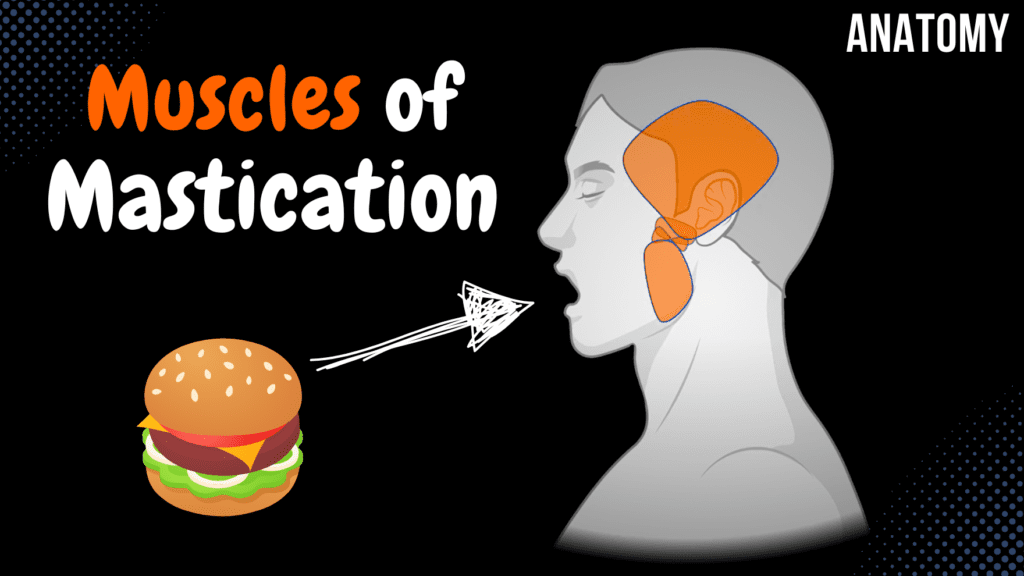
Muscles of Mastication (Origin, Insertion, Function) Official Links Instagram Youtube Jki-discord Notes & Illustrations Quizzes Summary & Transcript Notes ☆ Members Only Go to PDF Notes Illustrations ☆ Members Only Go to Illustrations 12345678910 Muscles of Mastication – QUIZ Test your understanding with 10 random multiple-choice questions from the question bank. You're in the preview mode. Note: All elements work correctly on the front end. 1 / 10 What structure forms the origin of the deep part of the masseter muscle? A) Deep surface of zygomatic arch B) Temporal fossa C) Lateral plate of pterygoid process D) Infratemporal crest The deep part of the masseter muscle originates from the deep surface of the zygomatic arch. 2 / 10 What is the insertion point of the temporal muscle? A) Coronoid process B) Masseteric tuberosity C) Mandibular angle D) Pterygoid fovea The temporal muscle inserts onto the coronoid process of the mandible. 3 / 10 Which muscle originates from the maxillary tuberosity and pterygoid fossa of the sphenoid bone? A) Medial pterygoid B) Lateral pterygoid C) Masseter D) Temporal muscle The medial pterygoid muscle originates from the maxillary tuberosity (superficial head) and the pterygoid fossa (deep head). 4 / 10 Which muscle has its origin on the temporal fossa and inserts onto the coronoid process of the mandible? A) Temporal muscle B) Lateral pterygoid C) Masseter D) Medial pterygoid The temporal muscle originates from the temporal fossa and inserts onto the coronoid process of the mandible. 5 / 10 What is the insertion of the masseter muscle? A) Mandibular angle B) Masseteric tuberosity C) Pterygoid fovea D) Coronoid process The masseter inserts into the masseteric tuberosity of the mandible. 6 / 10 What is the function of the medial pterygoid muscle during bilateral contraction? A) Retracts the mandible B) Elevates the mandible C) Depresses the mandible D) Protracts the mandible The medial pterygoid muscle elevates the mandible during bilateral contraction. 7 / 10 Which muscle originates from the zygomatic arch and inserts into the masseteric tuberosity? A) Masseter B) Medial pterygoid C) Temporal muscle D) Lateral pterygoid The masseter muscle originates from the zygomatic arch and inserts into the masseteric tuberosity. 8 / 10 Which muscle inserts into the pterygoid fovea on the neck of the mandible? A) Masseter B) Medial pterygoid C) Temporal muscle D) Lateral pterygoid The lateral pterygoid muscle inserts into the pterygoid fovea on the mandible’s neck. 9 / 10 Which nerve innervates all the muscles of mastication? A) Glossopharyngeal nerve (CN IX) B) Mandibular nerve (CN V3) C) Facial nerve (CN VII) D) Hypoglossal nerve (CN XII) All muscles of mastication are innervated by the mandibular nerve, a branch of the trigeminal nerve (CN V3). 10 / 10 What is the insertion point of the lateral pterygoid muscle? A) Mandibular angle B) Coronoid process C) Pterygoid fovea D) Masseteric tuberosity The lateral pterygoid muscle inserts into the pterygoid fovea of the mandible. Your score is The average score is 0% Description In this video, I go through the Muscles of Mastication, covering their origin, insertion, and function. Understanding these muscles is essential for comprehending jaw movements and chewing mechanics. Muscles of Mastication Temporal Muscle (Musculus Temporalis) Origin: Temporal fossa Parietal bone – Inferior temporal line Insertion: Coronoid process of the mandible (Processus coronoideus mandibulae) Function: Elevates mandible (Anterior fibers) Retracts mandible (Posterior fibers) Masseter Muscle (Musculus Masseter) Origin: Zygomatic Arch (Arcus zygomaticus) Insertion: Masseteric tuberosity of mandible (Tuberositas masseterica mandibulae) Function: Elevates mandible Medial Pterygoid Muscle (Musculus Pterygoideus Medialis) Origin: Superficial Part (Pars superficialis): Maxillary tuberosity (Tuberositas maxillae) Deep Part (Pars profunda): Pterygoid fossa of pterygoid process (Fossa pterygoidea ossis sphenoidalis) Function: Elevates mandible (Bilateral contraction) Frictional masticatory movement (Unilateral contraction) Lateral Pterygoid Muscle (Musculus Pterygoideus Lateralis) Origin: Superior Head (Caput superius): Infratemporal surface + Sphenoid Bone (Greater Wing) (Facies infratemporalis + alae majoris ossis sphenoidalis) Inferior Head (Caput inferius): Lateral lamina of pterygoid process (Sphenoidal bone) (Lamina lateralis processus pterygoidei) Insertion: Pterygoid fovea of the mandible (Fovea pterygoidea mandibulae) Function: Pushes mandible forward (Bilateral contraction) Frictional masticatory movement (Unilateral contraction) Transcript Introduction0:03Hey, what’s up. Meditay here and this.. is my first video of the muscular anatomy series.0:08So in this segment, we’re gonna cover all muscles of mastication. Which are a part0:13of the muscles of the head. Alright so All muscles of the head are divided into0:18two groups. The first group is the muscles of mastication. Mastication means to chew,0:23so those are the muscles responsible for chewing when you’re eating. And the second group is gonna0:28be fascial muscles or the muscles that are gonna be responsible for facial expression. So0:33we’re gonna focus on the mastication muscles here. So in this video, we’re gonna cover the origin and0:38insertion points of the 4 muscles of mastication, which are the Temporal, Masseter, Medial0:44Pterygoid, and Lateral Pterygoid muscles. And then in the next video, we’ll cover the facial muscles.0:50Alright. Before we start, I want you to have some basic understandingParts of Skeletal Muscle0:55of what makes up the different parts of a muscle. All the muscles in our body consist of an Origin1:01point, which is the part of the muscle that’s attached to a bone that does not move or move1:06a very slightly bit during contraction. It has a head, which is the proximal part of the muscle,1:11there’s a belly which is the widest part of the muscle. Then there’s a tail and an Insertion1:17point, which is the part of the muscle that’s attached to a bone that moves during contraction.1:22When you’re studying muscles in general, the origin and insertion points are what we1:27usually focus on. And again keep in mind that the origin point is the least movable part,1:32and the insertion point is the part of the bone that is moved during contraction.1:37In some locations you’ll also see the word belly, usually if the muscle is divided into two parts,1:42that’s when we mention this term. Awesome. Let’s now cover the muscles of mastication.Muscles of Mastication1:47The muscles of mastication again, consist of the temporal muscle. Masseter muscle, Medial Pterygoid1:53muscle, and Lateral