
Anatomy of the Heart – Layers & Conducting System
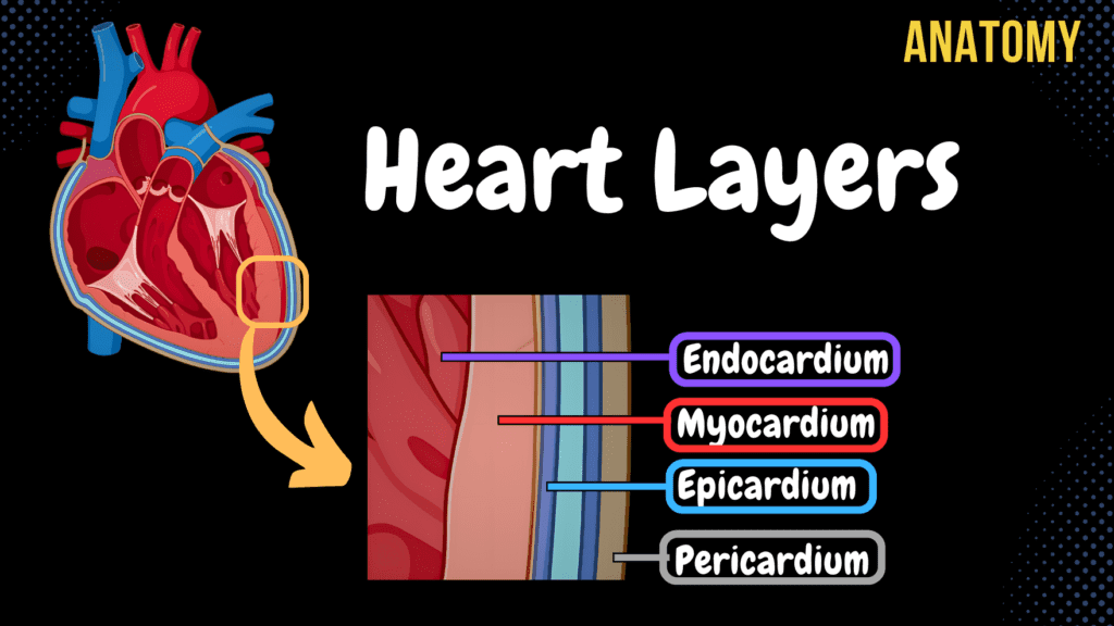
Anatomy of the Heart – Layers, Conducting System & Topography Official Links Instagram Youtube Jki-discord Notes & Illustrations Quizzes Summary & Transcript Notes ☆ Member Only Go to PDF Notes Illustrations ☆ Member Only Go to Illustrations 12345678910 Heart Anatomy (Layers) – QUIZ Test your understanding with 10 random multiple-choice questions from the question bank. You're in the preview mode. Note: All elements work correctly on the front end. 1 / 10 What structure lies laterally to the heart according to its syntopy? A) Esophagus B) Great blood vessels C) Diaphragm D) Pleura The pleura lies laterally to the heart according to its syntopy. 2 / 10 Which structure is located at the medial wall of the right atrium? A) Valve of coronary sinus B) Crista terminalis C) Fossa ovalis D) Interatrial septum The interatrial septum is located at the medial wall of the right atrium. 3 / 10 Which part of the myocardium contains longitudinal muscle fibers? A) Endocardial layer B) Epicardial layer C) Superficial and deep layers D) Middle layer The superficial and deep layers of the myocardium contain longitudinal muscle fibers. 4 / 10 Where is the mitral valve auscultated? A) Fifth intercostal space, right of sternum B) Fifth intercostal space, midclavicular line C) Fourth intercostal space, left of sternum D) Second intercostal space, right of sternum The mitral valve is auscultated at the fifth intercostal space at the midclavicular line. 5 / 10 What is the innermost layer of the heart? A) Endocardium B) Pericardium C) Epicardium D) Myocardium The endocardium is the innermost layer of the heart. 6 / 10 What is the visceral lamina of the serous pericardium also called? A) Parietal lamina B) Epicardium C) Myocardium D) Endocardium The visceral lamina of the serous pericardium is also called the epicardium. 7 / 10 What is the function of the fibrous pericardium? A) Aids in atrial contraction B) Facilitates electrical conduction C) Provides structural support D) Produces heart sounds The fibrous pericardium provides structural support and protects the heart. 8 / 10 Which pericardial sinus is located posterior to the ascending aorta and pulmonary trunk? A) Aortic sinus B) Transverse pericardial sinus C) Fibrous pericardial sinus D) Oblique pericardial sinus The transverse pericardial sinus is located posterior to the ascending aorta and pulmonary trunk. 9 / 10 What structure connects the myocardium and epicardium? A) Serous pericardium B) Fibrous pericardium C) Endocardium D) Chordae tendineae The serous pericardium connects the myocardium and the epicardium. 10 / 10 Which pericardial layer allows frictionless movement of the heart? A) Myocardium B) Fibrous pericardium C) Endocardium D) Serous pericardium The serous pericardium, with its visceral and parietal layers, allows frictionless movement of the heart. Your score is The average score is 0% Description This video covers the layers of the heart, its conducting system, and topography, including its holotopy, skeletopy, and syntopy. Understanding these aspects is crucial for medical students and professionals studying cardiovascular anatomy. Layers of the Heart: Serous Pericardium Fibrous Pericardium Endocardium Myocardium Epicardium Endocardium: Forms the cusps of the following valves: Bicuspid Valve Tricuspid Valve Aortic Valve Pulmonary Valve Valve of the Inferior Vena Cava (Vulva Vena Cava Inferioris) Valve for the Coronary Sinus (Vulva Sinus Coronarii) Myocardium: Forms fibrous rings and muscle layers: Myocardium of the Atria: Superficial Circular Muscle Fibers Deep Longitudinal Muscle Fibers Form Pectinate Muscle (Musculi Pectinati) Myocardium of the Ventricles: Deep Layer: Longitudinal Muscle Fibers Middle Layer: Circular Muscle Fibers Superficial Layer: Longitudinal Muscle Fibers Deep and Superficial layers form the Vortex of the Heart (Vortex Cordis) Left ventricle is thicker than the right ventricle Forms Trabeculae Carneae and Papillary Muscles (Musculi Papillares) Epicardium: It is the visceral lamina of the serous pericardium. Pericardium: Serous Pericardium (Pericardium Serosum) Visceral Lamina (Epicardium) Parietal Lamina Fibrous Pericardium (Pericardium Fibrosum) Pericardial Cavity (Cavitas Pericardiaca) Transverse Pericardial Sinus (Sinus Transversus Pericardii) Oblique Pericardial Sinus (Sinus Obliquus Pericardii) Conducting System of the Heart: Sinoatrial Node (SA Node) Atrioventricular Node (AV Node) Bundle of His Left Bundle Branch Right Bundle Branch Purkinje Fibers Topography of the Heart: Holotopy of the Heart: The heart lies in the mediastinum medium Skeletopy of the Heart: Superior Border: 3rd rib horizontally Right Border: Parallel to sternal margin (Linea Parasternalis) Lower Border: From the cartilage of the 5th rib to the 5th intercostal space Left Border: 5th intercostal space to the level of the 4th rib Openings of the Heart: Atrioventricular Openings: From the 3rd rib to the 6th sternal junction Aortic and Pulmonary Openings: From the 3rd sternal junction to the 4th sternal junction Topography of the Valves of the Heart: Aortic and Pulmonary Valve: Located at the 2nd intercostal space Tricuspid and Bicuspid (Mitral) Valve: Located at the 5th intercostal space Syntopy of the Heart: Anteriorly: Behind the sternum Posteriorly: Esophagus Laterally: Pleura Inferiorly: Diaphragm Superiorly: Great blood vessels Sources Used in This Video: Memorix Anatomy 2nd Edition by Hudák Radovan, Kachlík David, Volný Ondřej Biorender University Notes and Lectures Transcript Introduction0:03What’s up.0:04Meditay here.0:05Let’s talk about the heart again.0:07In the last video, we covered the circulation system and the general anatomy of the heart.0:12Now In this video, We’re going to cover the Layers of the heart, which include the Endocardium,0:16Myocardium, and Epicardium.0:19Then we’ll go through the conducting system.0:21And after that, we’re going to look at the general topography of the heart, which will0:25help you from a clinical perspective.0:27Let’s go through all of these starting with the layers of the heart.Layers of the Heart0:31So here you see a raw picture of the heart without any type of coverings.0:35In real life, the heart is covered by a wet surface called serous pericardium.0:40And then by another layer of dense connective tissue called fibrous pericardium.0:44We’ll now start by cut the heart like this, so see all the layers.0:48Then we’ll take a small segment, and zoom in.0:52Now we’re able to see all the layers of the heart, which include the endocardium,0:56myocardium and the Epicardium.0:59These three layers are what is considered a part of the actual heart.1:02The Serous and Fibrous pericardium we saw
Anatomy of the Heart – External & Internal Structures
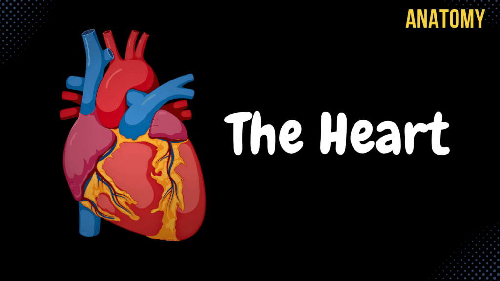
Anatomy of the Heart – External & Internal Structures Official Links Instagram Youtube Jki-discord Notes & Illustrations Quizzes Summary & Transcript Notes ☆ Member Only Go to PDF Notes Illustrations ☆ Member Only Go to Illustrations 12345678910 Heart Anatomy (Structures) – QUIZ Test your understanding with 10 random multiple-choice questions from the question bank. You're in the preview mode. Note: All elements work correctly on the front end. 1 / 10 What is the main function of the papillary muscles? A) Conduct electrical impulses B) Pump blood into the atria C) Open the heart valves D) Prevent valve prolapse Papillary muscles contract to tighten chordae tendineae, preventing valve prolapse during ventricular contraction. 2 / 10 Which groove is associated with the anterior interventricular branch? A) Posterior interventricular sulcus B) Coronary sulcus C) Sulcus terminalis D) Anterior interventricular sulcus The anterior interventricular sulcus contains the anterior interventricular artery and great cardiac vein. 3 / 10 Which structure carries impulses from the atrioventricular node to the ventricles? A) Internodal pathways B) SA node C) Bundle of His D) Purkinje fibers The Bundle of His transmits impulses from the AV node to the bundle branches, ensuring ventricular contraction. 4 / 10 What is the function of the nodules in the semilunar valves? A) Allow backflow of blood B) Assist chordae tendineae C) Facilitate blood ejection D) Ensure valve closure The nodules ensure complete closure of the semilunar valves by sealing the cusps. 5 / 10 What is the function of the chordae tendineae? A) Open the semilunar valves B) Facilitate atrial contraction C) Conduct electrical impulses D) Prevent valve prolapse The chordae tendineae anchor the atrioventricular valve cusps to papillary muscles, preventing valve prolapse. 6 / 10 Which arteries arise from the left coronary artery? A) Right coronary artery B) Circumflex and anterior interventricular C) Left marginal and diagonal arteries D) Posterior interventricular artery The left coronary artery gives rise to the circumflex artery and anterior interventricular artery. 7 / 10 What is the primary function of the pulmonary valve? A) Prevents ventricular inversion B) Prevents backflow into the ventricle C) Directs blood to coronary arteries D) Supplies oxygenated blood The pulmonary valve prevents backflow of blood into the right ventricle from the pulmonary trunk. 8 / 10 What is the function of the left auricle? A) Facilitates blood oxygenation B) Pumps blood into ventricles C) Increases atrial capacity D) Prevents valve regurgitation The left auricle increases the capacity of the left atrium and assists in blood collection. 9 / 10 What is the name of the opening between the right atrium and right ventricle? A) Pulmonary trunk B) Ostium of coronary sinus C) Right atrioventricular opening D) Left atrioventricular opening The right atrioventricular opening allows blood to flow from the right atrium to the right ventricle. 10 / 10 Which structure separates the right and left ventricles internally? A) Interatrial septum B) Crista terminalis C) Sulcus terminalis D) Interventricular septum The interventricular septum divides the right and left ventricles and consists of muscular and membranous parts. Your score is The average score is 0% Description This video covers the blood circulation in the body and the anatomy of the heart, including its external and internal structures. Understanding the pathway of blood through the heart and its anatomical components is essential for mastering cardiovascular physiology and anatomy. Blood Circulation in the Body: Pulmonary Circulation (Circulus Sanguid Minor): Deoxygenated blood enters the right atrium through the superior and inferior vena cava. Blood flows through the tricuspid valve into the right ventricle. The right ventricle pumps blood through the pulmonary valve into the pulmonary artery. The pulmonary artery carries blood to the lungs for oxygenation. Systemic Circulation (Circulus Sanguis Major): Oxygenated blood returns from the lungs into the left atrium. The left atrium sends blood through the bicuspid (mitral) valve into the left ventricle. The left ventricle pumps blood through the aortic valve into the aorta. The aorta distributes oxygenated blood throughout the body. External Structures of the Heart: Apex of the Heart (Apex Cordis) Base of the Heart (Basis Cordis) Pulmonary Surface (Facies Pulmonalis) Sternocostal Surface (Facies Sternocostalis) Diaphragmatic Surface (Facies Diaphragmatica) Right Border / Right Margin of the Heart (Margo Dexter Cordis) Coronary Sulcus (Sulcus Coronarius) Anterior Interventricular Sulcus (Sulcus Interventricularis Anterior) Posterior Interventricular Sulcus (Sulcus Interventriculare Posterior) Internal Structures of the Heart: Septum of the Heart (Septum Cordis): Interventricular Septum (Septum Interventriculare) Muscular Part (Pars Muscularis) Membranous Part (Pars Membranacea) Interatrial Septum (Septum Interatriale) Right Atrium: Anterior Wall: Right Auricle (Auricula Dextra) Superior Wall: Opening of Superior Vena Cava (Ostium Vena Cava Superioris) Posterior Wall: Opening of Inferior Vena Cava (Ostium Vena Cava Inferiores) Sinus of Vena Cava (Sinus Venarum) Terminal Crest (Crista Terminalis) Medial Wall: Interatrial Septum, Oval Fossa (Fossa Ovalis) Inferior Wall: Right Atrioventricular Opening (Ostium Atrioventriculare Dextrum), Tricuspid Valve Right Ventricle: Trabeculae Carneae Papillary Muscles (Musculi Papillares) Tricuspid Valve (Anterior, Posterior, and Septal Cusps) Pulmonary Valve Left Atrium: Anterior Wall: Left Auricle (Auricula Sinister) Posterior Wall: Pulmonary Veins and Openings of Pulmonary Veins (Ostia Venarum Pulmonalium) Medial Wall: Interatrial Septum Left Ventricle: Bicuspid (Mitral) Valve (Anterior, Posterior, and Commissural Cusps) Aortic Valve (Vulva Aortae) Trabeculae Carneae Papillary Muscles and Tendinous Chords Sources Used in This Video: Memorix Anatomy 2nd Edition by Hudák Radovan, Kachlík David, Volný Ondřej Complete Anatomy by 3D4Medical Biorender University Notes and Lectures Transcript Introduction0:03Hey, What’s up. Meditay here. Let’s talk about the anatomy of the heart. In this video,0:08We’re first going to look at how the blood circulates in the body.0:11After that, we’re going to cover the different external structures you’ll find on the surface0:16of the heart. Then we’re going to open up the heart and go through the internal structures of0:20each chamber, which include the right and left atrium and the right and left ventricles.0:25Then in the next video, we’ll talk about the layers of the heart’s wall,0:29the conducting system, and then the topography. Now, let’s start with the circulation.Blood Circulation System0:33So here is see the anterior view of the thorax, right?0:37If you