
Facial Nerve (CN VII)
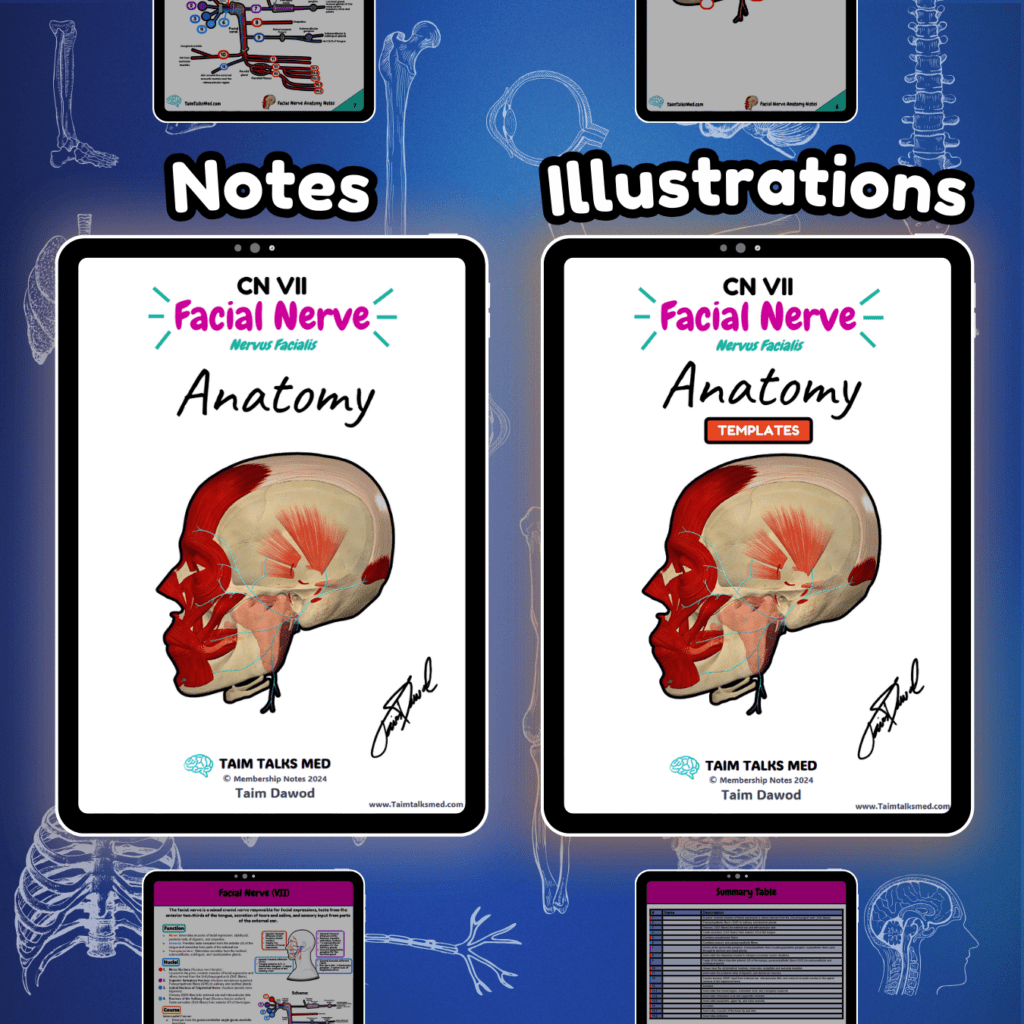
Facial Nerve (CN VII) Neurology Cranial Nerves Notes 📅 Last Updated: 01.01.2025 🔄 Version: 1.0 This PDF covers the facial nerve (CN VII) and its motor, sensory, and autonomic functions. The pathway is detailed but visually simplified, showing its brainstem origin, course through the skull, and branches to facial muscles and glands. A table at the end summarizes the pathway and nerves. Become a Member Buy this bundle Go To Notes (Preview) Go to Illustrations (Preview) Watch The Lecture Go To Notes Go to Illustrations Watch The Lecture Need it offline? Open PDF → Click the three-dot menu → Download PDF. 📖 Sources: Singh, I. (2017). Human Neuroanatomy (10th ed.). Kozlowski, T. (2017). Memorix Anatomy: The Complete Study Guide (2nd ed.). Thieme Medical Publishers. Helwany M, Bordoni B. Neuroanatomy, Cranial Nerve 7 (Facial). In: StatPearls [Internet]. Treasure Island (FL): StatPearls Publishing; 2022.
Abducens Nerve (CN VI)
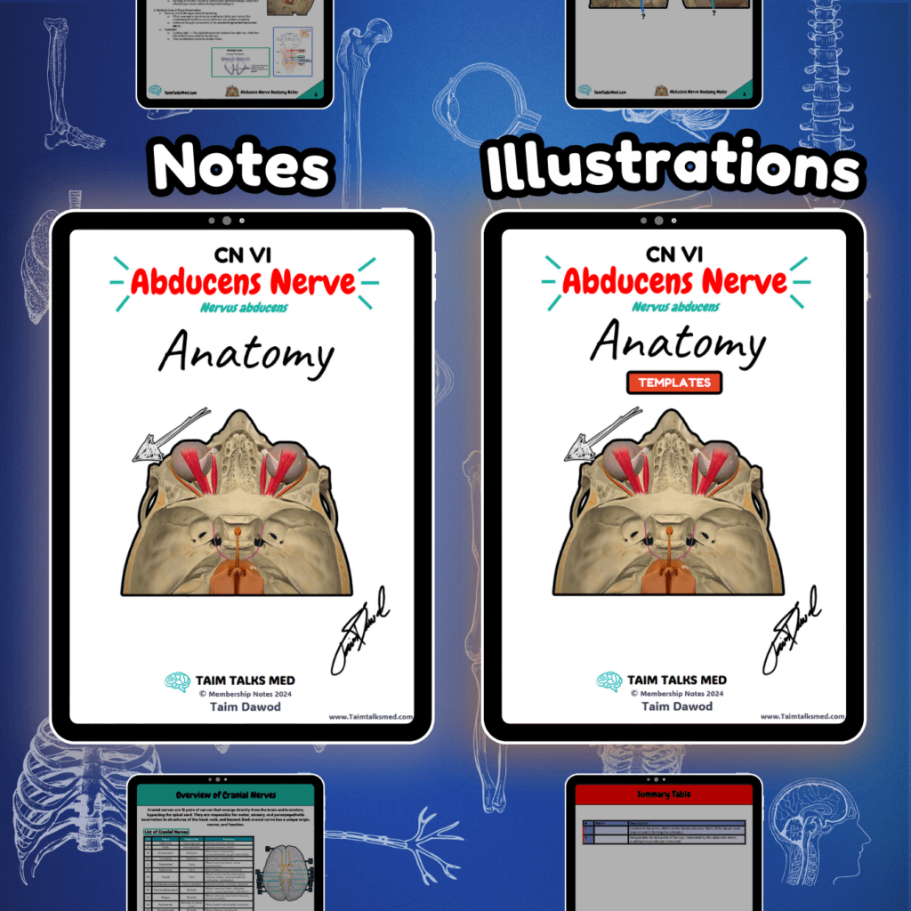
Abducens Nerve (CN VI) Neurology Cranial Nerves Notes 📅 Last Updated: 01.01.2025 🔄 Version: 1.0 This PDF covers the abducens nerve (CN VI) and its role in eye abduction by innervating the lateral rectus muscle. The pathway is detailed but visually simplified, showing its brainstem origin, course through the cavernous sinus, and entry into the orbit. A table at the end summarizes the pathway and nerves. Become a Member Buy this bundle Go To Notes (Preview) Go to Illustrations (Preview) Watch The Lecture Go To Notes Go to Illustrations Watch The Lecture Download the PDFs: [download_button url=”https://taimtalksmed.com/wp-content/uploads/2025/03/Abduncens-Nerve.zip”] Available downloads: (Your limit renews automatically each billing cycle based on your subscription plan). 📖 Sources: Singh, I. (2017). Human Neuroanatomy (10th ed.). Kozlowski, T. (2017). Memorix Anatomy: The Complete Study Guide (2nd ed.). Thieme Medical Publishers. Helwany M, Bordoni B. Neuroanatomy, Cranial Nerve 6 (Abducens) [Updated 2022 Aug 8]. In: StatPearls [Internet]. Treasure Island (FL): StatPearls Publishing; 2022 Jan-.
Trigeminal Nerve (CN V)
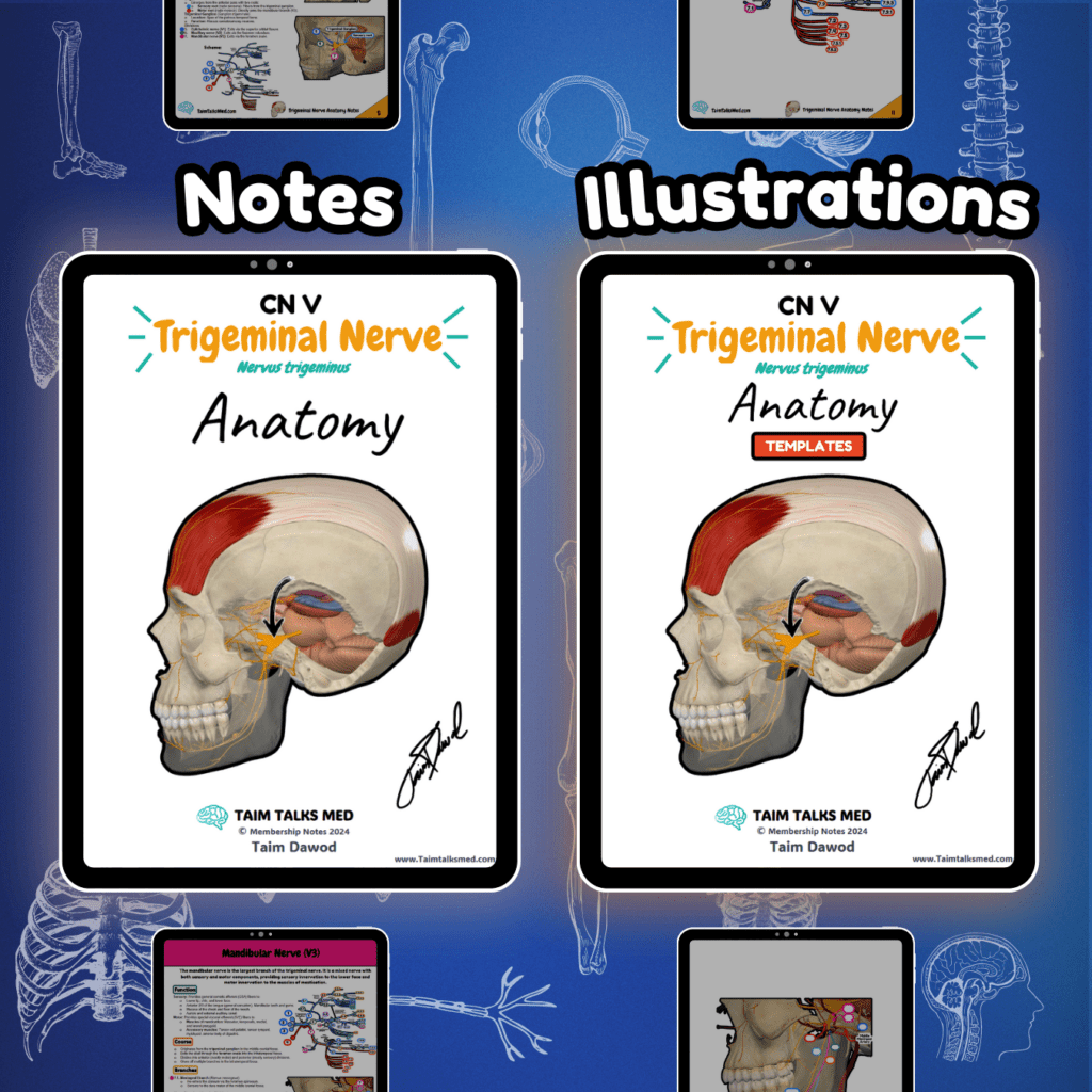
Trigeminal Nerve (CN V) Neurology Cranial Nerves Notes 📅 Last Updated: 01.01.2025 🔄 Version: 1.0 This PDF covers the trigeminal nerve (CN V) and its three main branches (V1, V2, V3). The pathway is detailed but visually simplified, showing sensory and motor functions, nuclei, and clinical conditions. A table at the end summarizes structures and innervation. Become a Member Buy this bundle Go To Notes (Preview) Go to Illustrations (Preview) Watch The Lecture Go To Notes Go to Illustrations Watch The Lecture Need it offline? Open PDF → Click the three-dot menu → Download PDF. 📖 Sources: Singh, I. (2017). Human Neuroanatomy (10th ed.). Kozlowski, T. (2017). Memorix Anatomy: The Complete Study Guide (2nd ed.). Thieme Medical Publishers. Helwany M, Bordoni B. Neuroanatomy, Cranial Nerve 5 (Trigeminal) [Updated 2022 Aug 8]. In: StatPearls [Internet]. Treasure Island (FL): StatPearls Publishing; 2022
Trochlear Nerve (CN IV)
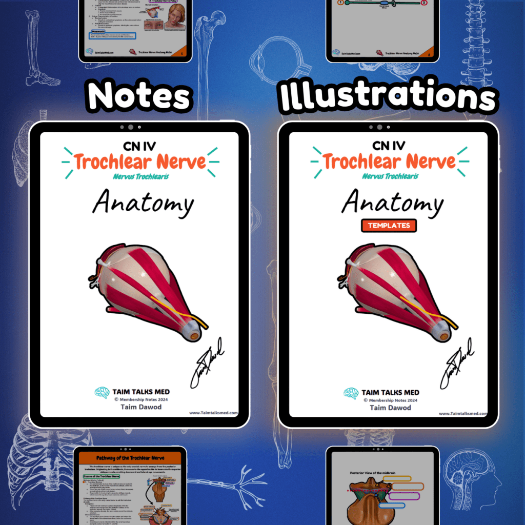
Trochlear Nerve (CN IV) Neurology Cranial Nerves Notes 📅 Last Updated: 01.01.2025 🔄 Version: 1.0 This PDF covers the trochlear nerve (CN IV) and its role in innervating the superior oblique muscle. The pathway is detailed but visually simplified, showing its midbrain origin, contralateral crossing, and orbital course. A table at the end summarizes the structures and functions. Become a Member Buy this bundle Go To Notes (Preview) Go to Illustrations (Preview) Watch The Lecture Go To Notes Go to Illustrations Watch The Lecture Need it offline? Open PDF → Click the three-dot menu → Download PDF. 📖 Sources: Singh, I. (2017). Human Neuroanatomy (10th ed.). Kozlowski, T. (2017). Memorix Anatomy: The Complete Study Guide (2nd ed.). Thieme Medical Publishers. Helwany M, Bordoni B. Neuroanatomy, Cranial Nerve 4 (Trochlear) [Updated 2022 Aug 8]. In: StatPearls [Internet]. Treasure Island (FL): StatPearls Publishing; 2022 Jan-. Visuals: Complete Anatomy, Biorender, PowerPoint, Camtasia 2021
Oculomotor Nerve (CN III)
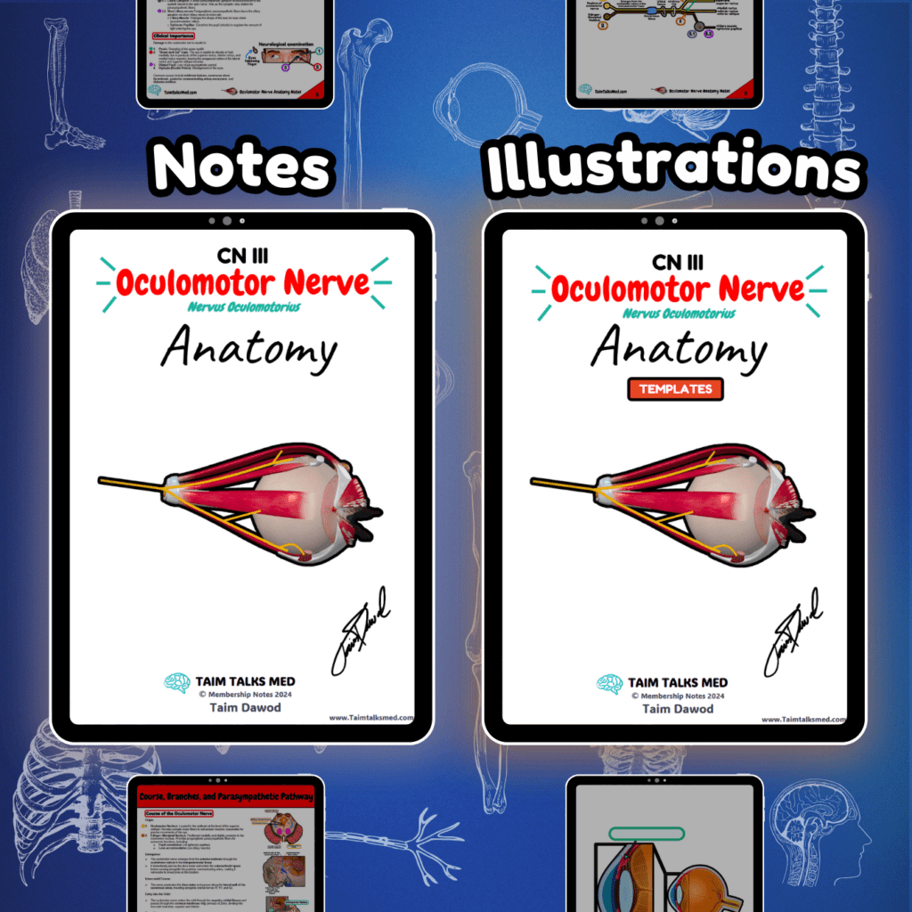
Oculomotor Nerve (CN III) Neurology Cranial Nerves Notes 📅 Last Updated: 01.01.2025 🔄 Version: 1.0 This PDF covers the oculomotor nerve (CN III) and its role in eye movement, eyelid elevation, and pupil constriction. The pathway is detailed but visually simplified, with diagrams of the midbrain, nerve course, extraocular muscles, and autonomic fibers. A table at the end summarizes key structures and clinical conditions. Become a Member Buy this bundle Go To Notes (Preview) Go to Illustrations (Preview) Watch The Lecture Go To Notes Go to Illustrations Watch The Lecture Need it offline? Open PDF → Click the three-dot menu → Download PDF. 📖 Sources: Singh, I. (2017). Human Neuroanatomy (10th ed.). Kozlowski, T. (2017). Memorix Anatomy: The Complete Study Guide (2nd ed.). Thieme Medical Publishers. Helwany M, Bordoni B. Neuroanatomy, Cranial Nerve 3 (Oculomotor) [Updated 2022 Aug 8]. In: StatPearls [Internet]. Treasure Island (FL): StatPearls Publishing; 2022 Jan-.
Optic Nerve (CN II)
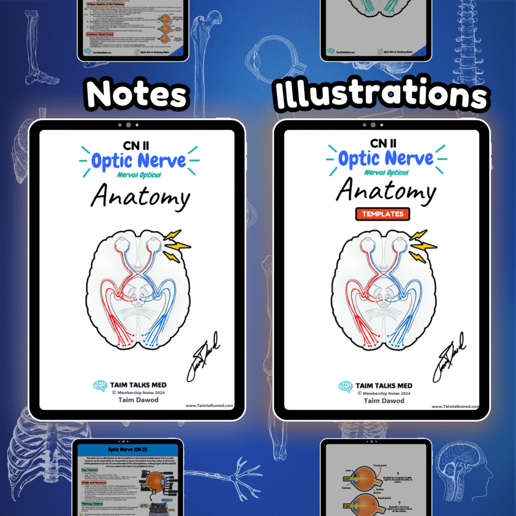
Optic Nerve (CN II) Neurology Cranial Nerves Notes 📅 Last Updated: 01.01.2025 🔄 Version: 1.0 This PDF covers the optic nerve (CN II) and provides an overview of how visual information is transmitted from the retina to the brain. Each part of the visual pathway is depicted in simplified diagrams, including the optic nerve, optic chiasma, optic tract, and optic radiations, with supporting diagrams explaining the neuronal order, visual fields, and functional connections. Topics include the course of the visual pathway, collateral reflex pathways, and clinical conditions such as hemianopia, quadrantanopia, and cortical blindness, supported by a stepwise breakdown of the entire pathway. Become a Member Buy this bundle Go To Notes (Preview) Go to Illustrations (Preview) Watch The Lecture Go To Notes Go to Illustrations Watch The Lecture Need it offline? Open PDF → Click the three-dot menu → Download PDF. 📖 Sources: Singh, I. (2017). Human Neuroanatomy (10th ed.). Kozlowski, T. (2017). Memorix Anatomy: The Complete Study Guide (2nd ed.). Thieme Medical Publishers. Helwany M, Bordoni B. Neuroanatomy, Cranial Nerve 2 (Optic) [Updated 2022 Aug 8]. In: StatPearls [Internet]. Treasure Island (FL): StatPearls Publishing; 2022.
Thoracolumbar Fascia
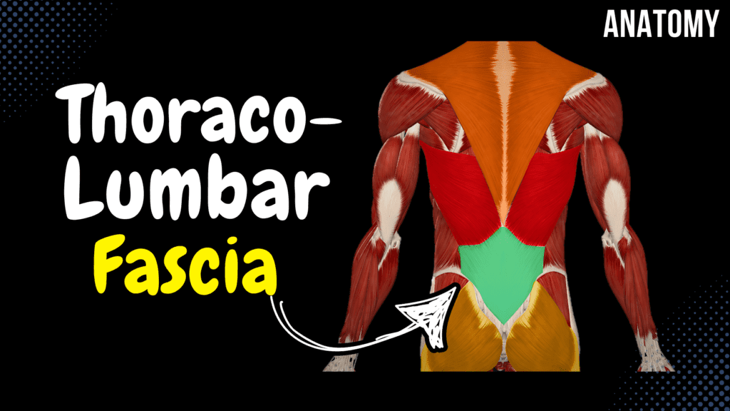
Thoracolumbar Fascia Official Links Instagram Youtube Jki-discord Notes & Illustrations Quizzes Summary & Transcript Notes ☆ Member Only Go to PDF Notes Illustrations ☆ Member Only Go to Illustrations 12345678910 Thoracolumbar Fascia – QUIZ Test your understanding with 10 random multiple-choice questions from the question bank. You're in the preview mode. Note: All elements work correctly on the front end. 1 / 10 Which muscle group does the posterior layer of the thoracolumbar fascia provide attachment for? A) Erector spinae B) Abdominal muscles C) Psoas major D) Transversospinal muscles The erector spinae group is attached to the posterior layer. 2 / 10 How many layers does the thoracolumbar fascia have? A) One B) Two C) Four D) Three The thoracolumbar fascia is divided into three layers: anterior, middle, and posterior. 3 / 10 Which fascia layer is directly involved in stabilizing the lumbar region during heavy lifting? A) Anterior layer B) Middle layer C) Deep layer D) Posterior layer The posterior layer plays a key role in stabilization during heavy lifting. 4 / 10 What is the anatomical relationship of the quadratus lumborum to the thoracolumbar fascia? A) Enclosed by the middle layer B) Unrelated to the thoracolumbar fascia C) Attached to the anterior layer D) Covered by the posterior layer The quadratus lumborum is enclosed by the middle layer. 5 / 10 Which muscles are supported by the posterior layer of the thoracolumbar fascia? A) Rectus abdominis B) Psoas major C) Erector spinae muscles D) Abdominal wall muscles The erector spinae muscles are supported by the posterior layer. 6 / 10 Which nerves pass close to the thoracolumbar fascia? A) Thoracic and cervical nerves B) Sciatic and pudendal nerves C) Iliohypogastric and ilioinguinal nerves D) Femoral and obturator nerves The iliohypogastric and ilioinguinal nerves pass close to the fascia. 7 / 10 What anatomical structure is supported by the thoracolumbar fascia during rotation? A) Lumbar vertebrae B) Sacrum C) Thoracic spine D) Pelvis The lumbar vertebrae are supported during rotation. 8 / 10 Which layer of the thoracolumbar fascia assists in lateral flexion of the spine? A) Anterior layer B) Superficial layer C) Posterior layer D) Middle layer The middle layer assists in lateral flexion. 9 / 10 Which nerve runs close to the thoracolumbar fascia in the lumbar region? A) Iliohypogastric nerve B) Subcostal nerve C) Genitofemoral nerve D) Femoral nerve The iliohypogastric nerve runs close to the thoracolumbar fascia. 10 / 10 Which muscle contributes to the formation of the posterior layer of the thoracolumbar fascia? A) Quadratus lumborum B) Transverse abdominis C) Psoas major D) Latissimus dorsi The latissimus dorsi contributes to the posterior layer. Your score is The average score is 0% Description This video is about the Thoracolumbar Fascia (Fascia Thoracolumbalis), its structure, and the muscles associated with its different layers. Thoracolumbar Fascia (Fascia Thoracolumbalis) Posterior Layer (Lamina Posterior) Associated with Deep Back Muscles Middle Layer (Lamina Media) Quadratus Lumborum Transverse Abdominal Internal Oblique External Oblique Anterior Layer (Lamina Anterior) Psoas Major Transcript Introduction0:03What’s up. Meditay here and in this video, we’ll be going through the thoracolumbar fascia.0:08The thoracolumbar fascia is a large roughly diamond shapes area of connective tissue inThoracolumbar Fascia0:13the lumbar region, as you see here. The good thing with this fascia is that it organizes0:18muscles in the lumbar region into groups and it also functions as a main attachment0:23point to large muscles like the trapezius, Latissimus Dorsi and the Gluteus Maximus.0:28And to go through this fascia, we need to look at a cross section of the back. So here’s aParts of the Thoracolumbar Fascia0:33superior view of one of the Lumbar vertebra. The thoracolumbar fascia is organized into0:39three layers. It has a Posterior Layer, which is the most superficial. It has a middle layer,0:44as you see here. And it has an anterior layer. Aight. So, the Posterior Layer, cover all0:50the deep back muscles, You know all the muscles of the transversospinal system,0:54of the spinotransverse system and the spinospinal system, this one is going to cover them.1:00Between the middle layer and the anterior layer, there’s the quadratus lumborum,1:04which is the posterior muscle of the abdomen. Then, from the anterior layer and the middle1:09layer, you’ll find the lateral abdominal muscles attached to them,1:12like the transverse abdominal muscle, and the Internal oblique muscle. The external oblique1:18is indirectly attached to the thoracolumbar fascia, not directly, as you see here.1:23And then the anterior layer is going to cover the psoas major from the posterior surface.1:28So that was everything I had for the Thoracolumabr fascia, and I hope that was helpful. Notes & Illustrations Quizzes Summary & Transcript Notes ☆ Member Only Go to PDF Notes Illustrations ☆ Member Only Go to Illustrations Thoracolumbar Fascia – QUIZ Test your understanding with 10 random multiple-choice questions from the question bank. Start Become a Member You have to become a member before you can access the Notes and the Quizzes. Membership Plans Description This video is about the Thoracolumbar Fascia (Fascia Thoracolumbalis), its structure, and the muscles associated with its different layers. Thoracolumbar Fascia (Fascia Thoracolumbalis) Posterior Layer (Lamina Posterior) Associated with Deep Back Muscles Middle Layer (Lamina Media) Quadratus Lumborum Transverse Abdominal Internal Oblique External Oblique Anterior Layer (Lamina Anterior) Psoas Major Transcript Introduction0:03What’s up. Meditay here and in this video, we’ll be going through the thoracolumbar fascia.0:08The thoracolumbar fascia is a large roughly diamond shapes area of connective tissue inThoracolumbar Fascia0:13the lumbar region, as you see here. The good thing with this fascia is that it organizes0:18muscles in the lumbar region into groups and it also functions as a main attachment0:23point to large muscles like the trapezius, Latissimus Dorsi and the Gluteus Maximus.0:28And to go through this fascia, we need to look at a cross section of the back. So here’s aParts of the Thoracolumbar Fascia0:33superior view of one of the Lumbar vertebra. The thoracolumbar fascia is organized into0:39three layers. It has a Posterior Layer, which is the most superficial. It has a middle layer,0:44as you see here. And it has an anterior layer. Aight. So, the
Deep Back Muscles
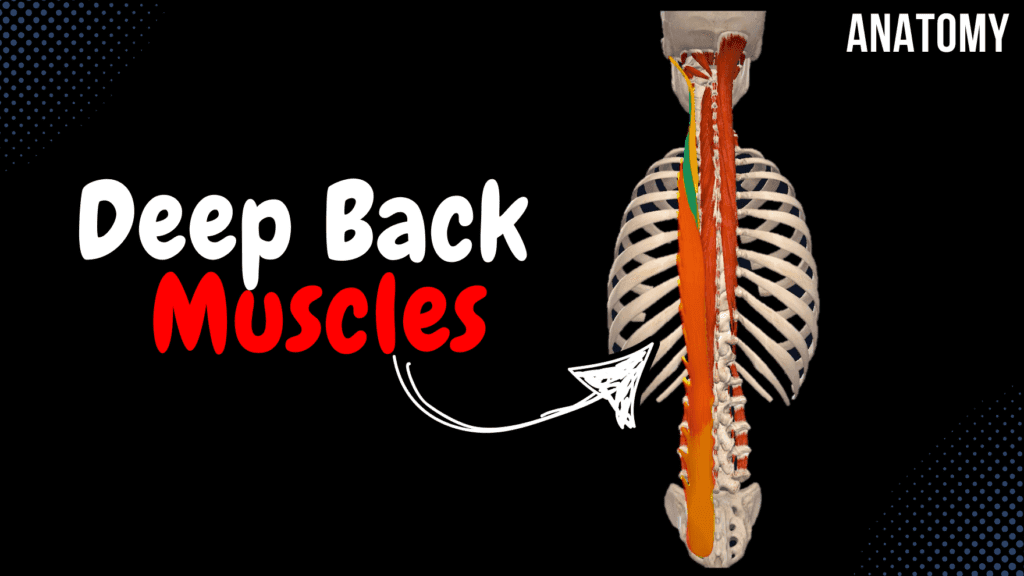
Deep Back Muscles (Division, Origin, Insertion, Function) Official Links Instagram Youtube Jki-discord Notes & Illustrations Quizzes Summary & Transcript Notes ☆ Member Only Go to PDF Notes Illustrations ☆ Member Only Go to Illustrations 12345678910 Deep Back Muscles – QUIZ Test your understanding with 10 random multiple-choice questions from the question bank. You're in the preview mode. Note: All elements work correctly on the front end. 1 / 10 Which muscle is part of the transversospinal system and inserts on the spinous process of the second vertebra above its origin? A) Intertransversarii B) Multifidi breves C) Rotatores breves D) Rotatores longi The rotatores longi muscles fit this description. 2 / 10 Which deep back muscle is the largest and most developed in the lumbar region? A) Multifidi B) Spinalis C) Interspinales D) Rotatores The multifidi muscles are largest in the lumbar region. 3 / 10 What is the function of the obliquus capitis inferior? A) Rotation of the atlas on the axis B) Extension of the thoracic spine C) Lateral flexion of the cervical spine D) Flexion of the cervical spine It rotates the atlas on the axis. 4 / 10 What is the function of the spinalis thoracis muscle? A) Extension of the thoracic spine B) Flexion of the lumbar spine C) Lateral flexion of the spine D) Rotation of the cervical spine It extends the thoracic spine. 5 / 10 What is the primary function of the semispinalis capitis? A) Rotation of the cervical spine B) Stabilization of the cervical spine C) Lateral flexion of the head D) Extension and contralateral rotation of the head and neck It extends the head and neck and rotates them contralaterally. 6 / 10 What is the function of the iliocostalis cervicis muscle? A) Stabilization of the thoracic spine B) Lateral flexion and extension of the cervical spine C) Flexion of the cervical spine D) Rotation of the cervical spine It assists with lateral flexion and extension of the cervical spine. 7 / 10 Which muscle extends and stabilizes the lumbar spine? A) Interspinales B) Longissimus C) Multifidi D) Rotatores The multifidi muscles extend and stabilize the lumbar spine. 8 / 10 Which muscle connects adjacent transverse processes in the cervical region? A) Interspinales B) Intertransversarii C) Rotatores D) Multifidi The intertransversarii muscles connect adjacent transverse processes. 9 / 10 Which muscle originates from the transverse process of C1 and inserts onto the occipital bone? A) Rectus capitis posterior minor B) Rectus capitis posterior major C) Obliquus capitis superior D) Obliquus capitis inferior This describes the obliquus capitis superior muscle. 10 / 10 What is the origin of the semispinalis thoracis? A) Transverse processes of C1-T5 B) Spinous processes of T10-L3 C) Spinous processes of T1-T8 D) Transverse processes of T6-T11 It originates from the transverse processes of T6-T11. Your score is The average score is 0% Description This video covers the deep muscles of the back, organized into different layers and muscle systems. Deep Muscles of the Back 3rd Layer Suboccipital Muscles System of Short Muscles 2nd Layer Transversospinal System 1st Layer (Superficial) Spinospinal System Spinotransverse System 1. Suboccipital Muscles System of deep muscles of the neck. Rectus Capitis Posterior Minor (Musculus Rectus Capitis Posterior Minor) Origin: Posterior Tubercle of Atlas (C1) Insertion: Occipital Bone below Inferior Nuchal Line Rectus Capitis Posterior Major (Musculus Rectus Capitis Posterior Major) Origin: Spinous Process of Axis (C2) Insertion: Occipital Bone – Inferior Nuchal Line Obliquus Capitis Superior (Musculus Obliquus Capitis Superior) Origin: Transverse Process of Atlas (C1) Insertion: Occipital Bone – Inferior Nuchal Line Obliquus Capitis Inferior (Musculus Obliquus Capitis Inferior) Origin: Spinous Process of Axis (C2) Insertion: Transverse Process of Atlas (C1) 2. System of Short Muscles Short muscles connecting adjacent vertebrae. Interspinales (Musculi Interspinales) Developed mainly in the cervical and lumbar regions. Origin: Spinous Process of vertebra below Insertion: Spinous Process of vertebra above Intertransversarii (Musculi Intertransversales) Mostly developed in the cervical region. Located between transverse processes. Origin: Transverse Process of vertebra below Insertion: Transverse Process of vertebra above 3. Transversospinal System Runs from the transverse process of a lower vertebra to the spinous process of an upper vertebra. Rotatores (Musculi Rotatores) Located in the thoracic vertebrae. Long Rotatores (Musculi Rotatores Longi) Origin: Transverse Process of vertebra below Insertion: Spinous Process of 2 vertebrae above Short Rotatores (Musculi Rotatores Breves) Origin: Transverse Process of vertebra below Insertion: Spinous Process of 1 vertebra above Multifidi (Musculi Multifidi) Fills space lateral to spinous processes. Most distinctive in the lumbar region. Short Multifidi (Musculi Multifidi Breves) Origin: Transverse Process of vertebra below Insertion: Spinous Process of 2 vertebrae above Long Multifidi (Musculi Multifidi Longi) Origin: Transverse Process of vertebra below Insertion: Spinous Process of 3 vertebrae above Semispinalis (Musculi Semispinales) Divided into thoracis, cervicis, and capitis. Semispinalis Thoracis (Musculi Semispinalis Thoracis) Origin: Transverse Process of T6-T11 Insertion: Spinous Process of C6-T4 Semispinalis Cervicis (Musculi Semispinalis Cervicis) Origin: Transverse Process of T1-T6 Insertion: Spinous Process of C2-C5 Semispinalis Capitis (Musculi Semispinalis Capitis) Origin: Transverse Process of C4-T6 Insertion: Occipital Bone – Between Inferior and Superior Nuchal Line 4. Spinospinal System Spinalis (Musculus Spinalis) Spinalis Cervicis (Musculus Spinalis Cervicis) Origin: Spinous Process of C6-T2 Insertion: Spinous Process of C2-C4 Spinalis Thoracis (Musculus Spinalis Thoracis) Origin: Spinous Process of T10-L3 Insertion: Spinous Process of T1-T8 5. Spinotransverse System Longissimus (Musculus Longissimus) Splenius (Musculus Splenius) Iliocostalis (Musculus Iliocostalis) Transcript Introduction0:03Hey What’s up. Meditay here and in this video, we’ll be covering the Deep muscles0:07of the back. Alright. Generally, the muscles of the back consist of superficial musclesDivision of the Back Muscles0:12and deep muscles. The superficial muscles consist of the The trapezius and Latissimus,0:17which are the 1st layer. The 2nd layer are the Rhomboid Major Minor and Levator0:22Scapula, and the 3rd layer of muscles consists of the Serratus Posterior superior and inferior.0:29And when you remove these three layers, you’ll finally get to the Deep muscles of the back.Division of the Deep Back Muscles0:34Now the deep muscles of the back are categorized based on their shape and structure and location.0:40Generally,
Superficial Back Muscles
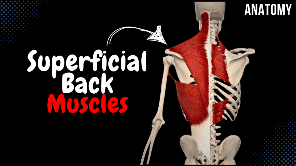
Superficial Back Muscles (Division, Origin, Insertion, Function) Official Links Instagram Youtube Jki-discord Notes & Illustrations Quizzes Summary & Transcript Notes ☆ Member Only Go to PDF Notes Illustrations ☆ Member Only Go to Illustrations 12345678910 Superficial Back Muscles – QUIZ Test your understanding with 10 random multiple-choice questions from the question bank. You're in the preview mode. Note: All elements work correctly on the front end. 1 / 10 Which muscle assists in respiration by elevating the ribs? A) Rhomboid major B) Latissimus dorsi C) Trapezius D) Serratus posterior superior The serratus posterior superior elevates the ribs, aiding in inspiration. 2 / 10 Which muscle depresses the ribs during expiration? A) Serratus posterior superior B) Rhomboid major C) Serratus posterior inferior D) Latissimus dorsi The serratus posterior inferior assists in expiration by pulling the ribs downward. 3 / 10 Which muscle forms the lower border of the posterior axillary fold? A) Serratus posterior inferior B) Rhomboid minor C) Latissimus dorsi D) Trapezius The latissimus dorsi forms the lower border of the posterior axillary fold. 4 / 10 Which superficial back muscle is innervated by the dorsal scapular nerve? A) Latissimus dorsi B) Rhomboid minor C) Serratus posterior superior D) Trapezius The rhomboids and levator scapulae are innervated by the dorsal scapular nerve. 5 / 10 Which muscle attaches to the acromion of the scapula? A) Latissimus dorsi B) Trapezius C) Rhomboid minor D) Serratus posterior inferior The trapezius inserts onto the acromion of the scapula. 6 / 10 What is the insertion point of the rhomboid minor? A) Medial border of scapula B) Spine of scapula C) Inferior angle of scapula D) Acromion process It inserts on the medial border of the scapula, above the rhomboid major. 7 / 10 Which muscle is part of the first layer of the superficial back muscles? A) Rhomboid minor B) Levator scapulae C) Serratus posterior inferior D) Trapezius The first layer includes large muscles like the trapezius. 8 / 10 Which superficial back muscle originates from the external occipital protuberance and inserts onto the clavicle? A) Trapezius B) Serratus posterior superior C) Rhomboid major D) Latissimus dorsi The trapezius originates from the external occipital protuberance and attaches to the clavicle. 9 / 10 Where does the rhomboid major originate? A) Spinous processes of T6-T12 B) Spinous processes of T1-T4 C) Lateral border of the scapula D) Superior nuchal line It originates from the spinous processes of T1-T4. 10 / 10 Which part of the trapezius is responsible for scapular elevation? A) Inferior B) Superior C) Middle D) All parts The superior part elevates the scapula. Your score is The average score is 0% Description This video is about the superficial muscles of the back, their anatomical layers, origins, insertions, and functions. Superficial Muscles of the Back Muscles of the 1st Layer Trapezius Latissimus Dorsi Muscles of the 2nd Layer Rhomboid Major Rhomboid Minor Levator Scapulae Muscles of the 3rd Layer Serratus Posterior Superior Serratus Posterior Inferior Trapezius (Musculus Trapezius) Superior Part Origin: Superior Nuchal Line External Occipital Protuberance Nuchal Ligament Insertion: Acromial End of Clavicle Acromion of Scapula Middle Part Origin: Spinous Process of C7-T3/T4 Insertion: Spine and Acromion of Scapula Inferior Part Origin: Spinous Process of T4-T12 Insertion: Spine of Scapula Latissimus Dorsi (Musculus Latissimus Dorsi) Origin: Spinous Process of T7-T12 Thoracolumbar Fascia Iliac Crest Inferior Surface of Ribs 9-12 Inferior Angle of Scapula Insertion: Crest of Lesser Tubercle (Humerus) (Crista Tuberculi Minoris Humeri) Rhomboid Major (Musculus Rhomboideus Major) Origin: Spinous Process of T1-T4 Insertion: Lower 2/3 of Medial Border of Scapula (Margo Medialis Scapulae) Function: Elevates and retracts the scapula Internal rotation of the scapula Rhomboid Minor (Musculus Rhomboideus Minor) Origin: Spinous Process of C6-C7 Insertion: Upper 1/3 of Medial Border of Scapula (Margo Medialis Scapulae) Function: Elevates and retracts the scapula Internal rotation of the scapula Levator Scapulae (Musculus Levator Scapulae) Origin: Spinous Process of C1-C4 Insertion: Superior Angle of Scapula (Angulus Superior Scapulae) Function: Elevates the scapula Serratus Posterior Superior (Musculus Serratus Posterior Superior) Origin: Spinous Process of C6-T2 Insertion: External Surface of 2nd-5th Ribs Function: Elevates the ribs (Inspiration) Serratus Posterior Inferior (Musculus Serratus Posterior Inferior) Origin: Spinous Process of T11-L2 Insertion: External Surface of 9th-12th Ribs Function: Pulls the ribs downward (Expiration) Transcript Introduction0:03Hey What’s up. Meditay here and in this video, we’ll be covering the Superficial muscles of0:08the back. Alright. Generally, the muscles of the back consist of superficial musclesDivision of the Superficial Muscles0:13and deep muscles. So let’s take a closer look at the superficial muscles.0:17These muscles are divided into layers. So, the 1st layer is the most superficial one.0:22It consist of the Trapezius, as you see here, and the Latissimus Dorsi.0:27The 2nd layer are the Rhomboid Major, Rhomboid Minor and Levator Scapula, and the 3rd layer of0:33muscles consists of the Serratus Posterior superior and Serratus Posterior inferior.0:39So these are the muscles we’re going to go through throughout this video.0:42We’ll start with the 1st Layer, the Trapezius. Now the trapezius Is this large muscleTrapezius0:48that take up the majority of your shoulders. They can also be classified as a cardiothoracic muscle,0:53in addition to being one of the superficial muscles of the back.0:57Now the Trapezius consists of 3 parts. There’s a Superior Part, a Middle part and an inferior part.1:04The superior part will originate from the Superior nuchal line, the external occipital protuberance,1:10and the nuchal ligament. It’s then going to insert1:13at the Acromial end of the clavicle, as you see here, as well as the acromion of the scapula.1:19Then we have the middle part, which originates from the spinous process of the C7-T3 or T4,1:26and it’s going to insert at the Spine of the Scapula as you see here, and the acromion.1:32Then we have the Inferior Part, which originates from the spinous process of T4-T121:38and insert at the spine of scapula as well. Ok. So what is the function of the Trapezius?1:44The Inferior part will pull the shoulder downwards, Middle and superior fibers1:49will pull the scapula towards the midline, as well as elevating the scapula. They may
Fascia of the Abdomen
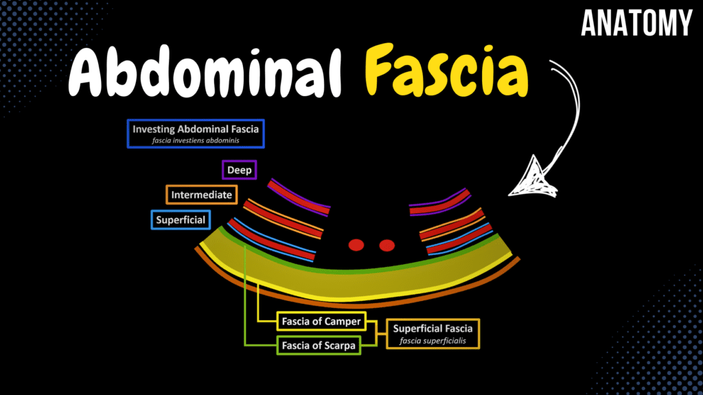
Fascia of the Abdomen (Superficial, Investing Abdominal, Endoabdominal) Official Links Instagram Youtube Jki-discord Notes & Illustrations Quizzes Summary & Transcript Notes ☆ Members Only Go to PDF Notes Illustrations ☆ Members Only Go to Illustrations 12345678910 Fascia of the Abdomen – QUIZ Test your understanding with 10 random multiple-choice questions from the question bank. You're in the preview mode. Note: All elements work correctly on the front end. 1 / 10 Which fascia surrounds the psoas major muscle? A) Psoas fascia B) Transversalis fascia C) Thoracolumbar fascia D) Iliac fascia The psoas fascia encloses the psoas major muscle. 2 / 10 Which layer of fascia forms the floor of the inguinal canal? A) Transversalis fascia B) Scarpa's fascia C) Iliac fascia D) Camper's fascia The transversalis fascia forms the floor of the inguinal canal. 3 / 10 What is the role of the vincula tendinum in tendon sheaths? A) Reduces friction B) Lubricates tendons C) Supplies nutrients to tendons D) Provides structural support Vincula tendinum supply nutrients to tendons within tendon sheaths. 4 / 10 What forms the outermost layer of the abdominal wall fascia? A) Investing abdominal fascia B) Thoracolumbar fascia C) Superficial fascia D) Endoabdominal fascia The superficial fascia forms the outermost layer of the abdominal wall fascia, comprising Camper’s and Scarpa’s layers. 5 / 10 What is the role of the transversalis fascia in hernia formation? A) Contributes to hernia formation B) Protects the lumbar spine C) Facilitates abdominal wall movement D) Stabilizes the iliac crest Weakness in the transversalis fascia can contribute to the development of inguinal hernias. 6 / 10 Which layer of fascia provides mechanical support and organization? A) Iliac fascia B) Camper's fascia C) Psoas fascia D) Scarpa's fascia Scarpa’s fascia lies beneath Camper’s fascia and provides mechanical support and structural organization. 7 / 10 What anatomical structure passes through the saphenous opening? A) Great saphenous vein B) Inferior vena cava C) Femoral artery D) Superficial epigastric vein The great saphenous vein passes through the saphenous opening in the fascia lata. 8 / 10 What is the primary anatomical location of the iliac fascia? A) Rectus sheath B) Thoracolumbar fascia C) Over the iliacus muscle D) Between internal oblique and external oblique The iliac fascia lies over the iliacus muscle, blending with the psoas fascia and extending to the pelvic brim. 9 / 10 What is the primary role of Camper’s fascia? A) Insulation and energy storage B) Stabilization of lumbar spine C) Protection from infections D) Structural support Camper’s fascia provides insulation, energy storage, and cushioning as the fatty layer of superficial abdominal fascia. 10 / 10 The transversalis fascia lies between which two structures? A) Transverse abdominal muscle and peritoneum B) Quadratus lumborum and iliac fascia C) Rectus sheath and internal oblique D) External oblique and Camper's fascia The transversalis fascia lies between the transverse abdominal muscle and the peritoneum. Your score is The average score is 0% Description This video is about the fascia of the abdomen, including its different layers and anatomical significance. Muscles of the Abdomen Lateral Group External Oblique Internal Oblique Transverse Abdominal Anterior Group Rectus Abdominis Pyramidalis Posterior Group Quadratus Lumborum Superficial Fascia (Fascia Superficialis) Fascia of Camper Fascia of Scarpa Investing Abdominal Fascia (Fascia Investiens Abdominis) Superficial Layer Intermediate Layer Deep Layer Endoabdominal Fascia (Fascia Endoabdominalis) Transversalis Fascia (Fascia Transversalis) Iliac Fascia (Fascia Iliaca) Transcript Introduction0:03What’s up. Meditay here and in this video, we’ll be going through0:06the Fascia you’ll find in the Abdomen. So the fascia of the abdomen vover theMuscles of the Abdomen0:11muscles of the abdomen from both the external and internal side. So here I’ve cut through all the0:16muscle layers of the abdomen. And remember, they consist of the external oblique, Internal Oblique0:22and Transverse Abdominal Muscle. These three at considered as the lateral abdominal muscles.0:27And we have the Anterior abdominal Muscles, like the Rectus Abdominis.0:30Our goal in this video is to go through the fascia that you’ll find wrapping around these muscles,0:36and separating them from the organs within the abdominal cavityContent0:39So In this video, we’re going to go through the Superficial Fascia,0:43we’ll go thrgouh the Investing Abdominal Fascia. And we’ll go thrgouh the Endoabdominal Fascia.0:48And to do that, we’ll have to make a transverse cut of the abdomen. Remove the upper half,0:53and look at it from this perspective. And now we’re gonna try to draw all0:57the structures and go through them as we do that. First we have the Skin.1:01And right underneath the skin layers, we’ll find the adipose tissue, or fat cells. Underneath the1:07fat cells, we can find the External Oblique. Internal Oblique, and Transverse Abdominal1:12Muscle. And in the middle here we can find the Rectus Abdominis muscle. So this is a very1:16schematic outline of the abdominal muscles. The first fascia we’re gonna go thrgouh isSuperficial Fascia1:21associated with the fat Layer. The fascia that cover the fat layer from the superficial side1:26is called Fascia of Camper. And the fascia that cover it from the inner side, is called1:31Fascia of Scarpa. The two layers of fascia together, form the so called Superficial Fascia.1:38So that’s the first one in our list. Next we have a Fascia called Investing Abdominal Fascia.Investing Abdominal Fascia1:45And as the name implies, this fascia is going to surround the abdominal muscles.1:49So the investing abdominal fascia si divided into three layers.1:54First is the Superficial investing abdominal fascia, aurrounding the external oblique.1:59Then there’s the Intermediate Investing abdominal fascia, surrounding the internal Oblique.2:04Then there’s the Deep investing abdominal fascia, surrounding the transverse abdominal muscle. Now.2:10Underneath the Deep investing abdominal fascia, that’s where you’ll find the third fascia we’reEndoabdominal Fascia2:15gonna talk about, called the endoabdominal fascia. And just underneath the endoabdominal fascia,2:21that’s where you’ll find the Parietal peritoneum, which over all of your internal organs.2:28Now, the endoabdominal fascia is actually divided into certain parts depending on the location of2:34it, So let’s look at that a little bit. So now we’re gonna draw the lateral view of he abdomen.2:38First we have the Skin, then the Fat cells, then The external oblique, Internal Oblique