
Portal Venous System
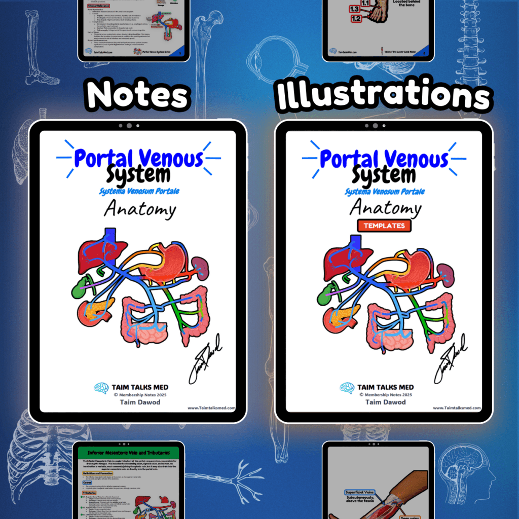
Portal Venous System Circulatory System Venous System Notes 📅 Last Updated: 01.01.2025 🔄 Version: 1.0 This PDF covers the portal vein and its tributaries, including the superior mesenteric, inferior mesenteric, and splenic veins. It also details porto-caval anastomoses, their clinical significance, and conditions such as portal hypertension, esophageal varices, and caput medusae. Become a Member Buy this bundle Go To Notes (Preview) Go to Illustrations (Preview) Watch The Lecture Go To Notes Go to Illustrations Watch The Lecture Need it offline? Open PDF → Click the three-dot menu → Download PDF. 📖 Sources: Singh, I. Human Neuroanatomy (10th ed.) Kozlowski, T. Memorix Anatomy (2nd ed.) Gray’s Anatomy: The Anatomical Basis of Clinical Practice (42nd ed.) Sinnatamby, C. S. Last’s Anatomy (12th ed.)
Veins of the Lower Limb
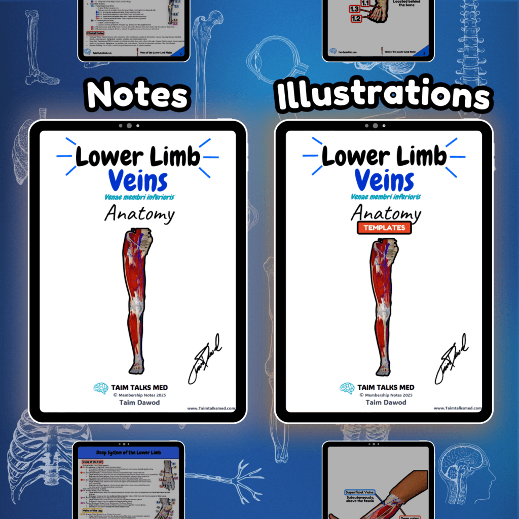
Veins of the Lower Limb Circulatory System Venous System Notes 📅 Last Updated: 01.01.2025 🔄 Version: 1.0 This PDF covers the deep and superficial venous systems of the lower limb, including the femoral, popliteal, tibial, and saphenous veins. It outlines venous drainage from the foot to the pelvis, highlighting connections, clinical importance, and anatomical landmarks. Become a Member Buy this bundle Go To Notes (Preview) Go to Illustrations (Preview) Watch The Lecture Go To Notes Go to Illustrations Watch The Lecture Need it offline? Open PDF → Click the three-dot menu → Download PDF. 📖 Sources: Singh, I. Human Neuroanatomy (10th ed.) Kozlowski, T. Memorix Anatomy (2nd ed.) Gray’s Anatomy: The Anatomical Basis of Clinical Practice (42nd ed.) Sinnatamby, C. S. Last’s Anatomy (12th ed.)
Iliac Veins
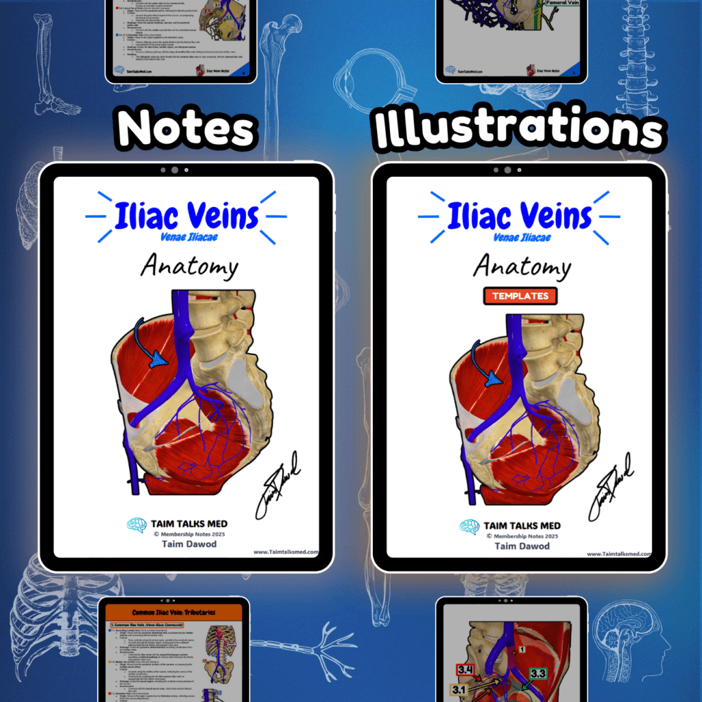
Iliac Veins Circulatory System Venous System Notes 📅 Last Updated: 01.01.2025 🔄 Version: 1.0 This PDF covers the common, external, and internal iliac veins, detailing their tributaries, drainage into the inferior vena cava, and roles in venous return. The internal iliac vein has parietal and visceral tributaries, draining pelvic structures and lower limbs. Become a Member Buy this bundle Go To Notes (Preview) Go to Illustrations (Preview) Watch The Lecture Go To Notes Go to Illustrations Watch The Lecture Need it offline? Open PDF → Click the three-dot menu → Download PDF. 📖 Sources: Singh, I. Human Neuroanatomy (10th ed.) Kozlowski, T. Memorix Anatomy (2nd ed.) Gray’s Anatomy: The Anatomical Basis of Clinical Practice (42nd ed.) Sinnatamby, C. S. Last’s Anatomy (12th ed.)
Inferior Vena Cava

Inferior Vena Cava Circulatory System Venous System Notes 📅 Last Updated: 01.01.2025 🔄 Version: 1.0 The inferior vena cava (IVC) is the largest vein in the human body, collecting deoxygenated blood from the lower half. It lacks valves and runs to the right of the abdominal aorta, passing through the caval opening in the diaphragm. The visceral tributaries include hepatic, renal, and genital veins, while the parietal tributaries consist of lumbar and inferior phrenic veins. Become a Member Buy this bundle Go To Notes (Preview) Go to Illustrations (Preview) Watch The Lecture Go To Notes Go to Illustrations Watch The Lecture Need it offline? Open PDF → Click the three-dot menu → Download PDF. 📖 Sources: Singh, I. Human Neuroanatomy (10th ed.) Kozlowski, T. Memorix Anatomy (2nd ed.) Gray’s Anatomy: The Anatomical Basis of Clinical Practice (42nd ed.) Sinnatamby, C. S. Last’s Anatomy (12th ed.)
Veins of the Upper Limb
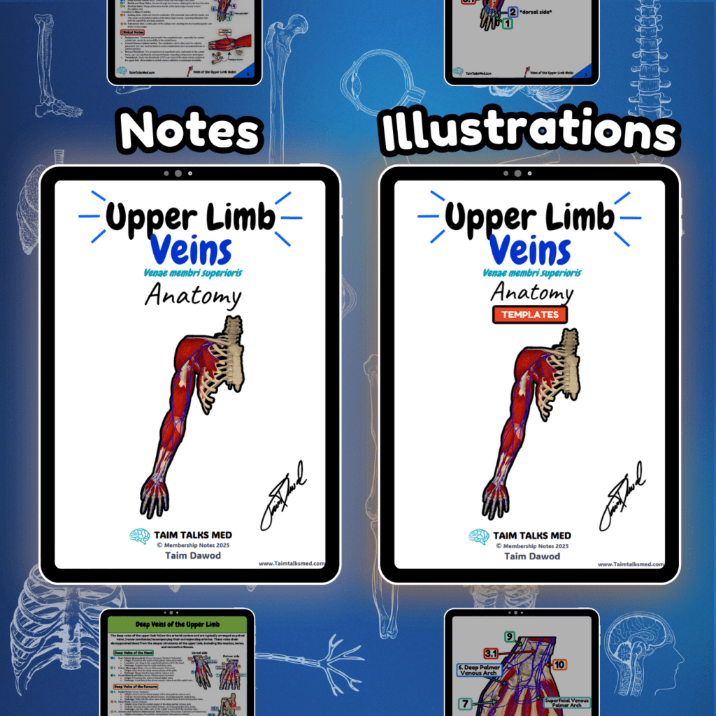
Veins of the Upper Limb Circulatory System Venous System Notes 📅 Last Updated: 01.01.2025 🔄 Version: 1.0 The veins of the upper limb are divided into superficial and deep systems. The superficial veins, such as the cephalic and basilic veins, run above the deep fascia, while the deep veins, including the brachial, radial, and ulnar veins, accompany arteries. The axillary vein serves as the major drainage conduit, continuing into the subclavian vein and subsequently the brachiocephalic vein. Become a Member Buy this bundle Go To Notes (Preview) Go to Illustrations (Preview) Watch The Lecture Go To Notes Go to Illustrations Watch The Lecture Need it offline? Open PDF → Click the three-dot menu → Download PDF. 📖 Sources: Singh, I. Human Neuroanatomy (10th ed.) Kozlowski, T. Memorix Anatomy (2nd ed.) Gray’s Anatomy: The Anatomical Basis of Clinical Practice (42nd ed.) Sinnatamby, C. S. Last’s Anatomy (12th ed.)
Veins of the Brain
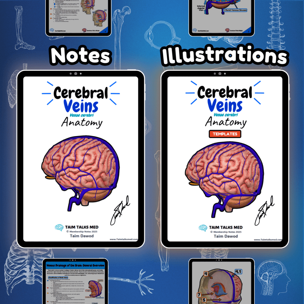
Veins of the Brain Circulatory System Venous System Notes 📅 Last Updated: 01.01.2025 🔄 Version: 1.0 The veins of the brain are valveless, thin-walled structures that drain deoxygenated blood from the brain, meninges, and cranial bones into the venous sinuses. They are divided into superficial cerebral veins, deep cerebral veins, dural venous sinuses, cerebellar veins, and other venous structures, such as diploic and emissary veins. These veins play a critical role in maintaining intracranial circulation and preventing venous congestion. Become a Member Buy this bundle Go To Notes (Preview) Go to Illustrations (Preview) Watch The Lectures Go To Notes Go to Illustrations Watch The Lectures Need it offline? Open PDF → Click the three-dot menu → Download PDF. 📖 Sources: Singh, I. Human Neuroanatomy (10th ed.) Kozlowski, T. Memorix Anatomy (2nd ed.) Gray’s Anatomy: The Anatomical Basis of Clinical Practice (42nd ed.) Sinnatamby, C. S. Last’s Anatomy (12th ed.)
Veins of the Head and Neck

Veins of the Head and Neck Circulatory System Venous System Notes 📅 Last Updated: 01.01.2025 🔄 Version: 1.0 The venous drainage of the head and neck includes the internal, external, and anterior jugular veins, which return deoxygenated blood to the superior vena cava. These veins receive tributaries from the face, scalp, brain, and neck muscles, forming crucial venous pathways. Become a Member Buy this bundle Go To Notes (Preview) Go to Illustrations (Preview) Watch The Lecture Go To Notes Go to Illustrations Watch The Lecture Need it offline? Open PDF → Click the three-dot menu → Download PDF. 📖 Sources: Singh, I. Human Neuroanatomy (10th ed.). Kozlowski, T. Memorix Anatomy (2nd ed.). Gray’s Anatomy: The Anatomical Basis of Clinical Practice (42nd ed.). Sinnatamby, C. Last’s Anatomy (12th ed.).
Superior Vena Cava
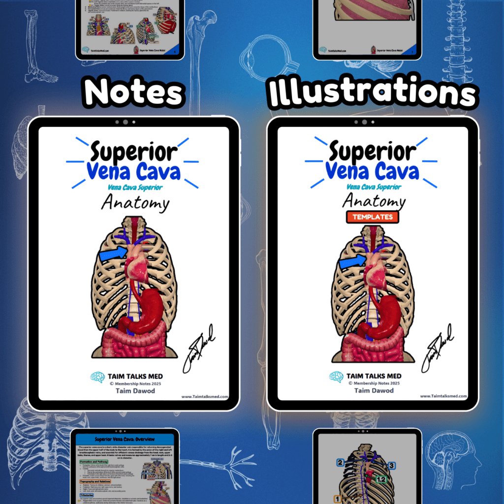
Superior Vena Cava Circulatory System Venous System Notes 📅 Last Updated: 01.01.2025 🔄 Version: 1.0 The superior vena cava (SVC) is a short, large-diameter vein with no valves, responsible for draining venous blood from the head, neck, upper limbs, and thorax into the right atrium. It receives major tributaries, including the brachiocephalic veins, azygos vein, and thoracic veins, forming a crucial part of systemic venous return. Become a Member Buy this bundle Go To Notes (Preview) Go to Illustrations (Preview) Watch The Lecture Go To Notes Go to Illustrations Watch The Lecture Need it offline? Open PDF → Click the three-dot menu → Download PDF. 📖 Sources: Singh, I. Human Neuroanatomy (10th ed.) Kozlowski, T. Memorix Anatomy (2nd ed.) Gray’s Anatomy: The Anatomical Basis of Clinical Practice (42nd ed.) Sinnatamby, C. S. Last’s Anatomy (12th ed.)
Lower Limb Arteries
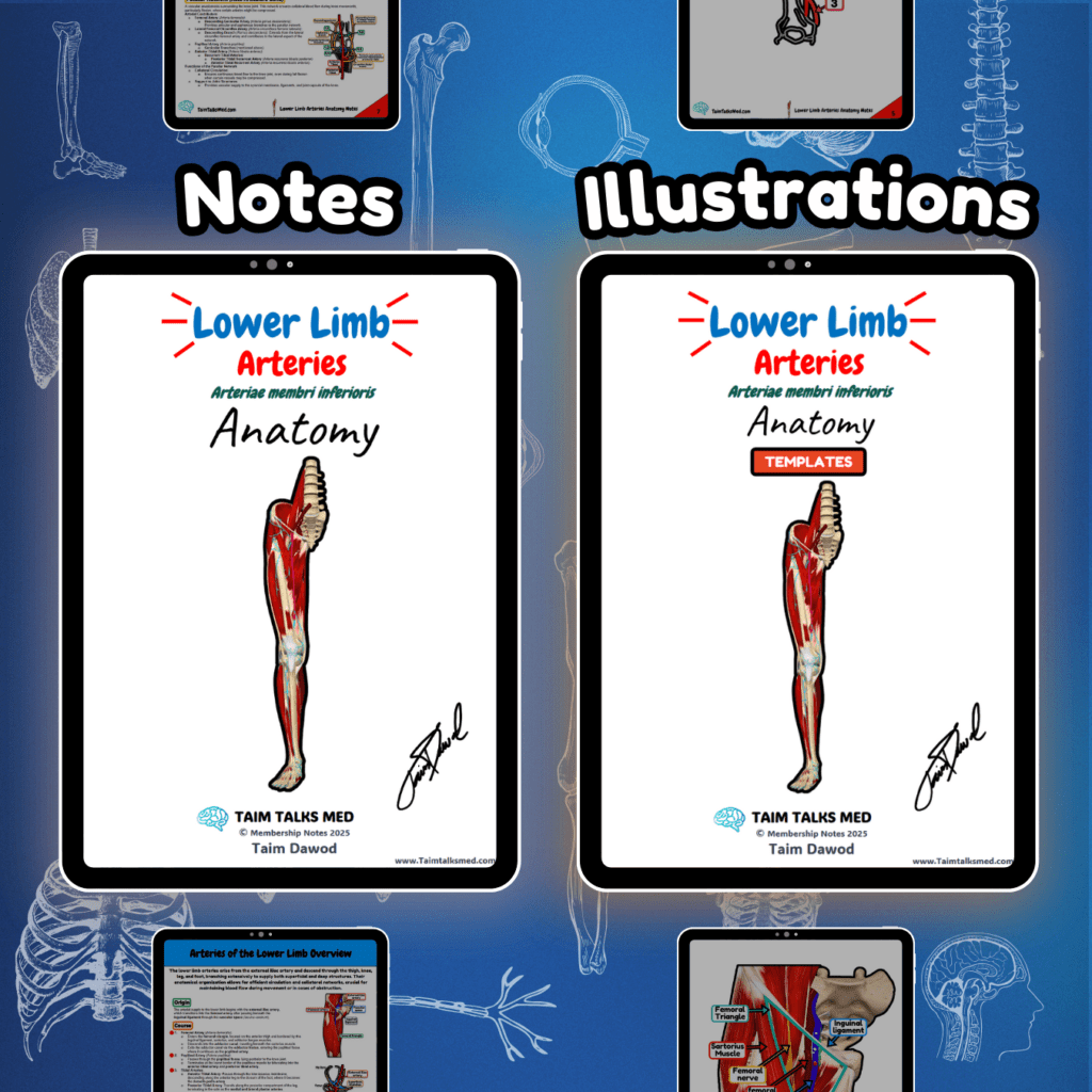
Lower Limb Arteries Circulatory System Arterial System Notes 📅 Last Updated: 01.01.2025 🔄 Version: 1.0 The lower limb arteries arise from the external iliac artery, transitioning into the femoral artery at the inguinal ligament. The system includes the popliteal artery, which bifurcates into the anterior and posterior tibial arteries, supplying the leg and foot. Collateral circulation is maintained via arterial anastomoses at the knee, ankle, and foot. Become a Member Buy this bundle Go To Notes (Preview) Go to Illustrations (Preview) Watch The Lectures Go To Notes Go to Illustrations Watch The Lectures Need it offline? Open PDF → Click the three-dot menu → Download PDF. 📖 Sources: Singh, I. Human Neuroanatomy (10th ed.) Kozlowski, T. Memorix Anatomy (2nd ed.) Gray’s Anatomy: The Anatomical Basis of Clinical Practice (42nd ed.) Sinnatamby, C. S. Last’s Anatomy (12th ed.)
Iliac Arteries
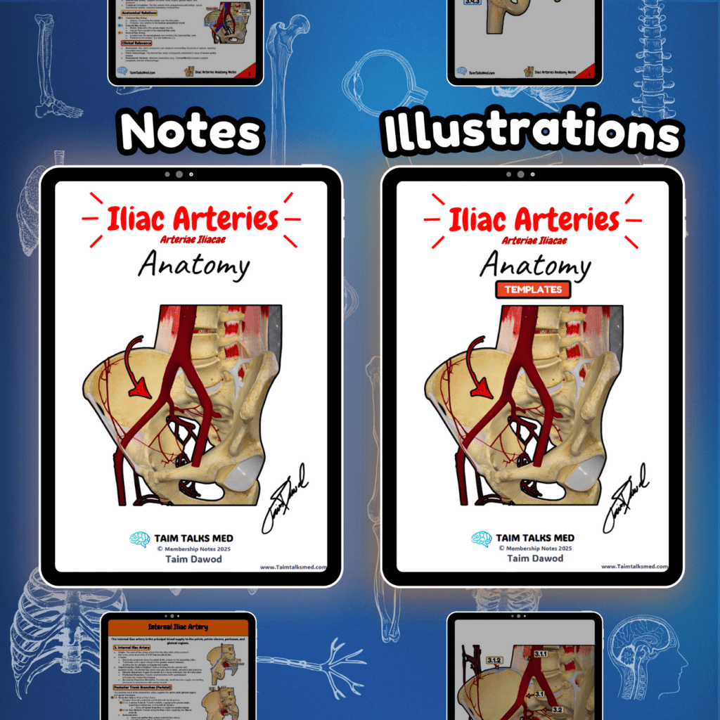
Iliac Arteries Circulatory System Arterial System Notes 📅 Last Updated: 01.01.2025 🔄 Version: 1.0 The iliac arteries originate from the abdominal aorta at L4, dividing into the common iliac arteries, which further split into the external iliac artery (supplying the lower limb) and the internal iliac artery (supplying the pelvis and perineum). Notable branches include the iliolumbar, obturator, superior gluteal, and internal pudendal arteries, forming critical anastomoses and ensuring blood supply to key regions. Become a Member Buy this bundle Go To Notes (Preview) Go to Illustrations (Preview) Watch The Lectures Go To Notes Go to Illustrations Watch The Lectures Need it offline? Open PDF → Click the three-dot menu → Download PDF. 📖 Sources: Singh, I. Human Neuroanatomy (10th ed.) Kozlowski, T. Memorix Anatomy (2nd ed.) Gray’s Anatomy: The Anatomical Basis of Clinical Practice (42nd ed.) Sinnatamby, C. S. Last’s Anatomy (12th ed.)