
Peripheral Nervous System Overview
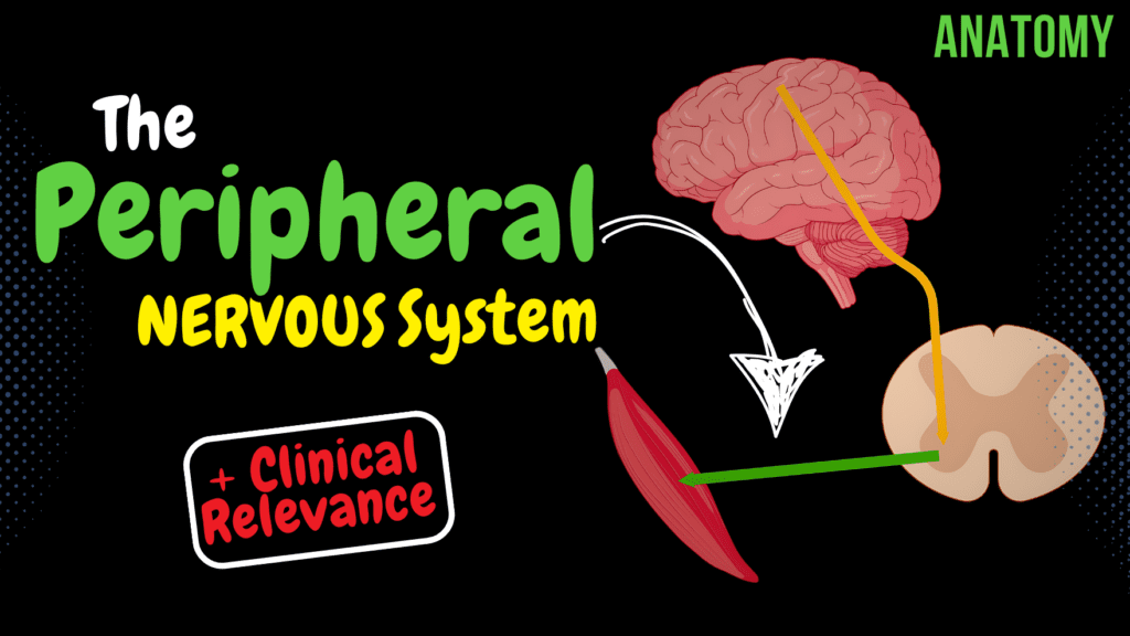
Peripheral Nervous System Overview (Classification, Spinal Nerve) + Clinical Relevance Official Links Instagram Youtube Jki-discord Notes & Illustrations Quizzes Summary & Transcript Notes ☆ Members Only Go to PDF Notes Illustrations ☆ Members Only Go to Illustrations 12345678910 Peripheral Nervous System Overview – QUIZ Test your understanding with 10 random multiple-choice questions from the question bank. You're in the preview mode. Note: All elements work correctly on the front end. 1 / 10 What is the difference between white and gray rami communicantes? A) White: sensory; Gray: motor B) White: pre-ganglionic; Gray: post-ganglionic C) Both carry motor fibers D) Both carry sensory fibers White rami communicantes carry pre-ganglionic fibers, while gray rami communicantes carry post-ganglionic fibers. 2 / 10 What is the function of proprioceptors? A) Respond to external stimuli B) Signal changes in temperature C) Detect changes in organ function D) Monitor body position and movement Proprioceptors detect body position and movement, providing sensory input from muscles, tendons, and joints. 3 / 10 What structure connects the posterior and anterior roots of the spinal nerve? A) White ramus communicans B) Dorsal root ganglion C) Sympathetic trunk D) Spinal nerve trunk The trunk of the spinal nerve (truncus nervi spinalis) is formed by the merging of the anterior and posterior roots. 4 / 10 What is the function of the posterior branch of the spinal nerve? A) Transmit sensory input B) Innervate deep back muscles C) Carry autonomic signals D) Innervate limbs The posterior branch innervates the deep back muscles and the overlying skin. 5 / 10 Which spinal nerve branch provides innervation to the meninges? A) Meningeal branch B) Sympathetic branch C) Anterior branch D) Posterior branch The meningeal branch innervates the meninges and vertebral column. 6 / 10 Where do pre-ganglionic sympathetic fibers originate? A) Lateral horn (C8-L2) B) Posterior horn C) Brainstem D) Anterior horn Pre-ganglionic sympathetic fibers originate in the lateral horn of the spinal cord from levels C8-L2. 7 / 10 Which cranial nerve is responsible for tongue movement? A) Hypoglossal nerve B) Trigeminal nerve C) Vagus nerve D) Glossopharyngeal nerve The hypoglossal nerve (cranial nerve XII) controls motor functions of the tongue. 8 / 10 What structure houses the sensory neuron cell bodies in the spinal nerve? A) Spinal ganglion B) White ramus communicans C) Sympathetic trunk ganglion D) Trunk of spinal nerve The spinal ganglion (ganglion sensorium nervi spinalis) is located in the dorsal root and houses sensory neuron cell bodies. 9 / 10 What is the anatomical name for the anterior root of the spinal nerve? A) Ramus posterior B) Radix posterior C) Ramus anterior D) Radix anterior The anterior root is also called the radix anterior and carries motor fibers. 10 / 10 How many nerve pairs form the brachial plexus? A) 6 B) 4 C) 3 D) 5 The brachial plexus is formed by the anterior branches of C5-T1 spinal nerves. Your score is The average score is 0% Description What is the Peripheral Nervous System? Cranial Nerves 12 nerve pairs Spinal Nerves 31 nerve pairs Classification of the PNS Sensory System Soamtic Sensory Fibers Visceral Sensory Fibers Exteroreceptors, Proprioreceptors, Periosteum Motor System Somatomotor System (Somatic system) Voluntary control Autonomic Nervous System / Visceromotor nervous system Sympathetic Nervous System (C8-L2 Lateral horn) Parasympahetic Nervous System (S2-S4 Lateral horn) Enteric Nervous System (myenteric and submucosal plexuses in walls of digestive organs) Clinical Significance Lower Motor Neuron damage Located in anterior horn of spinal cord and nuclei of cranial nerves Damage to lower motor neurons: Flaccid Paralysis Hypotonia Paresis/Plegia Hyporeflexia / areflexia Upper Motor Neuron damage May rise after a stroke or a perinatal hypoxia Spastic Paralysis Paresis / Plegia Hypertonia Hyperreflexia Babinski’s sign External Scheme of Spinal Nerve Rootlets (fila radicularia) Anterior root (radix anterior) Posterior root (radic posterior) Spinal Ganglion (ganglion sensorium nervi spinalis) Trunk of Spinal Nerve (truncus nervi spinalis) Posterior Branch (Ramus posterior) Anterior Branch (ramus anterior) Meningeal branch (ramus meningeus) Ganglion of sympathetic trunk (ganglion trunci sympathici) Gray ramus communicans White ramus communicans (ramus communicans albus) Internal Scheme of Spinal Nerve Somatomotor fibers Pre-ganglionic sympathetic fibers (Visceromotor fibers) Post-ganglionic Fibers Pre-ganglionic parasympathetic fibers Somatosensory fibers Viscerosensory fibers (baroreceptors, chemoreceptors, unspecific organ senstation) Posterior Branch: Suboccipital Nerve (nervus suboccipitalis) C1 Greater Occipital Nerve (nervus occipitalis major) C2 Occipital Nerve (nervus occipitalis tertius) C3 Superior Clunial Nerve (Nervi Clunium Superiores) L1-L3 Middle Clunial Nerves (Nervi Clunium Medii) S1-S3 Anterior Branch: Cervical Plexus (Plexus cervicalis) C1-C4 Brachial Plexus (Plexus brachialis) C4-T1 Thoracic Nerves (Nervi thoracici) T1-T12 Lumbar Plexus (Plexus Lumbalis) T12-L4 Sacral Plexus (Plexus Sacralis) L4-S4 Transcript Introduction0:06What’s up!0:07Taim Talks Med here.0:08Let’s talk about the Peripheral Nervous System!0:11This video is going to be an overview of the peripheral nervous system.0:15So all I’ve done is gather the information that will help you build a general mind map0:19around this topic.0:21So if you’re new to the PNS, I highly recommend you watch this video before you start studying0:27any plexuses or nerves.0:29With that being said, What we’re going to go through is the first0:32talk a little bit about what is considered the PNS0:36We’re gonna go through the classification of the PNS, where we will be talking a little0:41bit about the difference between motor and sensory nerves and the difference between0:46autonomic and somatic nerves.0:48Then we’ll make a simple outline of a spinal nerve.0:52We’re first gonna make an external scheme of the spinal nerve, then an internal scheme0:57of the spinal nerve.What is Considered the PNS?0:59Awesome.1:00So, what is considered the PNS?1:03I guess at this point, since you’re studying the PNS, you already know that the brain and1:08the spinal cord are considered the central nervous system.1:12That means that all the nerves that exit the brain and the spinal cord are considered the1:17PNS.1:19What do you call the nerves that exit the brain and the spinal cord?1:24Nerves exit the brain within the cranium.1:26They’re called cranial nerves.1:28Nerves exit the spinal cord; they’re called spinal nerves.1:32Easy.1:33These Nerves enable bidirectional communication between the central nervous system and the1:39rest of the body, which is referred to as the periphery.1:42There are 31 pairs of
Muscles of Mastication
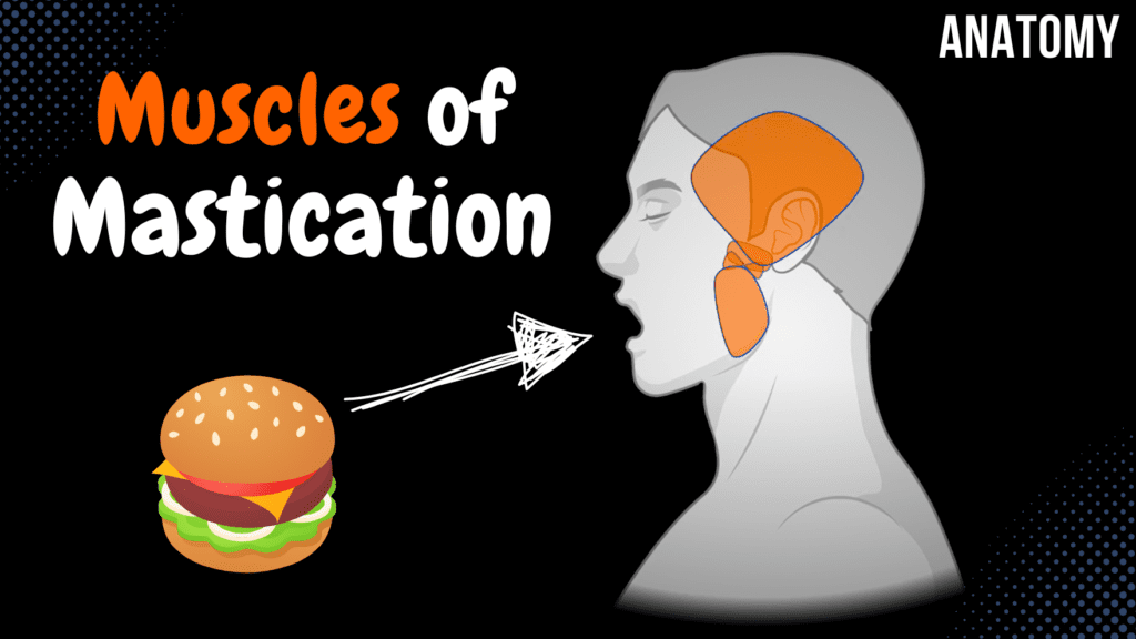
Muscles of Mastication (Origin, Insertion, Function) Official Links Instagram Youtube Jki-discord Notes & Illustrations Quizzes Summary & Transcript Notes ☆ Members Only Go to PDF Notes Illustrations ☆ Members Only Go to Illustrations 12345678910 Muscles of Mastication – QUIZ Test your understanding with 10 random multiple-choice questions from the question bank. You're in the preview mode. Note: All elements work correctly on the front end. 1 / 10 What structure forms the origin of the deep part of the masseter muscle? A) Deep surface of zygomatic arch B) Temporal fossa C) Lateral plate of pterygoid process D) Infratemporal crest The deep part of the masseter muscle originates from the deep surface of the zygomatic arch. 2 / 10 What is the insertion point of the temporal muscle? A) Coronoid process B) Masseteric tuberosity C) Mandibular angle D) Pterygoid fovea The temporal muscle inserts onto the coronoid process of the mandible. 3 / 10 Which muscle originates from the maxillary tuberosity and pterygoid fossa of the sphenoid bone? A) Medial pterygoid B) Lateral pterygoid C) Masseter D) Temporal muscle The medial pterygoid muscle originates from the maxillary tuberosity (superficial head) and the pterygoid fossa (deep head). 4 / 10 Which muscle has its origin on the temporal fossa and inserts onto the coronoid process of the mandible? A) Temporal muscle B) Lateral pterygoid C) Masseter D) Medial pterygoid The temporal muscle originates from the temporal fossa and inserts onto the coronoid process of the mandible. 5 / 10 What is the insertion of the masseter muscle? A) Mandibular angle B) Masseteric tuberosity C) Pterygoid fovea D) Coronoid process The masseter inserts into the masseteric tuberosity of the mandible. 6 / 10 What is the function of the medial pterygoid muscle during bilateral contraction? A) Retracts the mandible B) Elevates the mandible C) Depresses the mandible D) Protracts the mandible The medial pterygoid muscle elevates the mandible during bilateral contraction. 7 / 10 Which muscle originates from the zygomatic arch and inserts into the masseteric tuberosity? A) Masseter B) Medial pterygoid C) Temporal muscle D) Lateral pterygoid The masseter muscle originates from the zygomatic arch and inserts into the masseteric tuberosity. 8 / 10 Which muscle inserts into the pterygoid fovea on the neck of the mandible? A) Masseter B) Medial pterygoid C) Temporal muscle D) Lateral pterygoid The lateral pterygoid muscle inserts into the pterygoid fovea on the mandible’s neck. 9 / 10 Which nerve innervates all the muscles of mastication? A) Glossopharyngeal nerve (CN IX) B) Mandibular nerve (CN V3) C) Facial nerve (CN VII) D) Hypoglossal nerve (CN XII) All muscles of mastication are innervated by the mandibular nerve, a branch of the trigeminal nerve (CN V3). 10 / 10 What is the insertion point of the lateral pterygoid muscle? A) Mandibular angle B) Coronoid process C) Pterygoid fovea D) Masseteric tuberosity The lateral pterygoid muscle inserts into the pterygoid fovea of the mandible. Your score is The average score is 0% Description In this video, I go through the Muscles of Mastication, covering their origin, insertion, and function. Understanding these muscles is essential for comprehending jaw movements and chewing mechanics. Muscles of Mastication Temporal Muscle (Musculus Temporalis) Origin: Temporal fossa Parietal bone – Inferior temporal line Insertion: Coronoid process of the mandible (Processus coronoideus mandibulae) Function: Elevates mandible (Anterior fibers) Retracts mandible (Posterior fibers) Masseter Muscle (Musculus Masseter) Origin: Zygomatic Arch (Arcus zygomaticus) Insertion: Masseteric tuberosity of mandible (Tuberositas masseterica mandibulae) Function: Elevates mandible Medial Pterygoid Muscle (Musculus Pterygoideus Medialis) Origin: Superficial Part (Pars superficialis): Maxillary tuberosity (Tuberositas maxillae) Deep Part (Pars profunda): Pterygoid fossa of pterygoid process (Fossa pterygoidea ossis sphenoidalis) Function: Elevates mandible (Bilateral contraction) Frictional masticatory movement (Unilateral contraction) Lateral Pterygoid Muscle (Musculus Pterygoideus Lateralis) Origin: Superior Head (Caput superius): Infratemporal surface + Sphenoid Bone (Greater Wing) (Facies infratemporalis + alae majoris ossis sphenoidalis) Inferior Head (Caput inferius): Lateral lamina of pterygoid process (Sphenoidal bone) (Lamina lateralis processus pterygoidei) Insertion: Pterygoid fovea of the mandible (Fovea pterygoidea mandibulae) Function: Pushes mandible forward (Bilateral contraction) Frictional masticatory movement (Unilateral contraction) Transcript Introduction0:03Hey, what’s up. Meditay here and this.. is my first video of the muscular anatomy series.0:08So in this segment, we’re gonna cover all muscles of mastication. Which are a part0:13of the muscles of the head. Alright so All muscles of the head are divided into0:18two groups. The first group is the muscles of mastication. Mastication means to chew,0:23so those are the muscles responsible for chewing when you’re eating. And the second group is gonna0:28be fascial muscles or the muscles that are gonna be responsible for facial expression. So0:33we’re gonna focus on the mastication muscles here. So in this video, we’re gonna cover the origin and0:38insertion points of the 4 muscles of mastication, which are the Temporal, Masseter, Medial0:44Pterygoid, and Lateral Pterygoid muscles. And then in the next video, we’ll cover the facial muscles.0:50Alright. Before we start, I want you to have some basic understandingParts of Skeletal Muscle0:55of what makes up the different parts of a muscle. All the muscles in our body consist of an Origin1:01point, which is the part of the muscle that’s attached to a bone that does not move or move1:06a very slightly bit during contraction. It has a head, which is the proximal part of the muscle,1:11there’s a belly which is the widest part of the muscle. Then there’s a tail and an Insertion1:17point, which is the part of the muscle that’s attached to a bone that moves during contraction.1:22When you’re studying muscles in general, the origin and insertion points are what we1:27usually focus on. And again keep in mind that the origin point is the least movable part,1:32and the insertion point is the part of the bone that is moved during contraction.1:37In some locations you’ll also see the word belly, usually if the muscle is divided into two parts,1:42that’s when we mention this term. Awesome. Let’s now cover the muscles of mastication.Muscles of Mastication1:47The muscles of mastication again, consist of the temporal muscle. Masseter muscle, Medial Pterygoid1:53muscle, and Lateral
Sympathetic Nervous System
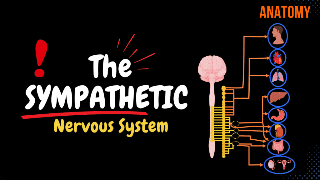
Sympathetic Nervous System (Ganglia, Neurons, Plexuses) Official Links Instagram Youtube Jki-discord Notes & Illustrations Quizzes Summary & Transcript Notes ☆ Members Only Go to PDF Notes Illustrations ☆ Members Only Go to Illustrations 12345678910 Sympathetic Nervous System – QUIZ Test your understanding with 10 random multiple-choice questions from the question bank. You're in the preview mode. Note: All elements work correctly on the front end. 1 / 10 What is the effect of sympathetic stimulation on the heart? A) Bronchoconstriction B) Vasodilation C) Decrease in heart rate D) Increase in heart rate Sympathetic stimulation increases heart rate and myocardial contractility via β1-adrenergic receptors. 2 / 10 Which plexus regulates bronchodilation? A) Hypogastric plexus B) Pulmonary plexus C) Cardiac plexus D) Celiac plexus The pulmonary plexus regulates bronchodilation during sympathetic activation. 3 / 10 Which nerve contributes to the pulmonary plexus? A) Greater splanchnic nerve B) Lumbar splanchnic nerve C) Thoracic splanchnic nerves D) Sacral splanchnic nerve Thoracic splanchnic nerves contribute to the pulmonary plexus. 4 / 10 What is the primary function of the superior cervical ganglion? A) Supplies the pelvis B) Supplies the head and neck C) Supplies the abdomen D) Supplies the thorax The superior cervical ganglion supplies sympathetic innervation to the head and neck. 5 / 10 What is the function of alpha-1 adrenergic receptors in the sympathetic system? A) Vasoconstriction B) Bronchoconstriction C) Vasodilation D) Bronchodilation Alpha-1 adrenergic receptors cause vasoconstriction to increase blood pressure. 6 / 10 Which nerve supplies sympathetic innervation to the kidneys? A) Least splanchnic nerve B) Greater splanchnic nerve C) Lumbar splanchnic nerve D) Lesser splanchnic nerve The least splanchnic nerve provides sympathetic innervation to the kidneys. 7 / 10 What is the role of grey rami communicantes? A) Carry postganglionic fibers to spinal nerves B) Connect the sympathetic trunk to the adrenal medulla C) Regulate neurotransmitter release D) Carry preganglionic fibers to ganglia Grey rami communicantes carry unmyelinated postganglionic fibers to spinal nerves. 8 / 10 Which ganglion is part of the cervical sympathetic trunk? A) Celiac ganglion B) Superior mesenteric ganglion C) Superior cervical ganglion D) Inferior hypogastric ganglion The superior cervical ganglion is located at the top of the sympathetic trunk in the neck. 9 / 10 Which sympathetic ganglion is associated with the cardiac plexus? A) Lumbar ganglion B) Thoracic ganglion C) Coccygeal ganglion D) Cervical ganglion The thoracic ganglia provide sympathetic fibers to the cardiac plexus, affecting heart function. 10 / 10 Which ganglia are part of the prevertebral sympathetic ganglia? A) Celiac, superior, and inferior mesenteric ganglia B) Sacral ganglia C) Cervical ganglia D) Thoracic ganglia The celiac, superior mesenteric, and inferior mesenteric ganglia are prevertebral ganglia. Your score is The average score is 0% Description Sympathetic Nervous System; A system of fight or flight, that increases energy expenditure and catabolism. It increases heart rate and myocardial contractility, dilates bronchi, increases blood pressure and indirectly increases blood glucose and lipid levels. It’s a part of the autonomic nervous system, which is under the motor division of peripheral nervous system. General Structures and terms: Group of cell bodies in CNS – Nucleus Group of cell bodies outside CNS – Ganglion Paravertebral ganglia Prevertebral ganglia Presynaptic neuron is a cholinergic neuron (Ach binds to nicotinic receptors, allowing an influx of cations) Postganglionic neurons are primarily adrenergic neurons (norepinephrine and epinephrine binds to adrenergic receptors, activating g-protein) Sympathetic Preganglionic Neurons: Arise from intermediolateral nuclei (C8/T1–L2/L3) White rami communicantes (rami communicantes albi) myelinated axons from the intermediolateral nucleus that enter the sympathetic trunk. Then: May terminate in the sympathetic ganglion at the same level May travel up or down the sympathetic trunk to terminate in ganglia at a higher or lower level May leave the sympathetic trunk to terminate in a peripheral ganglion Sympathetic Postganglionic neurons: Grey rami communicantes (rami communicantes grisei) Join spinal nerves and innervate cutaneous glands and erector pili muscle. Vascular branches Travel with arteries to provide sympathetic innervation of organs and limbs Visceral branches and splanchnic nerves (rami viscerales, nervi splanchnici) travel towards the organs Sympathetic Trunk: Cervical ganglia (ganglia cervicalia) Superior cervical ganglion Middle cervical ganglion Inferior cervical ganglion aka Cervicothoracic, stellate ganglion Thoracic ganglia (ganglia thoracica) Lumbar ganglia (ganglia lumbalia) Sacral ganglia (ganglia sacralia) Coccygeal ganglia, often fused to form ganglion impar (unpaired). Innervation of Head, Neck and Thorax: Superior Cervical postganglionic fibers: Grey rami communicans join the first 4 cervical nerves Internal carotid nerve (nervus caroticus internus) External carotid nerves (nervi carotici externi) Laryngopharyngeal branches (rami laryngopharyngei) Superior cervical cardiac nerve (nervus cardiacus cervicalis superior) Middle cervical postganglionic fibers: Grey rami communicans join 5th and 6th cervical nerves Visceral branches (rami viscerales) Middle cervical cardiac nerve (nervus cardiacus cervicalis medius) Inferior cervical postganglionic fibers Grey rami communicans join C7, C8 and T1 Vertebral nerve (nervus vertebralis) Inferior cervical cardiac nerve (nervus cardiacus cervicalis inferior) Thoracic ganglia Gray rami communicans join intercostal nerves Thoracic cardiac nerves (nervi cardiaci thoracici) form Cardiac plexus to increase contractility and heart rate Pulmonary branches (rami pulmonales) form pulmonary plexus to cause bronchodilation Oesophageal branches (rami oesophagei) form esophageal plexus to decrease peristalsis Innervation of abdomen and pelvis: Preganglionic fibers go towards prevertebral ganglia Greater splanchnic nerve (nervus splanchnicus major) travel towards celiac ganglia (ganglion ciliare), postganglionic fibers go through plexuses and reach proximal organs in abdomen Preganglionic fibers of greater splanchnic nerve go directly to renal medulla Lesser splanchnic nerve (nervus splanchnicus minor) towards superior mesenteric ganglion for the duodenum, small intestine, large intestine until proximal 2/3. Least splanchnic nerve (nervus splanchnicus imus) go towards the aorticorenal ganglion then towards the kidneys and the ureter Lumbar splanchnic nerves (nervi splanchnici lumbales) go towards the inferior mesenteric ganglia, then towards rest of large intestine, urinary bladder and genitalia Sacral splanchnic nerves (nervi splanchnici sacrales) Sources: Singh, I. (2017). Human neuroanatomy (10th ed) Kozlowski, T. (2017). Memorix Anatomy 2nd ed Gray’s Anatomy: The Anatomical Basis of Clinical Practice (42th ed.) Sinnatamby, Chummy S. (2011). Last’s Anatomy (12th ed.) Yan FL, Zhang JH. Role of the sympathetic nervous system
Parasympathetic Nervous System

Parasympathetic Nervous System (Craniosacral Outflow) Official Links Instagram Youtube Jki-discord Notes & Illustrations Quizzes Summary & Transcript Notes ☆ Members Only Go to PDF Notes Illustrations ☆ Members Only Go to Illustrations 12345678910 Parasympathetic Nervous System – QUIZ Test your understanding with 10 random multiple-choice questions from the question bank. You're in the preview mode. Note: All elements work correctly on the front end. 1 / 10 What is the main parasympathetic output of the vagus nerve? A) Body wall and limbs B) Lacrimal glands C) Thoracic and abdominal organs D) Salivary glands The vagus nerve (CN X) supplies parasympathetic fibers to thoracic and abdominal organs. 2 / 10 What is the primary neurotransmitter at parasympathetic ganglionic synapses? A) Dopamine B) Acetylcholine C) Serotonin D) Norepinephrine Acetylcholine (ACh) is the primary neurotransmitter at parasympathetic ganglionic synapses. 3 / 10 Which cranial nerve originates from the Edinger-Westphal nucleus? A) Vagus nerve (CN X) B) Facial nerve (CN VII) C) Oculomotor nerve (CN III) D) Glossopharyngeal nerve (CN IX) The oculomotor nerve (CN III) originates from the Edinger-Westphal nucleus and controls eye functions. 4 / 10 Which ganglion is involved in pupillary light reflex? A) Submandibular ganglion B) Pterygopalatine ganglion C) Otic ganglion D) Ciliary ganglion The ciliary ganglion mediates pupillary light reflex through parasympathetic pathways. 5 / 10 Which parasympathetic ganglion is involved in taste and salivation? A) Otic ganglion B) Ciliary ganglion C) Submandibular ganglion D) Pterygopalatine ganglion The submandibular ganglion is responsible for salivation from the sublingual and submandibular glands. 6 / 10 Which ganglion is associated with lacrimal gland secretion? A) Ciliary ganglion B) Pterygopalatine ganglion C) Otic ganglion D) Submandibular ganglion The pterygopalatine ganglion is associated with lacrimal gland innervation by the facial nerve. 7 / 10 Which nucleus is responsible for parasympathetic control of gastric motility? A) Posterior vagus nucleus B) Edinger-Westphal nucleus C) Superior salivatory nucleus D) Inferior salivatory nucleus The posterior nucleus of the vagus nerve controls gastric motility and secretion via parasympathetic output. 8 / 10 Which ganglion is responsible for salivation from the parotid gland? A) Submandibular ganglion B) Ciliary ganglion C) Otic ganglion D) Pterygopalatine ganglion The otic ganglion receives fibers from CN IX and controls the parotid gland’s salivary secretion. 9 / 10 Which ganglion provides parasympathetic control to the ciliary body? A) Submandibular ganglion B) Otic ganglion C) Pterygopalatine ganglion D) Ciliary ganglion The ciliary ganglion controls the ciliary body and the sphincter pupillae for accommodation and constriction. 10 / 10 What is the parasympathetic function of the vagus nerve? A) Innervates lacrimal glands B) Innervates thoracic and abdominal organs C) Innervates salivary glands D) Innervates the pupils It innervates thoracic and abdominal organs, regulating heart rate, digestion, and other autonomic functions. Your score is The average score is 0% Description Parasympathetic Nervous System Overview The parasympathetic nervous system is a division of the autonomic nervous system, which is part of the motor division of the peripheral nervous system. Neuroanatomy: General Structures and Terms Group of cell bodies in CNS: Nucleus Group of cell bodies outside CNS: Ganglion Presynaptic neuron: Cholinergic neuron (Acetylcholine binds to nicotinic receptors, allowing an influx of cations) Postganglionic neurons: Cholinergic neurons (Acetylcholine binds to muscarinic receptors of the target organ, activating a G-protein) Functional Aspects of Sympathetic and Parasympathetic Systems Territory: Sympathetic: All areas of the body Parasympathetic: No innervation in body walls and limbs Activity: Sympathetic: More generalized (1:15 ratio of pre- to postganglionic neurons) Parasympathetic: More specific (1:2 ratio of pre- to postganglionic neurons) Functions: Sympathetic: “Fight-or-Flight” – increased heart rate, bronchodilation, decreased gut motility Parasympathetic: “Rest-and-Digest” – decreased heart rate, bronchoconstriction, increased gut motility Cranial Outflow Oculomotor nerve (nervus oculomotorius) Edinger-Westphal nucleus → Ciliary ganglion → Ciliary body, sphincter pupillae Facial nerve (nervus facialis) Superior salivatory nucleus → Pterygopalatine ganglion → Lacrimal, nasal, and palatine glands Chorda tympani → Submandibular ganglion → Submandibular and sublingual glands Glossopharyngeal nerve (nervus glossopharyngeus) Inferior salivatory nucleus → Otic ganglion → Parotid gland Vagus nerve (nervus vagus) Posterior nucleus of the vagus → Smooth muscle, glands, internal organs Sacral Outflow Sacral parasympathetic nuclei (S2-S4) Sacral splanchnic nerves → Inferior hypogastric plexus → Pelvic organs Sources Used Singh, I. (2017). *Human Neuroanatomy (10th ed.)* Wineski, L. E. (2019). *Snell’s Clinical Anatomy by Regions (10th ed.)* Kozlowski, T. (2017). *Memorix Anatomy: The Complete Study Guide* Fryer, A. D., & Jacoby, D. B. (1998). *Muscarinic Receptors and Control of Airway Smooth Muscle* University lectures and notes Programs: Complete Anatomy, Biorender, PowerPoint, Camtasia Transcript Introduction0:06What’s up, Taim talks med here. In this video we’re gonna talk about the parasympathetic nervous0:10system. As you see from this brief diagram, the sympathetic and the parasympathetic parts of our0:16nervous system controls more or less all our internal organs. Sympathetic being the fight0:21or flight response, and parasympathetic being the rest and digest response. And they’re both0:27as you see here a part of the autonomic nervous system, which again is the motor0:32division of our peripheral nervous system. I did make an introductory video about the peripheral0:37nervous system, so if you guys have absolutely no clue what the peripheral nervous system is,0:42I urge you to watch that one first. But all in all I’ll try to simplify the parasympathetic0:48nervous system as much as I can so that it’ll make sense at a detailed level, within the0:54aspects of anatomy and physiology at least. So, in this video, we’re going detailed into0:59the parasympathetic nervous system. And we’re gonna do that by first going1:04through the general structure and terms associated with the parasympathetic aspect of the autonomic1:09nervous system. Basically talk a little bit about ganglia, the pre and post synaptic1:14neurons and their neurotransmitters, and basically how the parasympathetic1:18nervous system is built in general. Then we’re gonna talk about the cranial outflow,1:23and go quickly through the pathway of the cranial nerves involved and what structures1:27they innervate. And then run through the sacral outflow, where it originates from1:32and basically what it innervates and its function. Let’s go ahead and begin with some terms. Now theGanglion and Nucleus1:39autonomic nervous system –