
Kidneys
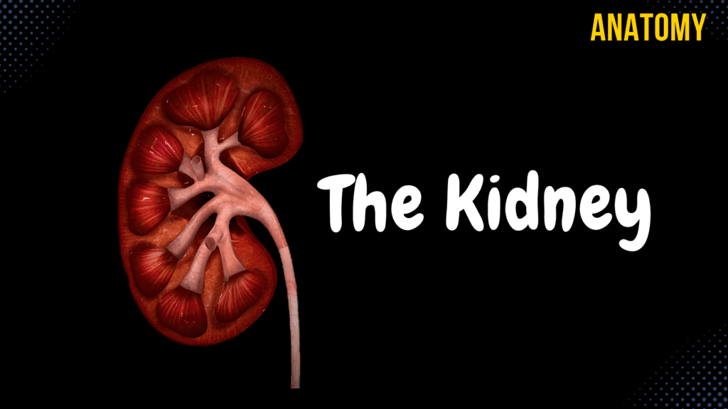
Kidneys (Functions, Structures, Coverings, Nephron) Official Links Instagram Youtube Jki-discord Notes & Illustrations Quizzes Summary & Transcript Notes ☆ Member Only Go to PDF Notes Illustrations ☆ Member Only Go to Illustrations 12345678910 Kidneys – QUIZ Test your understanding with 10 random multiple-choice questions from the question bank. You're in the preview mode. Note: All elements work correctly on the front end. 1 / 10 Which structure is found between the renal pyramids? A) Medullary rays B) Renal papillae C) Renal columns D) Renal lobes Renal columns are extensions of the cortex that separate the renal pyramids. 2 / 10 What is the apex of a renal pyramid called? A) Medulla B) Renal column C) Cortex D) Renal papilla The apex of a renal pyramid is called the renal papilla, where urine flows into the minor calyces. 3 / 10 What is the approximate thickness of the renal cortex? A) 15-20 mm B) 11-15 mm C) 4-11 mm D) 2-5 mm The renal cortex is approximately 4-11 mm thick. 4 / 10 Which anatomical structure collects urine from multiple collecting ducts? A) Renal pelvis B) Renal pyramid C) Renal papilla D) Minor calyx The minor calyx collects urine from multiple collecting ducts and funnels it into the major calyces. 5 / 10 Which structure anchors the kidneys to surrounding tissues? A) Perinephric fat B) Renal fascia C) Paranephric fat D) Fibrous capsule The renal fascia anchors the kidney and adrenal gland to surrounding structures. 6 / 10 What is the function of the renal pelvis? A) Filters plasma B) Anchors the kidney C) Produces hormones D) Funnels urine into the ureter The renal pelvis collects urine from the major calyces and funnels it into the ureter. 7 / 10 What is the terminal part of the nephron that delivers urine into the renal pelvis? A) Collecting duct B) Loop of Henle C) Renal corpuscle D) Proximal convoluted tubule The collecting duct collects urine from the nephron and empties into the renal pelvis. 8 / 10 Which structure of the kidney contains renal corpuscles? A) Renal medulla B) Renal cortex C) Renal sinus D) Renal pyramid The renal cortex contains renal corpuscles, proximal and distal convoluted tubules. 9 / 10 What forms the apex of the renal pyramid? A) Renal column B) Renal papilla C) Renal hilum D) Renal lobe The renal papilla forms the apex of the renal pyramid and drains urine into the minor calyces. 10 / 10 What is the primary site of plasma filtration in the kidney? A) Loop of Henle B) Collecting duct C) Bowman’s capsule D) Glomerulus Plasma filtration occurs in the glomerulus, a network of capillaries in the renal corpuscle. Your score is The average score is 0% Description Kidney Anatomy: Topography, Structures, and Function This video covers a comprehensive overview of the kidneys, including their topography, functions, external and internal structures, and nephron anatomy. 1. Topography of the Kidney The right kidney is slightly lower than the left. Both kidneys start at T12. Left kidney: Ends at L2. Right kidney: Ends at L3. 2. Functions of the Kidney Plasma Filtration: Through nephrons. Excretion: Removal of metabolic waste. Acid-Base Homeostasis: Regulates pH balance. Hormone Production: Erythropoietin, Renin. Vitamin D Metabolism: Activation of Vitamin D. 3. External Structures Weight: 120-200 grams. Size: 10-13 cm long, 5-6 cm wide, 4 cm thick. Inferior Pole (Extremitas Inferior). Superior Pole (Extremitas Superior): Suprarenal gland located on top. Lateral Border (Margo Lateralis). Medial Border (Margo Medialis): Contains the hilum (Hilum Renalis). 4. Coverings of the Kidney Fibrous Capsule (Capsula Fibrosa). Perinephric Fat (Capsula Adiposa). Renal Fascia (Fascia Renalis): Anterior Layer: Prerenal Layer (Lamina Prerenalis) / Fascia of Toldt. Posterior Layer: Retrorenal Layer (Lamina Retrorenalis) / Fascia of Zuckerkandl. Both layers fuse laterally. Peritoneum. 5. Internal Structures Renal Sinus Sinus Renalis: Fat-filled space inside the kidney. Renal Pelvis (Pelvis Renalis): Central collecting region. Renal Calyces (Calyces Renales): Divided into: Minor Renal Calyces (Calyx Renales Minor). Major Renal Calyces (Calyx Renales Major). Renal Medulla Renal Pyramids (Pyramides Renales). Renal Lobes (Lobi Renales). Renal Papillae (Papillae Renales): Tip of pyramids. Renal Medullary Rays (Radii Medullares): Contain nephrons’ loops. Loop of Henle and Collecting Ducts. Openings of Papillary Ducts (Foramina Papillaria). Renal Cortex Thickness: 4-11 mm. Renal Columns (Columna Renalis): Extension of the cortex into the medulla. Contains Renal Corpuscles, Proximal & Distal Convoluted Tubules. 6. Renal Lobe & Nephron Renal Corpuscle (Corpusculum Renis) Glomerulus: Network of capillaries. Afferent Arteriole (Vas Afferens): Brings blood in. Efferent Arteriole (Vas Efferens): Drains blood out. Glomerular Capsule (Capsula Glomeruli): Encases glomerulus. Nephron Tubules Proximal Convoluted Tubule (Tubulus Proximalis): Reabsorbs nutrients. Has a convoluted part and a straight part. Loop of Henle (Ansa Nephroni): Descending Limb. Ascending Limb. Distal Convoluted Tubule (Tubulus Distalis): Fine-tunes ion balance. Collecting Duct (Ductus Colligentes): Final urine concentration. Papillary Duct (Ductus Papillares): Empties into minor calyx. 7. Clinical Relevance Polycystic Kidney Disease (PKD): Genetic disorder causing multiple cysts in the kidney. Kidney Stones (Nephrolithiasis): Crystallized minerals causing obstruction. Acute Kidney Injury (AKI): Sudden loss of kidney function. Chronic Kidney Disease (CKD): Progressive decline in kidney function. Sources Used: Memorix Anatomy (2nd Edition) – Hudák Radovan, Kachlík David, Volný Ondřej. Complete Anatomy by 3D4Medical. Biorender. University notes and lectures. Transcript Introduction0:03What’s up.0:04Meditay here.0:05Let’s talk about the anatomy of the urinary system.0:07In this segment, we will be talking about the anatomy of the Kidneys.0:10Alright, so the Urinary system consists of all the organs involved in handling the urine.0:15These are the Kidneys, the Ureter, the Urinary bladder, and the urethra.0:20Our goal is to cover the anatomy of all these structures you see here, step by step, and0:25we’ll start with the Kidneys In this video, we’re first going to talk about0:28the functions of the kidneys.0:30Then we’ll talk about the external structures and the coverings of the kidneys.0:34After that, we’ll open up the kidney and cover the internal structures.0:39When we’re done with the kidneys, we’ll talk about the general anatomy of the nephron,0:42which is the functional unit of the kidney.Topography of Kidney0:45Alright, so here you see
Male Genital System
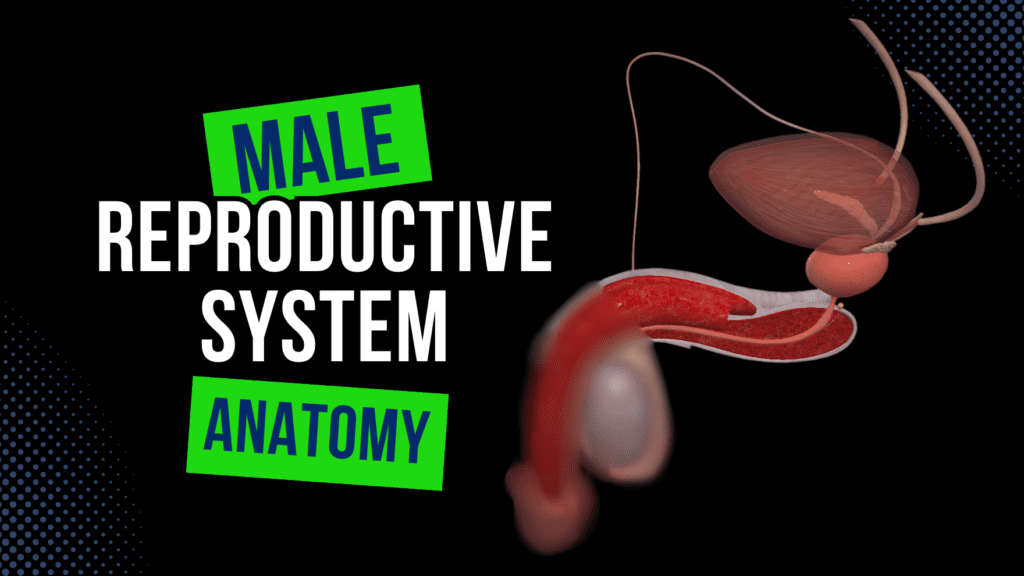
Male Genital System (Internal & External) Official Links Instagram Youtube Jki-discord Notes & Illustrations Quizzes Summary & Transcript Notes ☆ Member Only Go to PDF Notes Illustrations ☆ Member Only Go to Illustrations 12345678910 Male Genital System – QUIZ Test your understanding with 10 random multiple-choice questions from the question bank. You're in the preview mode. Note: All elements work correctly on the front end. 1 / 10 What structure carries sperm from the epididymis to the ejaculatory duct? A) Pampiniform plexus B) Ductus deferens C) Tunica vaginalis D) Prostate gland The ductus deferens transports sperm from the epididymis to the ejaculatory duct. 2 / 10 What structure anchors the penis to the pubic symphysis? A) Tunica albuginea B) Fundiform ligament C) Dartos fascia D) Suspensory ligament The suspensory ligament anchors the penis to the pubic symphysis. 3 / 10 What is the main role of Sertoli cells? A) Regulate blood supply B) Secrete testosterone C) Produce ejaculate fluid D) Regulate spermatogenesis Sertoli cells provide structural support and regulate spermatogenesis in the seminiferous tubules. 4 / 10 What is the name of the structure where the ductus deferens and seminal gland ducts join? A) Spongy urethra B) Prostatic urethra C) Membranous urethra D) Ejaculatory duct The ductus deferens and seminal gland ducts join to form the ejaculatory duct. 5 / 10 Which artery supplies blood to the ductus deferens? A) Artery to ductus deferens B) External iliac artery C) Pampiniform plexus D) Testicular artery The artery to the ductus deferens arises from the internal iliac artery and supplies the ductus deferens. 6 / 10 What is the function of the urethral crest in the prostate? A) Facilitates ejaculation B) Produces sperm C) Provides structural support D) Stores sperm The urethral crest provides structural support to the prostatic urethra. 7 / 10 What layer of the testis is responsible for the formation of septa and lobules? A) Cremaster muscle B) Tunica albuginea C) Vascular layer D) Internal spermatic fascia The tunica albuginea is a dense connective tissue layer that extends inward to form septa, dividing the testis into lobules. 8 / 10 Which artery primarily supplies the testis? A) Testicular artery B) Pampiniform plexus C) External iliac artery D) Internal iliac artery The testicular artery arises from the abdominal aorta and supplies the testis. 9 / 10 What is the primary function of the prostate gland? A) Facilitates sperm maturation B) Produces nourishing fluid for sperm C) Produces hormones D) Regulates testicular function The prostate gland produces a fluid that nourishes and protects sperm. 10 / 10 What does the seminal gland secrete? A) Mucus B) Spermatogenic fluid C) Testosterone D) Fructose-rich fluid The seminal gland secretes a fluid rich in fructose and prostaglandins to nourish sperm. Your score is The average score is 0% Description Complete Overview of Male Genital Organs (Internal & External) This video provides a comprehensive breakdown of male internal and external genital anatomy, covering structures, histology, functions, and clinical relevance. 1. Internal Genital Organs Testis Key Features: Male reproductive gland responsible for sperm production. Leydig Cells: Produce testosterone. Sertoli Cells: Structural support and aid spermatogenesis. Prenatal Development: Originates in the lumbar region. Descends and takes abdominal layers, forming scrotal layers. External Structures: Upper Pole (Extremitas Superior). Lower Pole (Extremitas Inferior). Anterior Border (Margo Anterior). Posterior Border (Margo Posterior). Lateral Surface (Facies Lateralis). Medial Surface (Facies Medialis). Internal Structures: Tunica Albuginea. Vascular Layer (Tunica Vasculosa). Septa of Testis (Septula Testis). Lobules of Testis (Lobuli Testis). Seminiferous Tubules (Tubuli Seminiferi Contorti & Recti). Rete Testis. Efferent Ductules (Ductuli Efferentes Testis). Epididymis Composed of tightly packed tubules; stores sperm. Head (Caput Epididymidis), Body (Corpus Epididymidis), Tail (Cauda Epididymidis). Coverings & Fixation of Testes & Epididymis Tunica Vaginalis: Visceral (Epiorchium) & Parietal Layer (Periorchium). Fascial Layers: Internal Spermatic Fascia, Cremaster, External Spermatic Fascia. Dartos Fascia (Tunica Dartos). Spermatic Cord (Funiculus Spermaticus) Contains Ductus Deferens, Testicular Artery, Pampiniform Plexus, Lymph Vessels. Ductus Deferens Pathway for sperm transport. Divided into Scrotal, Funicular, Inguinal, & Pelvic Parts. Seminal Gland (Glandula Vesiculosa) Produces ejaculate fluid. Joins ductus deferens to form Ejaculatory Duct. Prostate (Prostata) Structure: Base & Apex. Posterior & Anterior Surfaces. Prostatic Urethra. Histological Zones: Peripheral, Central, Transitional, & Anterior Fibromuscular Zones. Posterior Features: Urethral Crest, Seminal Colliculus, Prostatic Utricle (Fetal Remnant). Openings of Ejaculatory Ducts. Bulbo-Urethral Glands (Glandulae Bulbourethrales) Empties into the spongy urethra. Male Urethra (Urethra Masculina) Prostatic, Membranous, and Spongy Urethra. 2. External Genital Organs Penis External Structures: Root (Bulb & Crura). Body (Corpora Cavernosa & Corpus Spongiosum). Glans Penis (Corona, Collum, Prepuce, Frenulum). Internal Structures: Corpora Cavernosa & Corpus Spongiosum. Tunica Albuginea. Buck’s Fascia (Deep Fascia of Penis). Scrotum Contains layers derived from abdominal structures: Tunica Vaginalis, Internal & External Spermatic Fasciae, Cremaster Muscle, Dartos Fascia. 3. Clinical Relevance Testicular Torsion: Twisting of the spermatic cord leading to ischemia. Benign Prostatic Hyperplasia (BPH): Enlargement of the prostate affecting urination. Cryptorchidism: Failure of testis to descend. Sources Used: Memorix Anatomy (2nd Edition) – Hudák Radovan, Kachlík David, Volný Ondřej. Complete Anatomy by 3D4Medical. Biorender. Snell, Richard S. (2012). Clinical Anatomy by Regions (9th Edition). University notes and lectures. Transcript Introduction0:03What’s up.0:04Let’s go ahead and cover the male genital system.0:07We will cover the most important aspect of the male genitals, and hopefully, in the end,0:12you’ll have a pretty good understanding about this topic0:15Alright.0:16The male genital organs are divided into the internal genital organs and the external genital0:22organs.0:23We’ll cover both of these in detail, And we’ll start with the internal genitalInternal Genital Organs0:27organs So the internal genital organs consist of0:31the testes, epididymis, ductus deferens, seminal gland, Ejaculatory Duct, Prostate, Male urethra,0:40and the bulbourethral glands.0:43Alright, let’s follow this arrangement, starting with the testis.Testis0:47Now, of the whole male reproductive system, the primary functional cells the male has0:54are Leydig and Sertoli cells.0:56And both of them are found in the testes.1:00So the testicle or testis is the male reproductive gland or gonad in all animals, not just humans.1:07Leydig cells produce testosterone, which is a steroid hormone that binds to intracellular1:14receptors and regulates protein synthesis.1:18And what
Female Genital System
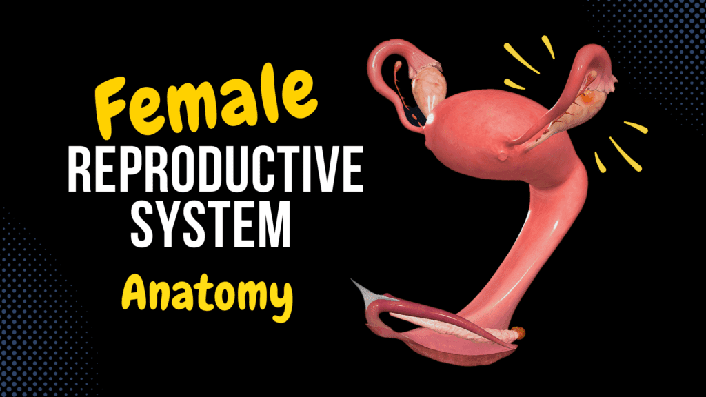
Female Genital System (Internal & External) Official Links Instagram Youtube Jki-discord Notes & Illustrations Quizzes Summary & Transcript Notes ☆ Member Only Go to PDF Notes Illustrations ☆ Member Only Go to Illustrations 12345678910 Female Genital System – QUIZ Test your understanding with 10 random multiple-choice questions from the question bank. You're in the preview mode. Note: All elements work correctly on the front end. 1 / 10 What is the purpose of the broad ligament in the female genital system? A) Facilitate uterine contractions B) Supply blood to ovaries C) Separate abdominal organs D) Support internal organs The broad ligament is a double fold of peritoneum that supports the uterus, uterine tubes, and ovaries, helping maintain their position. 2 / 10 Which lymph nodes drain the external genitalia in females? A) Para-aortic B) Lumbar C) Internal iliac D) Superficial inguinal The superficial inguinal lymph nodes primarily drain the external genitalia, including the labia majora and minora. 3 / 10 What is the function of the zona pellucida in oocyte development? A) Provides nutrients B) Supports the corpus luteum C) Facilitates sperm binding D) Produces follicular fluid The zona pellucida surrounds the oocyte and facilitates sperm binding, protecting the developing oocyte. 4 / 10 Which veins primarily drain the blood from the ovaries? A) Vaginal venous plexus B) Ovarian veins C) Uterine venous plexus D) Pudendal veins The right ovarian vein drains into the inferior vena cava, while the left ovarian vein drains into the left renal vein. 5 / 10 What is the histological structure of the vaginal epithelium? A) Stratified squamous B) Simple columnar C) Simple cuboidal D) Transitional epithelium The vaginal epithelium is composed of stratified squamous non-keratinized epithelium, which stores glycogen. 6 / 10 What is the histological lining of the uterine cavity? A) Transitional epithelium B) Simple columnar C) Simple cuboidal D) Stratified squamous The uterine cavity is lined by simple columnar epithelium that forms the endometrium. 7 / 10 What type of follicle is ready for ovulation? A) Primary follicle B) Graafian follicle C) Secondary follicle D) Primordial follicle The Graafian follicle, or tertiary follicle, contains the secondary oocyte and is ready for ovulation. 8 / 10 What are the main functions of the ovaries? A) Produce oocytes and hormones B) Transport gametes C) Regulate blood supply D) Provide structural support The ovaries are responsible for gametogenesis and the production of hormones such as estrogen and progesterone. 9 / 10 What are the structural layers of the uterine wall? A) Endometrium, myometrium, perimetrium B) Myocardium, epimysium C) Epimetrium, perimysium D) Serosa, stroma The uterine wall is composed of the endometrium (mucosa), myometrium (muscular layer), and perimetrium (serosa). 10 / 10 What are the functional layers of the endometrium during the menstrual cycle? A) Endometrial, stromal layers B) Mucosal, serosal layers C) Functional, basal layers D) Perimetrium, myometrium The functional layer of the endometrium undergoes cyclic changes and is shed during menstruation, while the basal layer regenerates it. Your score is The average score is 0% Description Complete Overview of Female Internal and External Genital Organs This video provides a detailed anatomical breakdown of the internal and external genital structures, including their histology, functions, and clinical relevance. Small Correction: At 7:06, the ovarian artery and vein run within the suspensory ligament of the ovary, not the ligament of the ovary as originally stated. 1. Internal Genital Organs Ovary Inner Structures: Tunica Albuginea (Tunica albuginea ovarii) Ovarian Cortex (Cortex ovarii) Ovarian Medulla (Medulla ovarii) Follicles External Structures: Mesovarian Border (Margo mesovaricus) Free Border (Margo liber) Tubal Extremity (Extremitas tubaria) Uterine Extremity (Extremitas uterina) Medial Surface (Facies medialis) Lateral Surface (Facies lateralis) Fixation of the Ovary: Ligament of Ovary (Ligamentum ovarii proprium) Suspensory Ligament of Ovary (Ligamentum suspensorium ovarii) Mesovarium Growth and Maturation of Follicles Primordial Follicle: Contains primary oocyte, single layer of follicular cells. Primary Follicles: After puberty (FSH stimulation), granulosa cells form. Secondary Follicle: Zona pellucida, follicular fluid, theca folliculi, estrogen production. Graafian Follicle: Mature follicle containing a secondary oocyte, released during ovulation. Corpus Luteum: Hormone-secreting structure post-ovulation. Corpus Albicans: Remnant of corpus luteum. Uterine Tube (Tuba Uterina) Parts of the Uterine Tube: Infundibulum (Infundibulum tubae uterinae) Abdominal Ostium (Ostium abdominale) Fimbriae (Fimbriae tubae uterina) Ovarian Fimbria (Fimbria ovarica) Ampulla (Ampulla tubae uterinae) Isthmus (Isthmus tubae uterinae) Uterine Part (Pars uterina) – Uterine Ostium (Ostium uterinum) Walls of Uterine Tube: Tunica Mucosa Tunica Muscularis Tunica Serosa Mesosalpinx Uterus Parts: Positioned in anteflexion. Fundus of Uterus (Fundus uteri). Body of Uterus (Corpus uteri). Intestinal (Posterior) Surface (Facies intestinalis). Vesical (Anterior) Surface. Cervix (Portio supravaginalis cervicis and vaginal part). Internal Structures (Histology): Endometrium: Functional and basal layers, simple columnar epithelium, uterine glands. Myometrium: Smooth muscle layers, vascularized middle layer. Perimetrium: Outer serous layer. Fixation of the Uterus Peritoneal Folds: Broad Ligament (Ligamentum latum uteri) Mesovarium Mesosalpinx Mesometrium Parametrial Ligaments: Cardinal Ligament (Ligamentum cardinale uteri). Vesico-uterine Ligament. Pubocervical Ligament. Uterosacral Ligaments. Recto-uterine Ligament. Round Ligament of Uterus (Ligamentum teres uteri). 2. Vagina Vaginal Orifice (Ostium Vaginae). Vestibule (Vestibulum Vaginae). Vaginal Canal (Canalis Vaginalis). Vaginal Fornices (Anterior, Posterior, and Lateral). Vaginal Wall (Histology): Mucosa – Forms transverse folds. Muscularis Layer. Clinical Relevance: Imperforate Hymen: May require medical intervention. Recto-uterine Pouch (Pouch of Douglas): Important for fluid accumulation. 3. External Genital Organs Vestibule: Lesser and Greater Vestibular Glands (Bartholin’s Glands). Labia Minora (Labia Minora Pudendi). Clitoris: Glans, Body, Crura, Prepuce. Bulb of Vestibule (Bulbus Vestibuli). Labia Majora (Labia Majora Pudendi). Mons Pubis. Sources Used: Memorix Anatomy (2nd Edition) – Hudák Radovan, Kachlík David, Volný Ondřej. Complete Anatomy by 3D4Medical. Biorender. Transcript Introduction0:03What’s up. Meditay Here. Let’s finally talk about the female genital system. Now, this is going to0:08be a longer video than usual. It’s not because the female genital system is necessarily difficult,0:14but it’s because I want to cover everything in detail so that you understand the fundamentals of the female genital system. I’ve put some timestamps on the bottom of the video,0:23so you can use that if it’s any specific topic you came here for. Alright. So the
Portal Venous System

Portal Venous System Official Links Instagram Youtube Jki-discord Notes & Illustrations Quizzes Summary & Transcript Notes ☆ Member Only Go to PDF Notes Illustrations ☆ Member Only Go to Illustrations 12345678910 Portal Venous System – QUIZ Test your understanding with 10 random multiple-choice questions from the question bank. You're in the preview mode. Note: All elements work correctly on the front end. 1 / 10 What is the main tributary of the inferior mesenteric vein? A) Left colic vein B) Paraumbilical veins C) Superior rectal vein D) Sigmoid veins The superior rectal vein is the main tributary of the inferior mesenteric vein. 2 / 10 Which vein drains the pancreas and is a tributary of the splenic vein? A) Left gastro-omental vein B) Short gastric vein C) Right gastro-omental vein D) Pancreatic veins The pancreatic veins drain the pancreas and empty into the splenic vein. 3 / 10 Which vein is responsible for caput medusae in portal hypertension? A) Left gastric vein B) Esophageal veins C) Paraumbilical veins D) Rectal veins Caput medusae occurs due to anastomoses between the paraumbilical veins and the superficial epigastric veins. 4 / 10 Which tributary of the portal vein drains the duodenum? A) Paraumbilical veins B) Pancreaticoduodenal veins C) Cystic vein D) Right colic vein The pancreaticoduodenal veins drain the duodenum and empty into the portal vein. 5 / 10 Which tributary of the portal vein drains the liver? A) Superior mesenteric vein B) Hepatic veins C) Splenic vein D) Paraumbilical veins The hepatic veins are tributaries of the inferior vena cava and indirectly drain blood processed by the liver. 6 / 10 Which condition results from an increase in pressure in the portal venous system? A) Pulmonary hypertension B) Right-sided heart failure C) Portal hypertension D) Varicose veins Portal hypertension occurs due to increased pressure in the portal venous system. 7 / 10 Which vein drains the small intestine into the portal system? A) Splenic vein B) Inferior mesenteric vein C) Superior mesenteric vein D) Portal vein The superior mesenteric vein drains the small intestine into the portal system. 8 / 10 Which vein is a tributary of the splenic vein and drains the greater curvature of the stomach? A) Right gastro-omental vein B) Short gastric vein C) Pancreatic vein D) Left gastro-omental vein The left gastro-omental vein is a tributary of the splenic vein and drains the greater curvature of the stomach. 9 / 10 Which two veins form the portal vein? A) Superior mesenteric vein and splenic vein B) Right gastric vein and left gastric vein C) Superior mesenteric vein and inferior mesenteric vein D) Inferior mesenteric vein and splenic vein The portal vein is formed by the union of the superior mesenteric vein and the splenic vein. 10 / 10 Which vein is responsible for draining the rectum into the inferior mesenteric vein? A) Superior rectal vein B) Middle rectal vein C) Inferior rectal vein D) Paraumbilical veins The superior rectal vein drains the rectum and empties into the inferior mesenteric vein. Your score is The average score is 0% Description This video provides a detailed breakdown of the Portal Venous System, including its tributaries, anatomical significance, and the clinical relevance of porto-caval anastomoses. 1. Portal Venous System The portal venous system drains blood from the unpaired visceral organs, including the: Large Intestine Small Intestine Pancreas Stomach Spleen Gall Bladder 2. Formation of the Portal Vein (Vena Portae Hepatis) The portal vein is formed by the union of the following major veins: Superior Mesenteric Vein (Vena Mesenterica Superior) Splenic Vein (Vena Splenica) Inferior Mesenteric Vein (Vena Mesenterica Inferior) 3. Tributaries of the Portal Vein Superior Mesenteric Vein Jejunal and Ileal Veins (Venae Jejunales et Ileales) Right Colic Vein (Vena Colica Dextra) Ileocolic Vein (Vena Ileocolica) Appendicular Vein (Vena Appendicularis) Middle Colic Vein (Vena Colica Media) Right Gastro-omental Vein (Vena Gastroomentalis Dextra) Inferior Mesenteric Vein Opens into either the splenic vein, superior mesenteric vein, or the portal vein. Superior Rectal Vein (Vena Rectalis Superior) Sigmoid Vein (Vena Sigmoideae) Left Colic Vein (Vena Colica Sinistra) Splenic Vein Starts from the splenic hilum. Left Gastro-omental Vein (Vena Gastroomentalis Sinistra) Short Gastric Veins (Venae Gastricae Breves) Pancreatic Veins (Venae Pancreaticae) Portal Vein Direct Tributaries Left Gastric Vein (Vena Gastrica Sinistra) Oesophageal Veins (Venae Oesophageae) Right Gastric Vein (Vena Gastrica Dextra) Prepyloric Vein (Vena Prepylorica) Pancreaticoduodenal Veins (Venae Pancreaticoduodenales) Cystic Vein (Vena Cystica) Paraumbilical Veins (Venae Paraumbilicales) 4. Porto-Caval Anastomoses Porto-caval anastomoses connect the portal system to the systemic (caval) venous system. These play a crucial role in cases of portal hypertension, where blood is redirected due to increased pressure in the portal system. Causes of Portal Hypertension Hepatic Causes: Liver cirrhosis (from alcoholic liver damage, non-alcoholic fatty liver disease, Hepatitis C infection, metabolic disorders). Pre-hepatic Causes: Portal vein thrombosis, tumors. Post-hepatic Causes: Right-sided heart failure. Major Porto-Caval Anastomoses 1. Esophageal Anastomoses Oesophageal veins drain into the azygos and hemiazygos veins, which empty into the superior vena cava. Clinical Relevance: Increased pressure in the portal system can lead to esophageal varices—a life-threatening condition. 2. Rectal Anastomoses Superior Rectal Vein (portal system) anastomoses with: Middle Rectal Vein → Internal Iliac Vein (caval system). Inferior Rectal Vein → Internal Pudendal Vein (caval system). Clinical Relevance: Increased portal pressure may lead to internal hemorrhoids. 3. Paraumbilical Anastomoses Paraumbilical veins (portal) anastomose with superficial epigastric veins and thoraco-epigastric veins (caval). Clinical Relevance: Causes Caput Medusae (appearance of engorged veins around the umbilicus). 4. Retroperitoneal Anastomoses Veins from the colon (portal) anastomose with lumbar veins (caval). 5. Clinical Relevance: Ascites Severe portal hypertension may also lead to ascites, the accumulation of fluid in the peritoneal cavity. Sources Used: Memorix Anatomy (2nd Edition) – Hudák Radovan, Kachlík David, Volný Ondřej. Complete Anatomy by 3D4Medical. Biorender. University Notes and Lectures. Transcript Introduction0:00[Music]0:03what’s up melte here0:05now now that we’re done with the0:06inferior vena cava and the superior vena0:09cava let’s finally cover the portal0:11venous system0:13now the portal venous system is a system0:15of veins that drain blood from the0:17unpaired organs of the abdominal cavity0:20these
Veins of the Lower Limb
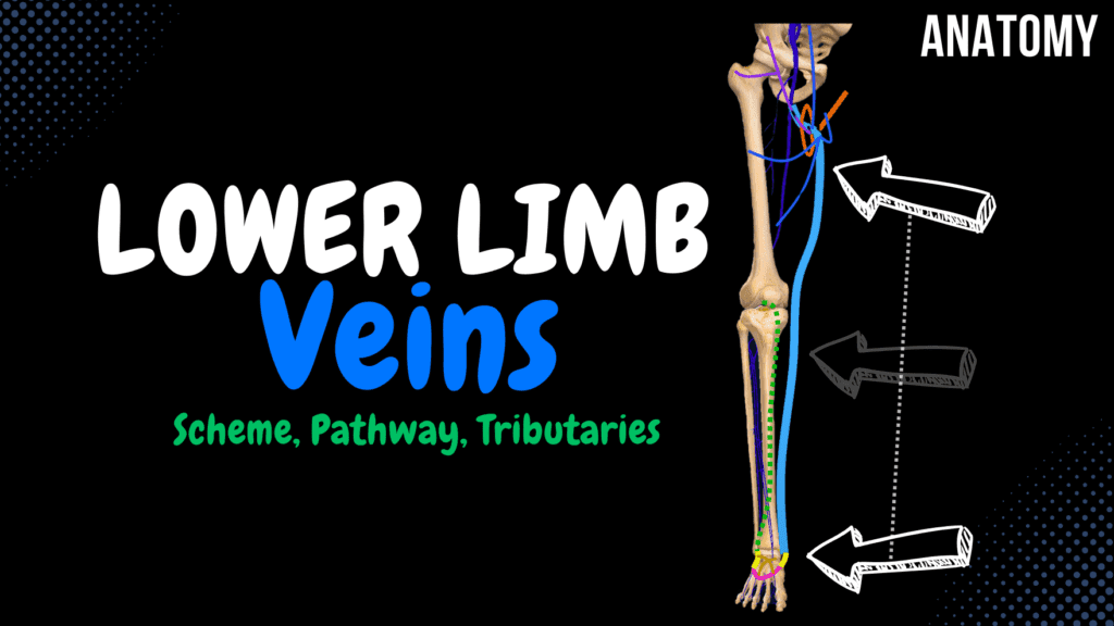
Veins of the Lower Limb Scheme (Division, Tributaries) Official Links Instagram Youtube Jki-discord Notes & Illustrations Quizzes Summary & Transcript Notes ☆ Members Only Go to PDF Notes Illustrations ☆ Members Only Go to Illustrations 12345678910 Lower Limb Veins – QUIZ Test your understanding with 10 random multiple-choice questions from the question bank. You're in the preview mode. Note: All elements work correctly on the front end. 1 / 10 Which perforating vein connects the medial malleolus to the posterior tibial veins? A) Popliteal perforators B) Medial malleolus perforators C) Dorsal perforators D) Calf perforators The perforating veins of the medial malleolus connect the great saphenous vein to the posterior tibial veins. 2 / 10 Which vein collects blood from the posterior thigh? A) Popliteal vein B) Small saphenous vein C) Great saphenous vein D) Deep vein of the thigh The deep vein of the thigh (profunda femoris vein) collects blood from the posterior thigh and drains into the femoral vein. 3 / 10 Which deep vein of the lower limb receives blood from the sole of the foot? A) Posterior tibial vein B) Fibular vein C) Plantar digital veins D) Dorsal venous arch The posterior tibial vein receives blood from the sole of the foot via the plantar veins. 4 / 10 Which vein drains the medial side of the thigh and leg? A) Great saphenous vein B) Small saphenous vein C) Posterior tibial vein D) Femoral vein The great saphenous vein drains the medial side of the thigh and leg, eventually emptying into the femoral vein. 5 / 10 Which vein drains the dorsal venous arch of the foot? A) Fibular vein B) Anterior tibial vein C) Plantar veins D) Great and small saphenous veins The great and small saphenous veins drain the dorsal venous arch of the foot. 6 / 10 Which venous condition involves dilated, tortuous superficial veins? A) Venous reflux B) Venous grafting C) Deep vein thrombosis D) Varicose veins Varicose veins occur due to valve insufficiency in superficial veins, leading to venous dilation. 7 / 10 Which vein originates from the medial marginal vein of the foot? A) Popliteal vein B) Great saphenous vein C) Fibular vein D) Small saphenous vein The great saphenous vein originates from the medial marginal vein of the foot and ascends along the medial leg. 8 / 10 Which vein forms the dorsal venous arch in the foot? A) Deep plantar veins B) Plantar venous arch C) Fibular veins D) Dorsal venous arch The dorsal venous arch is formed by the dorsal digital and metatarsal veins. 9 / 10 Which vein accompanies the fibular artery? A) Small saphenous vein B) Fibular vein C) Great saphenous vein D) Anterior tibial vein The fibular vein accompanies the fibular artery and drains into the posterior tibial vein. 10 / 10 Which vein drains the lateral dorsal part of the foot? A) Fibular vein B) Small saphenous vein C) Posterior tibial vein D) Great saphenous vein The small saphenous vein drains the lateral dorsal part of the foot. Your score is The average score is 0% Description This video provides a comprehensive overview of the veins of the lower limb, including their classification, deep and superficial systems, and key anatomical features. 1. Vein Classification Superficial System: Veins located in subcutaneous tissue; do not accompany arteries. Deep System: Veins accompanying arteries, usually in pairs. 2. Deep Veins of the Lower Limb Thigh: Femoral Vein: Originates from the external iliac vein and runs medially to the femoral artery. Deep Vein of the Thigh: Collects blood from the internal and posterior surfaces of the thigh. Popliteal Vein: Continuation of the femoral vein, located in the popliteal fossa. Leg: Anterior Tibial Vein: Originates from the popliteal vein and runs anteriorly. Posterior Tibial Vein: Also originates from the popliteal vein and runs posteriorly. Fibular Vein (Peroneal Vein): Descends behind the fibula and drains into the posterior tibial vein. 3. Veins of the Foot Dorsal Veins: Drain the dorsal side of the foot into the anterior tibial vein. Plantar Veins: Medial and Lateral Plantar Veins form the Deep Plantar Venous Arch. Deep Plantar Metatarsal and Digital Veins: Drain into the plantar venous arch. 4. Superficial Veins Great Saphenous Vein: Runs along the medial side of the leg and thigh, draining into the femoral vein. Small Saphenous Vein: Located at the posterior side of the leg and drains into the popliteal vein. Accessory Saphenous Veins: Tributaries that drain into the great saphenous vein. Superficial Epigastric Vein: Drains the anterior abdominal wall. Superficial Circumflex Iliac Vein: Drains the area around the anterior superior iliac spine. External Pudendal Vein: Drains external genitalia. 5. Dorsal Venous Network of the Foot A network of superficial veins that drains the dorsal side of the foot and connects to the great and small saphenous veins. 6. Veins Included in This Video Deep System of Veins: Femoral Vein Deep Vein of the Thigh Popliteal Vein Anterior Tibial Vein Posterior Tibial Vein Fibular Vein (Peroneal Vein) Dorsal Venous Arch of the Foot Dorsal Veins of the Foot Dorsal Digital Veins Dorsal Metatarsal Veins Medial Plantar Vein Lateral Plantar Vein Deep Plantar Venous Arch Deep Plantar Metatarsal Veins Deep Plantar Digital Veins Superficial Veins: Great Saphenous Vein Accessory Saphenous Veins Superficial Epigastric Vein Superficial Circumflex Iliac Vein External Pudendal Vein Small Saphenous Vein Dorsal Venous Network of the Foot Medial Marginal Vein Lateral Marginal Vein Sources Used: Memorix Anatomy (2nd Edition) – Hudák Radovan, Kachlík David, Volný Ondřej. Complete Anatomy by 3D4Medical. Biorender. University Notes and Lectures. Transcript Introduction0:00What’s up.0:03Meditay Here.0:04Let’s talk about the veins of the lower limb.Division0:07In order to structurize the veins of the lower limb, we generally divide them into 2 systems.0:13Simply, the superficial system and the deep system.0:16The deep system consists of veins that accompany arteries.0:20These are usually two veins, and they go together with the arteries.0:23The superficial system consists of veins in the subcutaneous tissue.0:28They’re unique for the venous system, so they don’t go with any arteries.0:32Simple, right?0:34Now let’s expand on this and
Iliac Veins
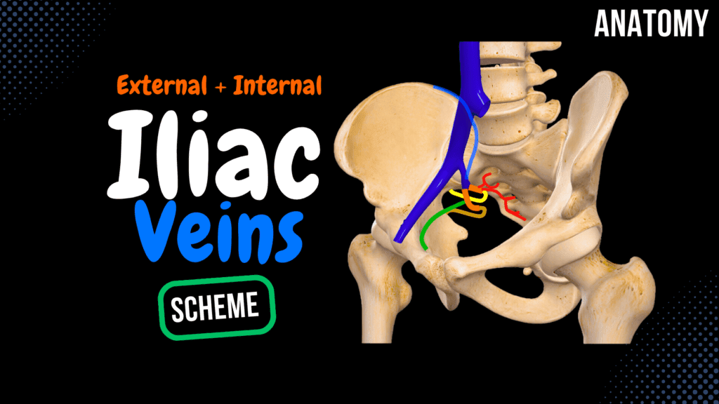
Iliac Veins Scheme (Visceral + Parietal Tributaries) Official Links Instagram Youtube Jki-discord Notes & Illustrations Quizzes Summary & Transcript Notes ☆ Member Only Go to PDF Notes Illustrations ☆ Member Only Go to Illustrations 12345678910 Iliac Veins – QUIZ Test your understanding with 10 random multiple-choice questions from the question bank. You're in the preview mode. Note: All elements work correctly on the front end. 1 / 10 Which visceral tributary of the internal iliac vein drains the uterus? A) Vaginal venous plexus B) Internal pudendal vein C) Vesical venous plexus D) Uterine venous plexus The uterine venous plexus drains the uterus into the internal iliac vein. 2 / 10 At what vertebral level do the common iliac veins unite to form the inferior vena cava? A) L3 B) L2 C) L4 D) L5 The common iliac veins unite to form the inferior vena cava at the L4 vertebral level. 3 / 10 What is the termination point of the common iliac veins? A) Median sacral vein B) Inferior vena cava C) External iliac vein D) Internal iliac vein The common iliac veins terminate at the inferior vena cava. 4 / 10 Which veins unite to form the common iliac vein? A) Obturator and iliolumbar veins B) External iliac and femoral veins C) Internal and external iliac veins D) Internal iliac and median sacral veins The internal and external iliac veins unite to form the common iliac vein. 5 / 10 Which vein drains the gluteus maximus and passes through the greater sciatic foramen? A) Superior gluteal vein B) Obturator vein C) Iliolumbar vein D) Inferior gluteal vein The inferior gluteal vein drains the gluteus maximus and passes through the greater sciatic foramen. 6 / 10 Which vein surrounds the prostate gland and communicates with the vesical venous plexus? A) Vaginal venous plexus B) Uterine venous plexus C) Vesical venous plexus D) Prostatic venous plexus The prostatic venous plexus surrounds the prostate gland and communicates with other pelvic venous plexuses. 7 / 10 Which tributary of the external iliac vein anastomoses with the superior epigastric vein? A) Median sacral vein B) Deep circumflex iliac vein C) Inferior epigastric vein D) Obturator vein The inferior epigastric vein anastomoses with the superior epigastric vein and drains into the external iliac vein. 8 / 10 Which parietal tributary of the internal iliac vein drains the sacral region? A) Inferior gluteal vein B) Superior gluteal vein C) Iliolumbar vein D) Lateral sacral vein The lateral sacral veins drain the sacral region and communicate with the vertebral venous plexus. 9 / 10 What forms the median sacral vein? A) Ascending lumbar vein B) Lateral sacral vein C) Iliolumbar vein D) Median sacral vein The median sacral vein originates from the posterior surface of the sacrum and terminates in the left common iliac vein. 10 / 10 Which tributary of the external iliac vein drains the anterior abdominal wall? A) Deep circumflex iliac vein B) Inferior epigastric vein C) Lateral sacral vein D) Superior gluteal vein The inferior epigastric vein drains the anterior abdominal wall and anastomoses with the superior epigastric vein. Your score is The average score is 0% Description This video provides an overview of the anatomy of the common, external, and internal iliac veins, their tributaries, and their roles in the vascular system. 1. Common Iliac Vein Formed by the confluence of the internal and external iliac veins at the level of L4-L5 vertebrae. No valves present in the common iliac veins. Tributaries: Ascending Lumbar Vein: Anastomoses with azygos and hemiazygos veins. Median Sacral Vein: Drains into the left common iliac vein. Iliolumbar Veins: May drain into the internal iliac vein. 2. External Iliac Vein Continuation of the common femoral vein starting at the inguinal ligament. Tributaries: Inferior Epigastric Vein: Drains the anterior abdominal wall, anastomoses with the superior epigastric vein. Deep Circumflex Iliac Veins: Collect blood from the iliac region. 3. Internal Iliac Vein Divided into two divisions with parietal and visceral tributaries. Parietal Tributaries: Superior and Inferior Gluteal Veins: Drain gluteal muscles. Obturator Vein: Drains medial thigh muscles. Lateral Sacral Veins: Form the sacral venous plexus. Visceral Tributaries: Rectal Venous Plexus: Drained by superior, middle, and inferior rectal veins. Vesical Venous Plexus: Drains the urinary bladder. Male: Fuses with the prostatic venous plexus. Female: Surrounds the bladder and drains the urethra. Uterine and Vaginal Venous Plexuses: Drain reproductive organs in females. Internal Pudendal Vein: Drains various structures near the pelvis. 4. Veins Included in This Video Common Iliac Vein External Iliac Vein Internal Iliac Vein Inferior Vena Cava Ascending Lumbar Vein Azygos Vein Hemiazygos Vein Median Sacral Vein Iliolumbar Veins Inguinal Ligament Inferior Epigastric Vein Superior Epigastric Vein Deep Circumflex Iliac Vein Superior Gluteal Vein Inferior Gluteal Vein Obturator Vein Lateral Sacral Vein Sacral Venous Plexus Rectal Venous Plexus Superior Rectal Vein Middle Rectal Vein Inferior Rectal Vein Internal Pudendal Vein Vesical Venous Plexus Prostatic Venous Plexus Deep Dorsal Vein of the Penis Dorsal Vein of the Clitoris Uterine Venous Plexus Vaginal Venous Plexus Sources Used: Memorix Anatomy (2nd Edition) – Hudák Radovan, Kachlík David, Volný Ondřej. Complete Anatomy by 3D4Medical. Biorender. University Notes and Lectures. Transcript Introduction0:02All right, guys, so now that we are done with the Inferior vena cava, let us go ahead and0:07cover the anatomy of the common, external, and internal iliac veins!Content0:11And we’re going to do that by breaking down their topography, visceral tributaries, and0:16parietal tributaries.0:17So, let us start with the common iliac vein.Common Iliac Vein0:20The common iliac veins originate at the confluence of the internal and external iliac veins;0:27confluence means where veins meet.0:29So, it is formed when these two veins meet.0:32And when the common iliac veins meet at the level of the vertebra L4-L5, they form the0:38Inferior Vena Cava.0:41And lastly, the common iliac veins have no valves.0:44You know I say this a lot, but usually, veins have valves to prevent the backflow of blood.0:50These large veins don’t have any valves.0:53Alright so the first tributary of the common iliac vein is the ascending lumbar vein.0:58The right
Inferior Vena Cava
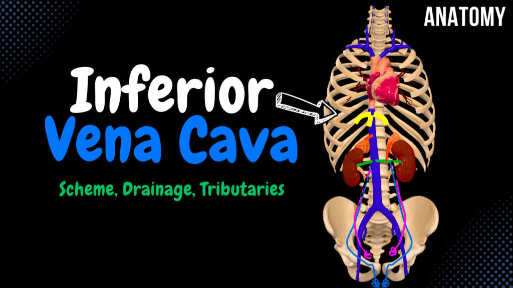
Inferior Vena Cava Scheme (Visceral + Parietal Tributaries) Official Links Instagram Youtube Jki-discord Notes & Illustrations Quizzes Summary & Transcript Notes ☆ Member Only Go to PDF Notes Illustrations ☆ Member Only Go to Illustrations 12345678910 Inferior Vena Cava – QUIZ Test your understanding with 10 random multiple-choice questions from the question bank. You're in the preview mode. Note: All elements work correctly on the front end. 1 / 10 What is the clinical significance of the hepatic veins? A) Regulate portal pressure B) Drain blood from the liver C) Form portocaval anastomoses D) Connect lumbar veins The hepatic veins drain the liver and serve as a key component in systemic venous circulation. 2 / 10 Which visceral tributaries drain into the left renal vein before reaching the IVC? A) Inferior phrenic veins B) Right gonadal veins C) Lumbar veins D) Left gonadal and left suprarenal veins The left suprarenal and left gonadal veins drain into the left renal vein before reaching the IVC. 3 / 10 Which vein drains the caudate lobe of the liver? A) Left hepatic vein B) Right hepatic vein C) Intermediate hepatic vein D) Portal vein The intermediate hepatic vein drains the caudate lobe of the liver into the IVC. 4 / 10 Which vein lies on the right side of the abdominal aorta? A) Hepatic vein B) Right renal vein C) Inferior vena cava D) Lumbar vein The inferior vena cava ascends on the right side of the abdominal aorta. 5 / 10 Which parietal tributaries communicate with the ascending lumbar veins? A) Lumbar veins B) Left renal veins C) Right hepatic veins D) Inferior phrenic veins The lumbar veins communicate with the ascending lumbar veins, forming connections with the azygos system. 6 / 10 What is the significance of the caval opening? A) Prevents IVC collapse B) Regulates blood flow C) Allows IVC passage through diaphragm D) Forms the azygos system The caval opening allows the IVC to pass through the diaphragm and into the thoracic cavity. 7 / 10 Which vein is the largest tributary of the inferior vena cava? A) Hepatic veins B) Renal veins C) Inferior phrenic veins D) Lumbar veins The hepatic veins are the largest tributaries of the inferior vena cava, draining blood from the liver. 8 / 10 Which vein passes posterior to the superior mesenteric artery? A) Right renal vein B) Left renal vein C) Left testicular vein D) Inferior phrenic vein The left renal vein passes posterior to the superior mesenteric artery and anterior to the abdominal aorta. 9 / 10 Which vein courses anterior to the abdominal aorta and posterior to the superior mesenteric artery? A) Left renal vein B) Hepatic vein C) Inferior phrenic vein D) Right renal vein The left renal vein courses anterior to the abdominal aorta and posterior to the superior mesenteric artery. 10 / 10 Which vein drains the left adrenal gland into the left renal vein? A) Left suprarenal vein B) Hepatic veins C) Right renal vein D) Lumbar veins The left suprarenal vein drains the left adrenal gland into the left renal vein. Your score is The average score is 0% Description This video covers the Inferior Vena Cava (IVC), its anatomical course, tributaries, and clinical significance. 1. Topography of the Inferior Vena Cava Largest vein in the human body. Drains blood from the lower half of the body. No valves. Lies on the right side of the abdominal aorta. Forms the right sagittal groove on the liver. Passes through the caval opening of the diaphragm at T8. 2. Visceral Tributaries Hepatic Veins (Venae Hepaticae): Right Hepatic Vein Intermediate Hepatic Vein Left Hepatic Vein Right Suprarenal Vein (Vena Suprarenalis Dextra) Left Renal Vein (Vena Renalis Sinistra) Left Suprarenal Vein Left Testicular Vein / Left Ovarian Vein Right Renal Vein (Vena Renalis Dexter) Right Testicular Vein / Right Ovarian Vein Genital tributaries drain the Pampiniform Plexus (Plexus Pampiniformis). 3. Parietal Tributaries Lumbar Veins (Venae Lumbales) Communicate with: External and Internal Vertebral Venous Plexuses Ascending Lumbar Veins (Vena Lumbalis Ascendens) Inferior Phrenic Veins (Venae Phrenicae Inferiores) 4. Clinical Relevance IVC Thrombosis: Can lead to lower limb edema and collateral circulation formation. IVC Compression: Common in pregnancy due to uterus expansion, leading to supine hypotension syndrome. Nutcracker Syndrome: Compression of the left renal vein between the aorta and superior mesenteric artery, causing hematuria and left flank pain. Sources Used: Memorix Anatomy (2nd Edition) – Hudák Radovan, Kachlík David, Volný Ondřej. Complete Anatomy by 3D4Medical. Biorender. University Notes and Lectures. Transcript Introduction0:03Now that we’re done with the superior vena cava,0:06let’s go ahead and cover the anatomy of the inferior vena cava0:09So in this video, we’re gonna cover topography. The visceral tributaries0:14And the parietal tributaries of it So let’s now start with the Topography.Topography of Inferior Vena Cava0:19The inferior Vena Cava is the largest vein in the human body.0:23It collects blood from the lower half of the body. So here’s the body.0:28Here’s the diaphragm And this is where this vein mainly drains.0:35And lastly, it has no valves. Veins usually have valves to prevent the backflow of blood.0:41The inferior vena cava, similar to some other large veins of the body, doesn’t have any valves.0:47Alright. So the inferior vena cava starts at the junction0:50of the right and left common iliac veins at the level of L5. So when the common iliac veins meet,0:57they form the inferior vena cava. It is runs on the right side of1:01the vertebral column, lying on the right of the abdominal aorta as you see here.1:07And if we add the liver. You’ll see that the Inferior vena cava forms a groove1:12on the posterior surface of the liver. And then it goes through the diaphragm1:17through the caval opening, together with the right phrenic nerve.1:21So that was it for the topography. Now finally Let’s go through the Visceral TributariesTributaries of Inferior Vena Cava1:28draining organs. And the Parietal Tributaries draining structures like bones and muscle.Visceral Tributaries1:33So, the first veins in our list that drain blood into the inferior vena cava are the
Veins of the Upper Limb
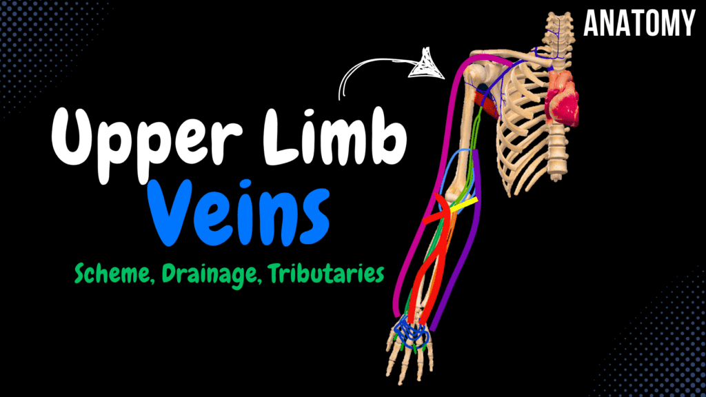
Veins of the Upper Limb (Subclavian, Axillary, Superficial and Deep Veins) Official Links Instagram Youtube Jki-discord Notes & Illustrations Quizzes Summary & Transcript Notes ☆ Member Only Go to PDF Notes Illustrations ☆ Member Only Go to Illustrations 12345678910 Veins of the Upper Limb – QUIZ Test your understanding with 10 random multiple-choice questions from the question bank. You're in the preview mode. Note: All elements work correctly on the front end. 1 / 10 Which vein connects the cephalic vein with the basilic vein at the wrist? A) Dorsal venous network B) Radial veins C) Intercapitular veins D) Median cubital vein The intercapitular veins connect the cephalic and basilic veins at the wrist. 2 / 10 Which vein connects the cephalic and basilic veins in the cubital fossa? A) Deep brachial vein B) Radial vein C) Median cubital vein D) Subclavian vein The median cubital vein connects the cephalic and basilic veins across the cubital fossa. 3 / 10 Which veins accompany the collateral arteries around the elbow? A) Collateral veins of the elbow B) Axillary vein C) Basilic vein D) Radial veins The collateral veins of the elbow accompany the collateral arteries around the elbow joint. 4 / 10 Which vein begins at the lower border of the teres major muscle? A) Axillary vein B) Subclavian vein C) Basilic vein D) Cephalic vein The axillary vein begins at the lower border of the teres major muscle, formed by the union of the brachial and basilic veins. 5 / 10 Which deep veins drain the anterior compartment of the arm? A) Ulnar veins B) Brachial veins C) Radial veins D) Basilic vein The brachial veins drain the anterior compartment of the arm and accompany the brachial artery. 6 / 10 Which veins form the deep system of veins in the forearm? A) Intercapitular veins B) Cephalic and basilic veins C) Radial and ulnar veins D) Median cubital vein The radial and ulnar veins form the deep system of veins in the forearm. 7 / 10 What is the clinical significance of the dorsal venous network? A) Deep venous drainage B) Collateral circulation C) Venous return D) Venous access The dorsal venous network serves as the origin for the cephalic and basilic veins and is commonly used for venous access. 8 / 10 What are venae comitantes? A) Superficial veins B) Connecting veins in the hand C) Paired veins accompanying arteries D) Tributaries of the axillary vein Venae comitantes are paired veins that accompany arteries, such as the brachial, radial, and ulnar veins. 9 / 10 What forms the dorsal venous network of the hand? A) Superficial veins of the hand B) Deep veins of the hand C) Intercapitular veins D) Median antebrachial vein The dorsal venous network is formed by superficial veins on the dorsum of the hand, including tributaries of the cephalic and basilic veins. 10 / 10 Which veins drain the medial forearm and join the brachial veins? A) Ulnar veins B) Median antebrachial vein C) Basilic vein D) Radial veins The ulnar veins drain the medial forearm and join the brachial veins in the cubital fossa. Your score is The average score is 0% Description This video covers the Venous Drainage of the Upper Limb, including the Subclavian Vein, Axillary Vein, Deep Venous System, and Superficial Venous System. 1. Subclavian Vein Passes within the groove for the subclavian vein (sulcus venae subclaviae). Receives blood from: Dorsal Scapular Vein (Vena Scapularis Dorsalis) External Jugular Vein (Vena Jugularis Externa) 2. Axillary Vein Begins at the lower border of the Teres Major Muscle (Musculus Teres Major). Receives tributaries from: Thoraco-Epigastric Vein (Venae Thoracoepigastricae) Lateral Thoracic Vein (Vena Thoracica Lateralis) Anterior Circumflex Humeral Vein (Vena Circumflexa Humeri Anterior) Posterior Circumflex Humeral Vein (Vena Circumflexa Humeri Posterior) Cephalic Vein (Vena Cephalica) 3. Deep Venous System Follows the course of arteries and runs as paired veins until the axilla. Brachial Veins (Venae Brachiales) Ulnar Veins (Venae Ulnares) Collateral Veins of the Elbow (Venae Collaterale Articulationis Cubiti) Deep Brachial Veins Superior Ulnar Collateral Vein Inferior Ulnar Collateral Vein Radial Recurrent Vein Radial Veins (Venae Radiales) Deep Venous Palmar Arch (Arcus Venosus Palmaris Profundus) 4. Superficial Venous System Basilic Vein (Vena Basilica) Cephalic Vein (Vena Cephalica) Median Cubital Vein (Vena Mediana Cubiti) – Common site for venipuncture. Median Antebrachial Vein (Vena Mediana Antebrachii) Intercapitular Vein (Venae Intercapitulares) Dorsal Venous Network of the Hand (Rete Venosum Dorsale Manus) Clinical Relevance: Venipuncture: The median cubital vein is the preferred site for drawing blood. Deep Vein Thrombosis (DVT): Can occur in the deep venous system, affecting the brachial, axillary, or subclavian veins. Subclavian Vein Compression: In thoracic outlet syndrome, the subclavian vein can be compressed, causing venous congestion in the upper limb. Sources Used: Memorix Anatomy (2nd Edition) – Hudák Radovan, Kachlík David, Volný Ondřej. Complete Anatomy by 3D4Medical. Biorender. University Notes and Lectures. Transcript Introduction0:03In the last video, we covered the tributaries of the superior vena cava and the brachiocephalic0:08veins.0:09Now let’s go ahead and follow these veins as we covert eh veins of the upper limb.0:14So in this video, we’re gonna gover the veins of the upper limb by first going through the0:19Subclavian Vein Then the Axillary Vein0:22And then covert he deep system of veins in the arm,0:25Followed by the superficial system of veins in the arm!Veins of the Systemic Circulation0:28Awesome So, the easiest way to understand the veins0:31of the systemic circulation is to divide them into their own systems.0:35So the veins of the heart form their own system We have the Veins of the Inferior Vena Cava,0:40which is responsible for supplying the lower half of the body0:44Veins of the superior vena cava for the upper half of the body0:48And the portal system, which drains nutrients from the intestines0:51and waste products from the spleen and dumps them into the liver to be processed,0:55which then lead the blood into the inferior vena cava again.0:58So these are the 4 main systems of veins we have in our bodies.Subclavian Vein1:02Let’s now talk briefly about the venous drainage
Cerebral Veins
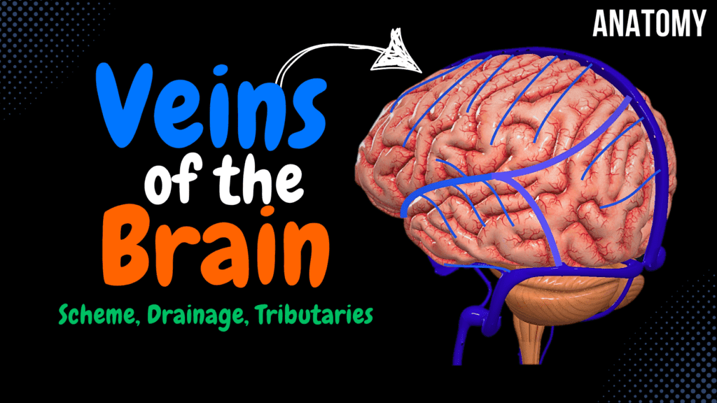
Cerebral Veins (Deep & Superficial + Diploic and Emissary Veins) Official Links Instagram Youtube Jki-discord Notes & Illustrations Quizzes Summary & Transcript Notes ☆ Members Only Go to PDF Notes Illustrations ☆ Members Only Go to Illustrations 12345678910 Cerebral Veins – QUIZ Test your understanding with 10 random multiple-choice questions from the question bank. You're in the preview mode. Note: All elements work correctly on the front end. 1 / 10 What is the function of the inferior cerebral veins? A) Drain superior hemispheres B) Drain lower hemispheres into transverse and cavernous sinuses C) Regulate CSF flow D) Facilitate venous outflow The inferior cerebral veins drain the lower surfaces of the cerebral hemispheres into the transverse and cavernous sinuses. 2 / 10 Which superficial cerebral veins primarily drain into the superior sagittal sinus? A) Inferior cerebral veins B) Superficial middle cerebral vein C) Diploic veins D) Superior cerebral veins The superior cerebral veins primarily drain into the superior sagittal sinus. 3 / 10 Which emissary vein connects the mastoid region with the sigmoid sinus? A) Condylar emissary vein B) Mastoid emissary vein C) Parietal emissary vein D) Occipital emissary vein The mastoid emissary vein (vena emissaria mastoidea) connects the mastoid region with the sigmoid sinus. 4 / 10 What is the role of diploic veins? A) Connect dural sinuses with extracranial veins B) Drain the diploe of the skull into venous sinuses C) Provide CSF reabsorption D) Drain the diencephalon Diploic veins lie within the diploe of the skull and drain into the venous sinuses. 5 / 10 Which vein connects the frontal and temporal regions to the superior sagittal sinus? A) Basal vein B) Diploic vein C) Inferior anastomotic vein of Labbe D) Superior anastomotic vein of Trolard The superior anastomotic vein of Trolard connects the frontal and temporal regions to the superior sagittal sinus. 6 / 10 What do the deep cerebral veins drain? A) Cerebral cortex B) Subarachnoid space C) Venous sinuses D) Diencephalon and deep hemispheres The deep cerebral veins drain the diencephalon and deep parts of the cerebral hemispheres. 7 / 10 Which cerebral vein drains the superior temporal gyrus? A) Superior cerebral vein B) Inferior cerebral vein C) Basal vein D) Superficial middle cerebral vein The superficial middle cerebral vein drains the superior temporal gyrus into the cavernous sinus. 8 / 10 What is the primary drainage pathway for the inferior cerebral veins? A) Transverse and cavernous sinuses B) Straight sinus C) Superior sagittal sinus D) Great cerebral vein The inferior cerebral veins primarily drain into the transverse and cavernous sinuses. 9 / 10 Which emissary vein connects the posterior cranial fossa to the sigmoid sinus? A) Occipital emissary vein B) Condylar emissary vein C) Mastoid emissary vein D) Diploic vein The condylar emissary vein connects the posterior cranial fossa to the sigmoid sinus. 10 / 10 Which deep vein runs below the hypothalamus and drains into the great cerebral vein? A) Superior cerebral vein B) Basal vein C) Superior choroid vein D) Internal cerebral vein The basal vein runs below the hypothalamus and drains into the great cerebral vein. Your score is The average score is 0% Description This video covers the Cerebral Veins, including their classification, course, and clinical significance. Key Characteristics of Cerebral Veins: No valves. Lie in the subarachnoid space. Drain into the venous sinuses. Divided into: Deep Cerebral Veins: Drain the diencephalon and deep parts of the hemispheres. Superficial Cerebral Veins: Drain the cerebral cortex. Deep Cerebral Veins: Drain blood from the diencephalon and deep structures of the hemispheres into the Great Cerebral Vein (Vena Magna Cerebri). Great Cerebral Vein (Vena Magna Cerebri): Main deep venous drainage of the brain. Basal Vein (Vena Basalis): Runs underneath the hypothalamus, formed by small anterior cerebral veins. Internal Cerebral Vein (Vena Interna Cerebri): Drains deep structures of the brain. Superior Thalamostriate Vein (Vena Thalamostriata Superior): Drains the thalamus and striatum. Superior Choroid Vein: Drains the choroid plexus. Anterior Vein of Septum Pellucidum: Drains the septum pellucidum. Superficial Cerebral Veins: Collect blood from the cerebral cortex and drain into the venous sinuses. Superficial Middle Cerebral Vein (Vena Cerebri Media Superficialis): Drains the frontal, temporal, and parietal lobes. Superior Anastomotic Vein of Trolard (Vena Anastomotica Superior): Connects the superficial middle cerebral vein with the superior sagittal sinus. Inferior Anastomotic Vein of Labbé (Vena Anastomotica Inferior): Connects the superficial middle cerebral vein with the transverse sinus. Superior Cerebral Veins (Venae Cerebri Superiores): Drain into the superior sagittal sinus. Inferior Cerebral Veins (Venae Cerebri Inferiores): Drain into the transverse and cavernous sinuses. Superior and Inferior Cerebellar Veins (Venae Superiores et Inferiores Cerebelli): Drain the cerebellum. Diploic Veins: Located within the diploë (spongy bone of the skull), these veins drain blood into the dural venous sinuses. Emissary Veins: These veins connect the dural venous sinuses with the extracranial veins, forming Cranio-Cerebral Anastomoses. Parietal Emissary Vein (Vena Emissaria Parietalis) Mastoid Emissary Vein (Vena Emissaria Mastoidea) Occipital Emissary Vein (Vena Emissaria Occipitalis) Condylar Emissary Vein (Vena Emissaria Condylaris) Clinical Relevance: Cavernous Sinus Thrombosis: Infection from the face (danger triangle) can spread to the cavernous sinus via emissary veins. Subdural Hematoma: Rupture of superficial cerebral veins leads to bleeding into the subdural space. Stroke: Blockage of deep cerebral veins can result in venous infarction. Sources Used: Memorix Anatomy (2nd Edition) – Hudák Radovan, Kachlík David, Volný Ondřej. Complete Anatomy by 3D4Medical. Biorender. University Notes and Lectures. Transcript Introduction0:03In the last video, we covered the anatomy of the dural sinuses;0:06Now let’s go ahead and cover the rest of the veins you’ll find supplying the structures0:10of the brain.0:11So, In this video, we’re first going to talk about the cerebral veins0:15Which include the deep cerebral veins and the superficial cerebral veins.0:20Then we’ll talk briefly about the diploic veins and then the emissary’s veins.0:25And again, in the previous video, we talked about the dural sinuses; so if that topic0:29is unfamiliar to you I’ll put a link to a playlist that covers0:33the venous drainage of different parts of our bodies.Veins of the Systemic
Dural Venous Sinuses
Dural Venous Sinuses (Location + Visual Scheme) Official Links Instagram Youtube Jki-discord Notes & Illustrations Quizzes Summary & Transcript Notes ☆ Member Only Go to PDF Notes Illustrations ☆ Member Only Go to Illustrations 12345678910 Dural Venous Sinuses – QUIZ Test your understanding with 10 random multiple-choice questions from the question bank. You're in the preview mode. Note: All elements work correctly on the front end. 1 / 10 Which sinus runs along the inferior free edge of the falx cerebri? A) Inferior sagittal sinus B) Cavernous sinus C) Superior sagittal sinus D) Sigmoid sinus The inferior sagittal sinus is located along the inferior free edge of the falx cerebri. 2 / 10 Which sinus receives blood from the superior sagittal sinus and the straight sinus? A) Sigmoid sinus B) Transverse sinus C) Cavernous sinus D) Inferior petrosal sinus The transverse sinus receives blood from the superior sagittal and straight sinuses. 3 / 10 Which sinus lies along the petrous ridge of the temporal bone? A) Inferior petrosal sinus B) Sigmoid sinus C) Straight sinus D) Superior petrosal sinus The superior petrosal sinus lies along the petrous ridge and drains into the transverse sinus. 4 / 10 What is the function of the arachnoid granulations in the superior sagittal sinus? A) Reabsorb CSF B) Collect blood from emissary veins C) Facilitate venous outflow D) Drain the cerebellum Arachnoid granulations reabsorb cerebrospinal fluid (CSF) into the venous system. 5 / 10 What structures run within the cavernous sinus? A) Superior petrosal sinus B) Internal carotid artery and cranial nerves C) Great cerebral vein D) Inferior petrosal sinus The cavernous sinus contains the internal carotid artery and cranial nerves III, IV, V1, V2, and VI. 6 / 10 What is the main drainage pathway for the occipital sinus? A) Confluence of sinuses B) Sigmoid sinus C) Cavernous sinus D) Transverse sinus The occipital sinus drains into the confluence of sinuses. 7 / 10 Which sinus is associated with the diaphragma sellae? A) Transverse sinus B) Superior sagittal sinus C) Sigmoid sinus D) Cavernous sinus The cavernous sinus is associated with the diaphragma sellae and surrounds the pituitary gland. 8 / 10 What is the primary tributary of the straight sinus? A) Great cerebral vein B) Transverse sinus C) Superior sagittal sinus D) Cavernous sinus The straight sinus receives blood from the inferior sagittal sinus and the great cerebral vein. 9 / 10 What is the primary tributary of the cavernous sinus? A) Superior ophthalmic vein B) Basilar plexus C) Transverse sinus D) Inferior petrosal sinus The superior ophthalmic vein is the primary tributary of the cavernous sinus. 10 / 10 Which sinus is located on either side of the sella turcica? A) Cavernous sinus B) Transverse sinus C) Superior sagittal sinus D) Inferior petrosal sinus The cavernous sinus is located on either side of the sella turcica and surrounds the internal carotid artery and cranial nerves. Your score is The average score is 0% Description This video covers the Veins of the Systemic Circulation, including: Veins of the Superior Vena Cava Veins of the Inferior Vena Cava Veins of the Heart The Portal System Topography of the Dural Venous Sinuses The Dural Venous Sinuses are located between the Periosteal and Meningeal layers of the Dura Mater. They drain venous blood from the brain into the Internal Jugular Veins. Classification of Dural Venous Sinuses: Midline Sinuses: Superior Sagittal Sinus (Sinus Sagittalis Superior): Drains into the confluence of sinuses. Inferior Sagittal Sinus (Sinus Sagittalis Inferior): Runs along the lower border of the falx cerebri and drains into the straight sinus. Straight Sinus (Sinus Rectus): Formed by the union of the inferior sagittal sinus and the great cerebral vein (Vein of Galen). Anterior Intercavernous Sinus (Sinus Intercavernosus Anterior): Connects the two cavernous sinuses anteriorly. Posterior Intercavernous Sinus (Sinus Intercavernosus Posterior): Connects the two cavernous sinuses posteriorly. Basilar Plexus (Plexus Basilaris): Communicates with the internal vertebral venous plexus, allowing venous drainage between the cranial cavity and vertebral column. Lateral Sinuses: Transverse Sinus (Sinus Transversus): Drains the confluence of sinuses into the sigmoid sinus. Sigmoid Sinus (Sinus Sigmoideus): Continuation of the transverse sinus that empties into the internal jugular vein. Superior Petrosal Sinus (Sinus Petrosus Superior): Drains the cavernous sinus into the transverse sinus. Inferior Petrosal Sinus (Sinus Petrosus Inferior): Drains the cavernous sinus into the internal jugular vein. Posterior Sinuses: Occipital Sinus (Sinus Occipitalis): Located along the margin of the foramen magnum, drains into the confluence of sinuses. Confluence of Sinuses (Torcular Herophili): Meeting point of the superior sagittal sinus, straight sinus, and occipital sinus. Anterior Sinuses: Cavernous Sinus (Sinus Cavernosus): Large venous plexus around the pituitary gland that communicates with the facial vein via the ophthalmic veins. Sphenoparietal Sinus (Sinus Sphenoparietalis): Drains the superficial middle cerebral vein into the cavernous sinus. Clinical Relevance: Cavernous Sinus Thrombosis: Infection from the face (danger triangle) can spread to the cavernous sinus via the ophthalmic veins, leading to severe complications. Venous Sinus Thrombosis: Can occur in conditions like hypercoagulability disorders, causing headaches, seizures, and stroke-like symptoms. Basilar Plexus and Vertebral Veins: Provide a route for metastases to spread between the brain and lower body. Sources Used: Memorix Anatomy (2nd Edition) – Hudák Radovan, Kachlík David, Volný Ondřej. Complete Anatomy by 3D4Medical. Biorender. University Notes and Lectures. Transcript Introduction0:00[Music]0:08let’s go ahead and talk about the0:09anatomy of the dural venous sinuses and0:12we’re going to do that by first going0:14through the location of the dural0:16sinuses basically where they are in0:18comparison to the meninges0:20and then we’re going to visualize the0:22dural venous sinuses schematically so0:25that it’s easier to remember the dural0:27sciences and then in the next video0:29we’re going to go through the cerebral0:31veins and the emissary veins and the0:33diploic veins and then the cerebral0:36circulation in generalSystemic Veins0:37all right0:38so the veins of the systemic circulation0:41consist of four kind of systems0:44you have the veins of the heart0:46then you have the veins of the inferior0:48vena cava0:49and the veins of the superior vena cava0:52and then you have the portal system0:54so the superior venous sinuses we’re0:57going to